




Volume 210 - Issue 1
Radiotherapy and immunotherapy: a synergistic effect in cancer care
Authors: Guy‐Anne Turgeon, Andrew Weickhardt, Arun A Azad, Benjamin Solomon and Shankar Siva
Med J Aust 2019; 210 (1): 47-53. || doi: 10.5694/mja2.12046
Published online: 14 January 2019
Published online: 14 January 2019
At present, high level evidence for the safety and efficacy of radiotherapy and immunotherapy still need to be demonstrated clinically; therefore, the combination cannot be recommended outside of clinical trials. It is also currently unclear which patients will benefit from the combination. However, due to the increased number of clinical trials investigating radiotherapy as a mean to antitumour immunity, it is likely that the evidence will emerge shortly.40,41 In 2016, a study presented a list of 93 active trials combining radiotherapy with an antibody inhibiting CTLA-4, PD-1 and PD-L1, transforming growth factor β-cytokine, or other immune molecules.40 ClinicalTrials.gov now reports at least 200 completed or recruiting trials evaluating the new combination. Box 1 lists investigator-initiated clinical trials combining radiotherapy and immunotherapy in Australia. If positive, the results of these trials are likely to lead to a paradigm shift in the use of radiotherapy, which would certainly be a cost-effective and low toxicity mean to enhance the response to immune agents in addition to its well established role of killing tumour cells.
Trial name
(trial registration no.)
Disease
(stage)
SABRSeq
(NCT03307759)
- 18-20 Gy/1 fraction to 1-3 metastases, 7 days before ICIs
- 18-20 Gy/1 fraction to 1-3 metastases, 7 days after ICIs
RAPPORT
(NCT02855203)
- 18-20 Gy/1 fraction to 1-5 metastases, 5 days before ICIs
BOSTON-II (closed)
(NCT02303366)
- 20 Gy/1 fraction to 1-5 metastases
AZTEC
(TROG 17.05)
- 20 Gy/1 fraction to 1-4 metastases
- 24 Gy/3 fractions to 1-4 metastases
- Delivered before ICIs
ICE-PAC
(ACTRN12618000954224)
NIVORAD (TROG 16.01) SABR-IMPACT I (ACTRN12616001064493) (U1111-1185-5624) PCR-MIB (NCT02662062) ABC-X Study (NCT03340129) Ave-Rec (NCT03299660)
ICIs = immune checkpoint inhibitors. 5FU = fluorouracil. mCRPC = metastatic castration-resistant prostate cancer. NSCLC = non-small cell lung cancer. PFS = progression-free-survival. RCC = renal cell carcinoma. rPFS = radiological progression-free survival. SABR = stereotactic ablative radiotherapy. TURBT = transurethral resection of bladder tumour. ◆
Much needs to be understood to optimise the complex immunological effect of radiotherapy and its interaction with ICIs. Histological subtype, target organ (bone, visceral, brain), immunotherapy molecule, volume of irradiation, radiotherapy dose and sequence are hypothesised to have different effects. Radiotherapy pulsing, in which doses of radiation are delivered regularly (eg, monthly) throughout the ICI therapy, has also been proposed.64In conclusion, ICIs result in impressive clinical responses in select groups of patients, but optimal results in larger populations will require combination with other therapies. Radiotherapy will certainly be part of this arsenal, but fundamental questions about mechanisms of treatment non-redundancy, tumour resistance and patients’ tolerance are to be answered. Preclinical studies to direct informed clinical trial design are needed to determine how best to integrate these modalities and optimise their synergistic effect.
Summary
- Radiotherapy is an effective treatment modality commonly used in efforts to cure many localised cancers and in the palliation of symptoms in metastatic cancers.
- Immunotherapy has revolutionised cancer care by increasing the disease control and overall survival of patients in several cancer types; however, the majority of patients do not respond to currently available therapies based on immune checkpoint inhibitors (ICIs). The benefit of those agents is limited to patients who have a pre‐existing active immune microenvironment that can be reactivated by ICIs.
- It is now recognised that radiotherapy does not only directly kill tumour cells but it also changes the tumour microenvironment, enhancing tumour cell recognition by the immune system and, therefore, acting as an in situ vaccine.
- Radiotherapy increases expression of tumour‐associated antigens, causes the release of cytokines, stimulates recruitment of dendritic cells and, most importantly, stimulates the proliferation and priming of cytotoxic CD8+ T cells in the tumour microenvironment. This immunological cascade specifically generates activated T cells able to induce immunogenic cell death directed against cancer cells bearing those antigens.
- By its ability to overcome some tumour immune escape mechanisms, radiation provides a non‐pharmacological and cost‐effective approach to potentially improve the systemic response to immune checkpoints inhibitors.
In Australia, 50% of patients diagnosed with cancer will receive radiotherapy during their disease course, either for curative or palliative intent.1 The effectiveness of radiotherapy has traditionally been solely explained by the irreparable DNA damage to tumour cells targeted within the treated volume, resulting in cell death or loss of replicative potential. However, a growing body of preclinical and clinical data suggests that the immune system is also a critical determinant of response to radiotherapy and that a combination of radiotherapy and immunotherapy may be synergistic.
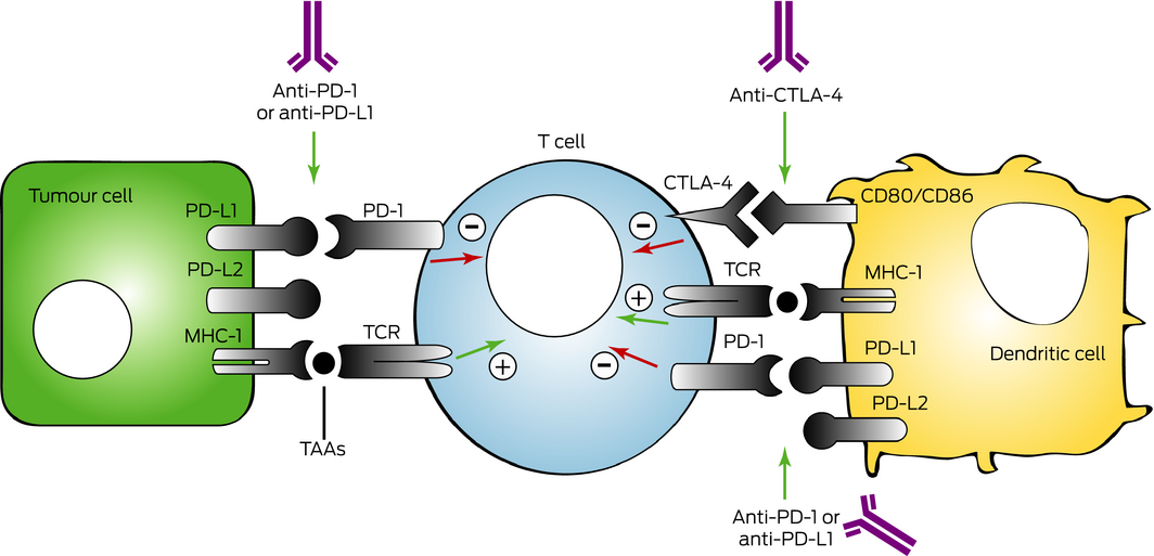
In the past 5 years, immunological approaches to cancer therapy using immune checkpoint inhibitors (ICIs) have revolutionised cancer care in a range of malignancies such as metastatic melanoma, renal cell carcinoma, non‐small cell lung cancer (NSCLC), Merkel cell carcinoma, Hodgkin lymphoma and head, neck and bladder cancers.2,3,4,5,6,7,8,9,10,11 These ICIs — in the form of antibodies to checkpoint proteins involved in self‐regulation of the immune response to cancer (Box 1), such as the cytotoxic T‐lymphocyte‐associated protein 4 (CTLA‐4), the programmed cell death protein 1 (PD‐1) or its main ligand PD‐L1 — have led to significant improvements in outcomes. The addition of local radiation to immunotherapy has the potential to enhance the efficacy of ICIs by upregulating the tumour neoantigen expression and enhancing tumour cell recognition by the immune system. This combination may provide a non‐pharmacological, low toxicity and cost‐effective approach to increase the systemic response and maximise the death of tumour cells.
For this review, we performed a search in MEDLINE and PubMed of original research, review articles and meta‐analyses relevant to the combination of radiotherapy and immunotherapy published between January 2000 and June 2018.
Cancer development: tumour and host immune interaction
The immune system is the first line of defence against cancer. As pathological agents, cancer cells express antigens distinguishing them from normal tissue and marking them for elimination.12 The dynamic states between the tumour cells and the host's immune system — in which the tumour can be directly eliminated, maintained in a state of equilibrium, or escape — are called immunosurveillance and immunoediting.12
During neoplastic transformation, before the adaptive immunity is elicited, the innate immune system recognises tissue changes leading to the production of cytokines, which are key mediators of immunosurveillance. The cellular damage caused by this innate activity then releases tumour‐associated antigens. These antigens are presented to dendritic cells, which process the antigens into peptides that can be loaded into their surface major histocompatibility complex (MHC) class 1 and 2 molecules for recognition by the adaptive immune T cells, CD8+ and CD4+ respectively.12 The action of these innate and acquired immune effectors leads to the destruction of the tumour — the killing being mainly caused by the actions of tumour‐specific effector T cells.12 However, if the tumour elimination is incomplete and the equilibrium is lost, cellular adaptation through clonal evolution enables immune evasion. This resistance to immune rejection is recognised as a requirement for immune escape and allows the tumour to grow and become clinically detectable.13
The clinical importance of effector T cells is demonstrated by the presence of tumour‐infiltrating lymphocytes and their relation to outcome. Greater tumour‐infiltrating lymphocytes correlate with better survival in many types of cancers.14,15,16 This finding supports the hypothesis that strategies to increase T cell infiltrates can be an immunogenic way to offer opportunities to recover effective immune reactivity.
Radiation as a vaccine: clinical anecdotes and biological mechanisms
Mitotic catastrophe is well known to be the main mechanism of tumour cell lethality following conventionally fractionated radiotherapy, while apoptosis and necrosis are observed after a higher dose per fraction. In addition, a frequently overlooked byproduct of this radiation damage to the tumour cells is the release of a large amount of tumour‐associated antigens, leading to the potential of radiotherapy to act as an in situ vaccine. This potential is clinically supported by the rare phenomenon of the “abscopal effect”, observed when ionising radiation is administered alone.17 The abscopal effect — first described in 1953,18 derived from the Latin “ab” (away from) and “scopus” (target) — defines the regression or disappearance of a tumour outside of the radiation field but within the same organism. An example of this effect was published in 2012,19 in which a patient treated with ipilimumab for metastatic melanoma had very slow progression on the immunotherapy agent. After 16 months of treatment, a paraspinal lesion causing back pain was treated with radiotherapy (three daily treatments of 9.5 Gy) and the ipilimumab was continued. Three months after radiotherapy, the irradiated lesion had regressed significantly as well as other metastatic sites away from the radiation field. This response persisted for at least 10 months and was supported by plasma immunological markers. It is interesting that, with the advent of immunotherapy, this phenomenon has been increasingly observed in patients and in preclinical models.20,21,22,23
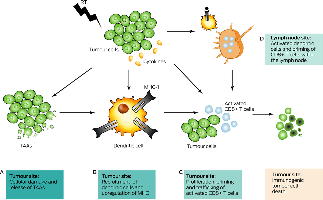
Pathophysiologically, the effect of ionising radiation on the tumour microenvironment can be summarised as follows (Box 2):
-
radiotherapy causes tumour cell damage, resulting in a microenvironment replete with necrotic and apoptotic tumour cells and debris referred to as tumour‐associated antigens; these antigens lead to the generation of activated T cells specifically able to induce immunogenic cell death directed against the tumour bearing those antigens;24
-
radiotherapy promotes antigen recognition by activating the dendritic cells25 and stimulating the MHC‐1 molecules,26,27 leading to improved tumour‐directed cell kill by the CD8+ T cells;25,27
-
radiotherapy promotes the release of chemokines that attract activated T cells and enhance the trafficking of antitumour immune effector cells to the tumour site;24,28 and
-
radiotherapy increases the number of activated antigen‐presenting cells (mainly dendritic cells) carrying tumour‐derived peptides within tumour draining lymph nodes.24
However, the host response to radiation is complex. Radiation can also activate immunosuppressive components as the transforming growth factor β‐cytokine and promote the accumulation of regulatory T cells and pro‐tumorigenic macrophages.29 It is believed that, overall, the pro‐immunogenic effects of radiotherapy outweigh the immunosuppressive effects, but further studies are necessary to improve our understanding and predict and optimise this treatment strategy. Nevertheless, radiotherapy has certainly the ability to overcome some immunosuppressive mechanisms, potentially improving the systemic response of immunotherapy agents.
Immunotherapy: a revolution in oncology
The immune system is poised delicately in its role of surveillance and subsequent destruction for foreign versus problematic overactivation and subsequent autoimmunity. There are several key autoregulatory checkpoints that serve to prevent the overactivation of the immune system when foreign antigens are detected. Examples of key immune checkpoints include the CTLA‐4 protein and PD‐1 and its ligand PD‐L1, which are negative regulators of T cell immune function,30 acting differently within the immune system. CTLA‐4 stops autoreactive T cells at the initial stage of naive T cell activation, typically in lymph nodes, whereas the PD‐1 pathway suppresses previously activated T cells at the later stages of an immune response, primarily in peripheral tissues. In other words, CTLA‐4 and PD‐1 promote self‐tolerance and prevent autoimmune diseases, but they can also prevent the immune system from killing cancer cells. Cancer cells may overexpress ligands for these proteins, in particular PD‐L1 (Box 1) interacting with PD‐1, as well as other proteins that allow the cancer cell to avoid host immune checkpoints and surveillance as a means to promote their continued survival. The success of immunotherapies results in the inhibition of these targets leading to an effect of increased activation of the immune system.
Long term analysis of survival data across several CTLA‐4 inhibitor phase 2 and phase 3 trials in melanoma showed that the survival curve begins to plateau at about 3 years, with 3‐year survival rates of 22% indicating the presence of long term responders to treatment.6 The PD‐1 pathway blockade has also impressive clinical trial results, with 10–40% response rates to single agent PD‐1/PD‐L1 inhibitors in a broad range of tumours, including melanoma, renal cell carcinoma, bladder and NSCLC, with 20–40% of the responding patients developing durable control of their cancer and superiority over alternative therapies.2,3,4,5,6,7 Furthermore, the effects of the CTLA‐4 and PD‐1 antibodies do not appear to be redundant and, in some cancers, their combination has already shown clinical superiority to single agents.31,32
However, the benefit of these agents seems limited to a minority of cancers that have a high tumour mutational burden or to cancers with pre‐existing T cell infiltrate that can be reactivated by ICIs. For the majority of patients, additional interventions are needed to overcome primary or acquired resistance to ICIs, and there is an urgent need to increase responses in this group of patients.
Predictive biomarkers of response to immunotherapy
Methods to identify prospectively patients who are more likely to benefit from ICIs are presently suboptimal. For CTLA‐4, the low level of inducible CTLA‐4 expression and the widespread expression of its ligands (mainly CD80 and CD86) are not useful as predictive biomarkers. An association between melanoma mutational load and clinical benefit with CTLA‐4 blockade has been shown, but it was insufficient alone to predict which patients are likely to respond to treatment.33 The identification of a neoantigen signature present in the tumour shows promise, but much work is still to be done.33
For anti‐PD‐1/PD‐L1 antibodies, the molecular factors and receptors necessary to make a tumour responsive to treatment also remains unclear. The protein expression of PD‐L1 has been widely examined as a potential biomarker and has been associated with more frequent responses to PD‐1 and PD‐L1 inhibitors.7,34 However, patients with PD‐L1‐negative tumours also respond to anti‐PD‐1/PD‐L1 antibodies, indicating that PD‐L1 expression in itself is not an absolute determinant of the effectiveness of therapy and, thus, it cannot be used to exclude patients from treatment.3,11 In addition, variations in PD‐L1 immunohistochemistry methodology and scoring have led to discrepant results.35 Upregulation of PD‐L1 not only on tumour cells but on tumour‐infiltrating immune cells may identify additional patients responsive to PD‐1 and PD‐L1 blockade.7,34 Similar to CTLA‐4 blockade, a higher mutational burden in the tumour (eg, melanoma, bladder and gastric cancers) has been correlated with a greater effect of anti‐PD‐1 antibodies treatment.36 Other factors also appear important and could be used as biomarkers, such as mismatch‐repair defects, interferon‐γ‐related gene expression signatures, and presence of tumour‐infiltrating lymphocytes34 or CD8+ T cells.7
Radiation and immunotherapy: preclinical evidence
Strategies to increase exposure of tumour antigens and upregulation of PD‐L1 are conceptually advantageous in combination with ICIs. One such strategy is the use of ionising radiation. In 2005, early evidence showed that local radiation can synergise with anti‐CTLA‐4 antibody to achieve tumour rejection using a breast carcinoma murine model.37 The study also found that the response of the combined treatment was mediated by CD8+ T cells. Following these findings, a number of preclinical studies showed that radiotherapy delivered alone upregulated tumour PD‐L1 expression, while the combination of radiotherapy and ICIs enhanced the antitumour effect of radiation consistent with the synergistic effect of both modalities.38,39 These preclinical data together with clinical observations of abscopal effects19,20,21,22,23 have led to several clinical trials evaluating the addition of radiotherapy to ICIs with the objective to improve tumour response, progression‐free survival and, ultimately, overall survival.40,41
Radiation and immunotherapy: clinical evidence
Radiotherapy and CTLA‐4 inhibitor
Case reports and small case series of patients treated with radiotherapy and ipilimumab reported striking examples of abscopal synergistic effects, with increased response rates and shrinkage of metastases compared with patients treated with ipilimumab alone.19,20,21,22,23
A preliminary analysis of a phase 2 trial in NSCLC (ClinicalTrials.gov identifier: NCT02221739) evaluating the combination of ipilimumab with radiotherapy reported that seven of 12 evaluable patients (planned accrual, 39 patients) achieved a complete or partial abscopal response (ie, complete or partial resolution of all metastases, radiated or not) compared with historical absence of response to ipilimumab monotherapy.42 In 35 patients with metastatic solid tumours, a phase 1 study (NCT02239900) combining ipilimumab and radiotherapy to one liver metastasis and/or one lung metastasis reported a clinical response rate (partial response or stable disease lasting ≥ 6 months) of 23%, and a greater immune T‐cell activation after liver radiotherapy compared with lung radiotherapy.43
In metastatic castration‐resistant prostate cancer, a phase 1/2 trial (NCT00323882) was negative. Evaluating ipilimumab with or without low dose palliative radiotherapy (1 × 8 Gy) in 33 patients with bone metastasis, the study showed no significant difference in prostate‐specific antigen decline (≥ 50% from baseline) or disease control with or without radiotherapy.44
Radiotherapy and PD‐1 and PD‐L1 inhibitors
PD‐1 and PD‐L1 inhibitors have lower toxicity than CTLA‐4 inhibitors and, with their effectiveness in multiple tumour streams, are attractive agents to combine with radiotherapy. A recent phase 2 trial including a variety of heavily pre‐treated solid tumours reported the combination to be safe, with 13% of patients having complete or partial response.45 In stage III NSCLC, the PACIFIC study (NCT02125461) found that after chemoradiation, adjuvant anti‐PD‐L1 antibody durvalumab versus placebo significantly improved progression‐free survival: 16.8 months (95% CI, 13.0–18.1) versus 5.6 months (95% CI, 4.6–7.8), respectively.46 In metastatic NSCLC treated with pembrolizumab, a post hoc subgroup analysis of KEYNOTE‐001 (NCT01295827) included 97 patients, of whom 43% had received prior radiotherapy.47 Patients who previously received any radiotherapy had significantly longer progression‐free survival (hazard ratio [HR], 0.56; 95% CI, 0.34–0.91; P = 0.019) compared with patients without previous radiotherapy, the 6‐month progression‐free survival was 49% (95% CI, 32–63) versus 23% (95% CI, 13–35), respectively. The overall survival was also significantly longer for patients who previously received radiotherapy than for patients without previous radiotherapy (HR, 0.58; 95% CI, 0.36–0.94; P = 0.026), the 6‐month overall survival being 73% (95% CI, 56–84) versus 45% (95% CI, 32–57), respectively.
Those findings are published in parallel to interesting retrospective series. A study in a metastatic melanoma cohort of 59 patients treated with a PD‐1 inhibitor concluded that patients who had received radiotherapy had a significantly better rate of objective response (64.7% v 33.3%; P = 0.02).48 However, another retrospective analysis including 164 patients with lung cancer reported that the overall survival rate of patients previously treated with radiotherapy was not significantly increased compared with patients who never received radiotherapy (HR, 0.66; 95% CI, 0.42–1.01; P = 0.06).49
Tolerances and toxicities
By their mechanism of action, the adverse events of ICIs are associated with inflammation and/or immune‐related events, including pneumonitis, hepatitis, colitis, pancreatitis, diabetes mellitus, hypo‐ or hyperthyroidism, adrenal insufficiency and hypophysitis.50 The currently available multi‐institutional experiences of the radiotherapy and ICIs combination report independent toxicities of each treatment, with no additional effect with concomitant regimens.44,45,46,47,48,49,51,52,53 Furthermore, the combination of radiotherapy with ICIs seems better tolerated than radiotherapy combined with targeted or chemotherapy agents.54,55 Grade 3 and grade 4 adverse events — that is, toxicity necessitating medical support (grade 3) or which is life threatening (grade 4) — occur in 15–30% of patients receiving immunotherapy alone,2,3,4,46 whereas radiotherapy toxicities are variable, as they depend entirely on dose, volume and location of treatment.56
Radiation and immunotherapy: timing and dose
Sequencing
At present, two unanswered questions are the optimal sequencing and the dose of radiotherapy when combined with immunotherapy. Investigating the optimal sequencing, a recent phase 1 clinical trial evaluating pembrolizumab with radiotherapy in metastatic urothelial carcinoma found better responses when radiotherapy was delivered after two cycles of nivolumab (response rate, 44%) compared with before the first cycle of nivolumab (response rate, 0%).57 In 88 patients with melanoma treated with ipilimumab, patients who received ipilimumab before radiotherapy had a longer irradiated tumour response compared with patients receiving ipilimumab after radiotherapy (74.7% v 44.8% at 12 months, P = 0.01).53
A 2014 study showed that concomitant administration of anti‐PD‐L1 antibodies with radiotherapy was superior to sequential radiotherapy followed by anti‐PD‐L1 antibodies.58 Analysing other agents, a 2016 study concluded that the ideal timing of administration of immunotherapy with radiation depends on the mechanism of action of the immunotherapy used.59
Dose and fractionation
Preclinical evidence supports the notion that different dose and fractionation schedules have differing pro‐immunogenic effects. Ablative doses (8–30 Gy per daily treatment) are the source of most reported abscopal effect cases17,19,20 and seem to have the potential for greater tumouricidal immune response than conventional radiotherapy (1.5–2 Gy per daily treatment). High doses result in greater stromal and vascular damage and increased apoptosis of tumour cells,60 and, therefore, create a tumour microenvironment that is highly enriched with tumour‐associated antigens. However, it is unclear which dose and fractionation are best. A 2017 study suggested that a single‐dose of 12–18 Gy (single treatment day) could possibly attenuate the immunological response of the cell, while three to five daily doses below 12 Gy were more immunogenic.61 This group also reported that when combining radiotherapy with an anti‐CTLA‐4 antibody, fractionated (five daily doses of 6 Gy or three daily doses of 8 Gy), but not single‐dose (one dose of 20 Gy), radiotherapy can induce an abscopal effect.62 However, these data are at odds with a meta‐analysis that identified the rate of abscopal effects to be proportional to the total equivalent biological dose (BED10) delivered.63 It is increasingly evident that the recipe for optimal radiotherapy delivery for immune activation is complex.
Conclusion
At present, high level evidence for the safety and efficacy of radiotherapy and immunotherapy still need to be demonstrated clinically; therefore, the combination cannot be recommended outside of clinical trials. It is also currently unclear which patients will benefit from the combination. However, due to the increased number of clinical trials investigating radiotherapy as a mean to antitumour immunity, it is likely that the evidence will emerge shortly.40,41 In 2016, a study presented a list of 93 active trials combining radiotherapy with an antibody inhibiting CTLA‐4, PD‐1 and PD‐L1, transforming growth factor β‐cytokine, or other immune molecules.40 ClinicalTrials.gov now reports at least 200 completed or recruiting trials evaluating the new combination. Box 3 lists investigator‐initiated clinical trials combining radiotherapy and immunotherapy in Australia. If positive, the results of these trials are likely to lead to a paradigm shift in the use of radiotherapy, which would certainly be a cost‐effective and low toxicity mean to enhance the response to immune agents in addition to its well established role of killing tumour cells.
Much needs to be understood to optimise the complex immunological effect of radiotherapy and its interaction with ICIs. Histological subtype, target organ (bone, visceral, brain), immunotherapy molecule, volume of irradiation, radiotherapy dose and sequence are hypothesised to have different effects. Radiotherapy pulsing, in which doses of radiation are delivered regularly (eg, monthly) throughout the ICI therapy, has also been proposed.64
In conclusion, ICIs result in impressive clinical responses in select groups of patients, but optimal results in larger populations will require combination with other therapies. Radiotherapy will certainly be part of this arsenal, but fundamental questions about mechanisms of treatment non‐redundancy, tumour resistance and patients’ tolerance are to be answered. Preclinical studies to direct informed clinical trial design are needed to determine how best to integrate these modalities and optimise their synergistic effect.
Provenance:
Commissioned; externally peer reviewed.
Box 1 – Mechanisms of action of the programmed cell death protein 1 (PD‐1) and its main ligand (PD‐L1) and cytotoxic T‐lymphocyte‐associated protein 4 (CTLA‐4) immune checkpoint inhibitors
The test involves timing how long it takes a person to rise from a chair, walk 3 metres, turn, and then sit again. ◆
Box 2 – Effect of ionising radiation on the tumour microenvironment
The test involves timing how long it takes a person to rise from a chair, walk 3 metres, turn, and then sit again. ◆
Box 3 – Investigator‐initiated trials combining radiation and immunotherapy which are currently recruiting in Australia
|
Trial name (trial registration no.)
|
Phase |
Disease (stage)
|
Radiotherapy dose |
Immunotherapy |
Question |
||||||||||
|
|
|||||||||||||||
|
SABRSeq (NCT03307759) |
1b (randomised) |
NSCLC (IV) |
|
Pembrolizumab |
Safety profile of SABR with ICIs when given before or after SABR |
||||||||||
|
RAPPORT (NCT02855203) |
1b/2 (single arm) |
RCC (oligometastatic IV) |
|
Pembrolizumab |
Safety profile of SABR with pembrolizumab |
||||||||||
|
BOSTON‐II (closed) (NCT02303366) |
1b (single arm) |
Breast (oligometastatic IV) |
|
Pembrolizumab |
Safety profile of SABR with pembrolizumab |
||||||||||
|
AZTEC (TROG 17.05) |
2 (randomised) |
Breast triple negative (IV with ≥ 2 metastases) |
|
Atezolizumab |
Efficacy and safety of two SABR fractionation with atezolizumab |
||||||||||
|
ICE‐PAC (ACTRN12618000954224) |
2 (single arm) |
Prostate (mCRPC) (IV) |
|
Avelumab |
rPFS at 24 weeks |
||||||||||
|
NIVORAD (TROG 16.01) |
2 (randomised) |
NSCLC (IV) |
|
Nivolumab |
PFS at 6 months |
||||||||||
|
SABR‐IMPACT I (ACTRN12616001064493) (U1111‐1185‐5624) |
1 (multilevel) |
Melanoma (non‐oligometastatic IV) |
|
Ipilimumab, nivolumab or pembrolizumab |
Maximum tolerated dose of SABR with ICIs |
||||||||||
|
PCR‐MIB (NCT02662062) |
2 (single arm) |
Bladder (II–III post‐TURBT) |
|
Pembrolizumab |
Grade 3 or 4 acute toxicities (excluding urinary) |
||||||||||
|
ABC‐X Study (NCT03340129) |
2 (single arm) |
Melanoma (brain metastasis IV) |
|
Ipilimumab and nivolumab |
Intracranial response to immunotherapy |
||||||||||
|
Ave‐Rec (NCT03299660) |
2 (single arm) |
Rectal carcinoma (III) |
|
Avelumab |
Pathological response rate |
||||||||||
|
|
|||||||||||||||
|
ICIs = immune checkpoint inhibitors. 5FU = fluorouracil. mCRPC = metastatic castration‐resistant prostate cancer. NSCLC = non‐small cell lung cancer. PFS = progression‐free‐survival. RCC = renal cell carcinoma. rPFS = radiological progression‐free survival. SABR = stereotactic ablative radiotherapy. TURBT = transurethral resection of bladder tumour. ◆ |
|||||||||||||||
Competing interests
Andrew Weickhardt has received consultancy fees from MSD, Novartis and Ipsen. Arun Azad is conducting an investigator‐initiated trial supported by Merck Serono. Benjamin Solomon has an advisory role for AstraZeneca, Roche‐Genentech, Bristol‐Myers Squibb, Merck, Pfizer and Novartis. Shankar Siva has an advisory role for Astellas Pharma and Janssen Pharmaceuticals, and receives research funding from Varian Medical Systems and Merck‐Sharp and Dohme Pharmaceuticals, and received speakers’ bureau, travel and accommodations expenses from Bristol‐Myers Squibb pharmaceuticals and Astellas Pharma.
References
- Barton MB, Jacob S, Shafiq J, et al. Estimating the demand for radiotherapy from the evidence: A review of changes from 2003 to 2012. Radiother Oncol 2014; 112: 140–144.
- Reck M, Rodríguez‐Abreu D, Robinson AG, et al. Pembrolizumab versus chemotherapy for PD‐L1–positive non–small‐cell lung cancer. N Engl J Med 2016; 375: 1823–1833.
- Rittmeyer A, Barlesi F, Waterkamp D, et al. Atezolizumab versus docetaxel in patients with previously treated non‐small‐cell lung cancer (OAK): a phase 3, open‐label, multicentre randomised controlled trial. Lancet 2017; 389: 255–265.
- Bellmunt J, de Wit R, Vaughn DJ, et al. Pembrolizumab as second‐line therapy for advanced urothelial carcinoma. N Engl J Med 2017; 376: 1015–1026.
- Motzer RJ, Escudier B, McDermott DF, et al. Nivolumab versus everolimus in advanced renal‐cell carcinoma. N Engl J Med 2015; 373: 1803–1813.
- Schadendorf D, Hodi FS, Robert C, et al. Pooled analysis of long‐term survival data from phase II and phase III trials of ipilimumab in unresectable or metastatic melanoma. J Clini Oncol 2015; 33: 1889–1894.
- Teng F, Meng X, Kong L, Yu J. Progress and challenges of predictive biomarkers of anti PD‐1/PD‐L1 immunotherapy: a systematic review. Cancer Lett 2018; 414: 166–173.
- Kaufman HL, Russell J, Hamid O, et al. Avelumab in patients with chemotherapy‐refractory metastatic Merkel cell carcinoma: a multicentre, single‐group, open‐label, phase 2 trial. Lancet Oncol 2016; 17: 1374–1385.
- Ansell SM, Lesokhin AM, Borrello I, et al. PD‐1 blockade with nivolumab in relapsed or refractory Hodgkin's lymphoma. N Engl J Med 2015; 372: 311–319.
- Ferris RL, Blumenschein G, Fayette J, et al. Nivolumab for recurrent squamous‐cell carcinoma of the head and neck. N Engl J Med 2016; 375: 1856–1867.
- Robert C, Long GV, Brady B, et al. Nivolumab in previously untreated melanoma without BRAF mutation. N Engl J Med 2015; 372: 320–330.
- Schreiber RD, Old LJ, Smyth MJ. Cancer immunoediting: integrating immunity's roles in cancer suppression and promotion. Science 2011; 331: 1565–1570.
- Hanahan D, Weinberg RA. Hallmarks of cancer: the next generation. Cell 2011; 144: 646–674.
- Galon J, Costes A, Sanchez‐Cabo F, et al. Type, density, and location of immune cells within human colorectal tumors predict clinical outcome. Science 2006; 313: 1960–1964.
- Loi S, Sirtaine N, Piette F, et al. Prognostic and predictive value of tumor‐infiltrating lymphocytes in a phase III randomized adjuvant breast cancer trial in node‐positive breast cancer comparing the addition of docetaxel to doxorubicin with doxorubicin‐based chemotherapy: BIG 02‐98. J Clini Oncol 2013; 31: 860–867.
- Nguyen N, Bellile E, Thomas D, et al. Tumor infiltrating lymphocytes and survival in patients with head and neck squamous cell carcinoma. Head Neck 2016; 38: 1074–1084.
- Siva S, MacManus MP, Martin RF, Martin OA. Abscopal effects of radiation therapy: a clinical review for the radiobiologist. Cancer Lett 2015; 356: 82–90.
- Mole R. Whole body irradiation: radiobiology or medicine? Br J Radiol 1953; 26: 234–241.
- Postow MA, Callahan MK, Barker CA, et al. Immunologic correlates of the abscopal effect in a patient with melanoma. N Engl J Med 2012; 366: 925–931.
- Reynders K, Illidge T, Siva S, et al. The abscopal effect of local radiotherapy: using immunotherapy to make a rare event clinically relevant. Cancer Treat Rev 2015; 41: 503–510.
- Golden EB, Demaria S, Schiff PB, et al. An abscopal response to radiation and ipilimumab in a patient with metastatic non‐small cell lung cancer. Cancer Immunol Res 2013; 1: 365–372.
- Chandra RA, Wilhite TJ, Balboni TA, et al. A systematic evaluation of abscopal responses following radiotherapy in patients with metastatic melanoma treated with ipilimumab. Oncoimmunology 2015; 4: e1046028.
- Koller KM, Mackley HB, Liu J, et al. Improved survival and complete response rates in patients with advanced melanoma treated with concurrent ipilimumab and radiotherapy versus ipilimumab alone. Cancer Biol Ther 2017; 18: 36–42.
- Lugade AA, Moran JP, Gerber SA, et al. Local radiation therapy of B16 melanoma tumors increases the generation of tumor antigen‐specific effector cells that traffic to the tumor. J Immunol 2005; 174: 7516–7523.
- Gupta A, Probst HC, Vuong V, et al. Radiotherapy promotes tumor‐specific effector CD8+ T cells via dendritic cell activation. J Immunol 2012; 189: 558–566.
- Wan S, Pestka S, Jubin RG, et al. Chemotherapeutics and radiation stimulate MHC class I expression through elevated interferon‐beta signaling in breast cancer cells. PLoS One 2012; 7: e32542.
- Reits EA, Hodge JW, Herberts CA, et al. Radiation modulates the peptide repertoire, enhances MHC class I expression, and induces successful antitumor immunotherapy. J Exp Med 2006; 203: 1259–1271.
- Matsumura S, Wang B, Kawashima N, et al. Radiation‐induced CXCL16 release by breast cancer cells attracts effector T cells. J Immunol 2008; 181: 3099–3107.
- Formenti SC, Demaria S. Combining radiotherapy and cancer immunotherapy: a paradigm shift. J Natl Cancer Inst 2013; 105: 256–265.
- Buchbinder EI, Desai A. CTLA‐4 and PD‐1 pathways: similarities, differences, and implications of their inhibition. Am J Clin Oncol 2016; 39: 98–106.
- Postow MA, Chesney J, Pavlick AC, et al. Nivolumab and ipilimumab versus ipilimumab in untreated melanoma. N Engl J Med 2015; 372: 2006–2017.
- Antonia SJ, López‐Martin JA, Bendell J, et al. Nivolumab alone and nivolumab plus ipilimumab in recurrent small‐cell lung cancer (CheckMate 032): a multicentre, open‐label, phase 1/2 trial. Lancet Oncol 2016; 17: 883–895.
- Snyder A, Makarov V, Merghoub T, et al. Genetic basis for clinical response to CTLA‐4 blockade in melanoma. N Engl J Med 2014; 371: 2189–2199.
- Taube JM, Klein A, Brahmer JR, et al. Association of PD‐1, PD‐1 ligands, and other features of the tumor immune microenvironment with response to anti‐PD‐1 therapy. Clin Cancer Res 2014; 20: 5064–5074.
- Ratcliffe MJ, Sharpe A, Midha A, et al. Agreement between programmed cell death ligand‐1 diagnostic assays across multiple protein expression cutoffs in non‐small cell lung cancer. Clin Cancer Res 2017; 23: 3585–3591.
- Rizvi NA, Hellmann MD, Snyder A, et al. Mutational landscape determines sensitivity to PD‐1 blockade in non–small cell lung cancer. Science 2015; 348: 124–128.
- Demaria S, Kawashima N, Yang AM, et al. Immune‐mediated inhibition of metastases after treatment with local radiation and CTLA‐4 blockade in a mouse model of breast cancer. Clin Cancer Res 2005; 11: 728–734.
- Deng L, Liang H, Burnette B, et al. Irradiation and anti–PD‐L1 treatment synergistically promote antitumor immunity in mice. J Clin Invest 2014; 124: 687–695.
- Twyman‐Saint Victor C, Rech AJ, Maity A, et al. Radiation and dual checkpoint blockade activate non‐redundant immune mechanisms in cancer. Nature 2015; 520: 373–377.
- Kang J, Demaria S, Formenti S. Current clinical trials testing the combination of immunotherapy with radiotherapy. J Immunother Cancer 2016; 4: 51.
- Vacchelli E, Bloy N, Aranda F, et al. Trial watch: immunotherapy plus radiation therapy for oncological indications. Oncoimmunology 2016; 5: e1214790.
- Golden EB, Chachoua A, Fenton‐Kerimian MB, et al. Abscopal responses in metastatic non‐small cell lung cancer (NSCLC) patients treated on a phase 2 study of combined radiation therapy and ipilimumab: evidence for the in situ vaccination hypothesis of radiation. Int J Radiat Oncol Biol Phys 2015; 93: S66–S67.
- Tang C, Welsh JW, de Groot P, et al. Ipilimumab with stereotactic ablative radiation therapy: phase I results and immunologic correlates from peripheral T cells. Clin Cancer Res 2017; 23: 1388–1396.
- Slovin SF, Higano CS, Hamid O, et al. Ipilimumab alone or in combination with radiotherapy in metastatic castration‐resistant prostate cancer: results from an open‐label, multicenter phase I/II study. Ann Oncol 2013; 24: 1813–1821.
- Luke JJ, Lemons JM, Karrison TG, et al. Safety and clinical activity of pembrolizumab and multisite stereotactic body radiotherapy in patients with advanced solid tumors. J Clini Oncol 2018; 36: 1611–1618.
- Antonia SJ, Villegas A, Daniel D, et al. Durvalumab after chemoradiotherapy in stage III non–small‐cell lung cancer. N Engl J Med 2017; 377: 1919–1929.
- Shaverdian N, Lisberg AE, Bornazyan K, et al. Previous radiotherapy and the clinical activity and toxicity of pembrolizumab in the treatment of non‐small‐cell lung cancer: a secondary analysis of the KEYNOTE‐001 phase 1 trial. Lancet Oncol 2017; 18: 895–903.
- Aboudaram A, Modesto A, Chaltiel L, et al. Concurrent radiotherapy for patients with metastatic melanoma and receiving anti‐programmed‐death 1 therapy: a safe and effective combination. Melanoma Res 2017; 27: 485–491.
- Hwang WL, Niemierko A, Hwang KL, et al. Clinical outcomes in patients with metastatic lung cancer treated with PD‐1/PD‐L1 inhibitors and thoracic radiotherapy. JAMA Oncol 2018; 4: 253–255.
- Weber JS, Yang JC, Atkins MB, Disis ML. Toxicities of immunotherapy for the practitioner. J Clini Oncol 2015; 33: 2092–2099.
- Kwon ED, Drake CG, Scher HI, et al. Ipilimumab versus placebo after radiotherapy in patients with metastatic castration‐resistant prostate cancer that had progressed after docetaxel chemotherapy (CA184‐043): a multicentre, randomised, double‐blind, phase 3 trial. Lancet Oncol 2014; 15: 700–712.
- Bang A, Wilhite TJ, Pike LRG, et al. Multicenter evaluation of the tolerability of combined treatment with PD‐1 and CTLA‐4 immune checkpoint inhibitors and palliative radiation therapy. Int J Radiat Oncol Biol Phys 2017; 98: 344–351.
- Qin R, Olson A, Singh B, et al. Safety and efficacy of radiation therapy in advanced melanoma patients treated with ipilimumab. Int J Radiat Oncol Biol Phys 2016; 96: 72–77.
- Kroeze SG, Fritz C, Hoyer M, et al. Toxicity of concurrent stereotactic radiotherapy and targeted therapy or immunotherapy: a systematic review. Cancer Treat Rev 2017; 53: 25–37.
- Sun XS, Sire C, Tao Y, et al. A phase II randomized trial of pembrolizumab versus cetuximab, concomitant with radiotherapy (RT) in locally advanced (LA) squamous cell carcinoma of the head and neck (SCCHN): first results of the GORTEC 2015‐01 “PembroRad” trial. J Clini Oncol 2018; 36: 6018.
- Hartsell WF, Scott CB, Bruner DW, et al. Randomized trial of short‐ versus long‐course radiotherapy for palliation of painful bone metastases. J Natl Cancer Inst 2005; 97: 798–804.
- Sundahl N, Rottey S, Decaestecker K, et al. OC‐0682: Phase 1 trial of pembrolizumab with SBRT in metastatic urothelial carcinoma. Radiother Oncol 2018; 127: S357–S358.
- Dovedi SJ, Adlard AL, Lipowska‐Bhalla G, et al. Acquired resistance to fractionated radiotherapy can be overcome by concurrent PD‐L1 blockade. Cancer Res 2014; 74: 5458–5468.
- Young KH, Baird JR, Savage T, et al. Optimizing timing of immunotherapy improves control of tumors by hypofractionated radiation therapy. PLoS One 2016; 11: e0157164.
- Park HJ, Griffin RJ, Hui S, et al. Radiation‐induced vascular damage in tumors: implications of vascular damage in ablative hypofractionated radiotherapy (SBRT and SRS). Radiat Res 2012; 177(3): 311–327.
- Vanpouille‐Box C, Alard A, Aryankalayil MJ, et al. DNA exonuclease Trex1 regulates radiotherapy‐induced tumour immunogenicity. Nat Commun 2017; 8: 15618.
- Dewan MZ, Galloway AE, Kawashima N, et al. Fractionated but not single‐dose radiotherapy induces an immune‐mediated abscopal effect when combined with anti–CTLA‐4 antibody. Clin Cancer Res 2009; 15: 5379–5388.
- Marconi R, Strolin S, Bossi G, Strigari L. A meta‐analysis of the abscopal effect in preclinical models: is the biologically effective dose a relevant physical trigger? PLoS One 2017; 12: e0171559.
- Kordbacheh T, Honeychurch J, Blackhall F, et al. Radiotherapy and anti‐PD‐1/PD‐L1 combinations in lung cancer: building better translational research platforms. Ann Oncol 2018; 29: 301–310.
From Ambition to Action: Closing Policy, Equity and Implementation Gaps in the Australian Cancer Plan's ‘Enhanced Consumer Experience’
Carla Thamm, Elise Button, Oluwaseyifunmi A. Agbejule, Reegan Knowles, Karen van Gorp, Mark B. Pinkham, Meinir Krishnasamy, Alexandra L. McCarthy, Raymond J. Chan
Sovereignty and Survival: Understanding Lung Cancer Outcomes for Aboriginal and Torres Strait Islander People in Australia, a Case–Control Study
Alice R. T. Bergin, Luc te Marvelde, Roger L. Milne, Benjamin J. Solomon, Kris Ivanova, Peter Savas, Stephen J. Luen, Jay Hamann, Andrea Casey, Nicole Watt, Sue M. Evans, Gail Garvey, Sherene Loi
The Changing Indications for Adult Liver Transplantation in Australia: A Narrative Review
Fadak Mohammadi, Alan J. Wigg, Ken Liu, Simone I. Strasser
Uneven Ground: Survival Differences Among Victorian Lung Cancer Patients by Location of Residence (2011–2023): A Retrospective Cohort Study
Evangeline Samuel, Eldho Paul, Mike Lloyd, Sanuki Tissera, Craig Underhill, Sagun Parakh, Phillip Parente, Inger Olesen, Javier Torres, Katharine See, Gavin M. Wright, David Langton, Thomas John, Matthew Conron, James Bartlett, Nicola Atkin, Nikolajs Zeps, Susan V. Harden, Wasek Faisal, John R. Zalcberg, Rob G. Stirling
Early-Onset Colorectal Cancer With Liver-Only Metastases: A Retrospective Cohort Study Integrating Prospectively Collected Real-World Clinical and Molecular Data From an Australian National Database (2009–2024) to Guide Treatment Planning
Savio G. Barreto, Christos S. Karapetis, Shahid Ullah, Matthew Burge, Susan Caird, Angus Campbell, Azim Jalali, Ross Jennens, Muhammad A. Khattak, Belinda Lee, Stephanie H. Lim, Shehara Mendis, Louise Nott, Timothy J. Price, Jeremy D. Shapiro, Jeanne Tie, Javier Torres, Colin Williams, Rachel Wong, Vanessa Wong, Peter Gibbs
Australian Pathways for Specialist Pain Management and Early Palliative Care for People With Pancreatic Cancer: Developed Using a Community Consensus Approach
Jennifer Philip, Melanie R. Lovell, Kylee Bellingham, Gail Garvey, Gregory B. Crawford, Nicole M. Rankin, Kara Burns, Isabel Young, Vivienne Milch, Dorothy Keefe, Katrina Anderson, James Lawson, Meinir Krishnasamy