




Medical education
Lessons from practice
Volume 210 - Issue 1
Methanol toxicity: a case of blindness treated with adjunctive steroids
Authors: Tanya Kowalski, Jitin Verma, Shaun L Greene and Jeremy Curtin
Med J Aust 2019; 210 (1): 14-15. || doi: 10.5694/mja2.12040
Published online: 14 January 2019
Published online: 14 January 2019
A 42-year-old man presented to our emergency department with sudden bilateral visual loss
Clinical record
A 42‐year‐old man presented to our emergency department with sudden bilateral visual loss. Background history included treated hepatitis C virus infection, heavy tobacco and alcohol use, and previous LSD and ecstasy use. Visual loss developed within 48 hours of consuming home‐distilled liquor from fermentation of molasses. He reported seeing a “whitewash” on awakening from sleep, but was able to differentiate shapes and shadows. At presentation, best corrected visual acuity (BCVA) was counting fingers only in both eyes. Cranial nerve and neurological examinations were otherwise unremarkable.
Initial blood gas measurements revealed metabolic acidosis: pH 7.12 (reference range [RR], 7.35–7.45), partial pressure of carbon dioxide 21 mmHg (RR, 35–45 mmHg), bicarbonate 6 mmol/L (RR, 22–29 mmol/L), base excess −23 mmol/L (RR, −3 to 3 mmol/L), and lactate 1.6 mmol/L (RR, 0.5–1.6 mmol/L). The patient was admitted to the intensive care unit and managed as per guidelines1 for presumed methanol toxicity: continuous haemodialysis, ethanol infusion titrated to a serum concentration of 1.0–1.5 mg/mL, intravenous bicarbonate, and intravenous folinic acid 50 mg 6‐hourly. Serum toxicology confirmed a methanol concentration of 10.8 mmol/L (toxicity > 3.12 mmol/L; severe toxicity > 6.2 mmol/L). Urine toxicology screen was negative.
On Day 2, BCVA deteriorated to no light perception bilaterally, with a left relative afferent pupillary defect detected. Ophthalmological examination was otherwise grossly normal — both optic nerves appeared pink and not swollen. On Day 3, metabolic acidosis had improved, with pH 7.4, partial pressure of carbon dioxide 38 mmHg, bicarbonate 23 mmol/L, base excess −1 mmol/L, and lactate 2.2 mmol/L. Ethanol infusion was ceased following confirmation of decreased serum methanol concentration (0.38mmol/L).
Haemodialysis was ceased on Day 4. Intravenous dexamethasone 4 mg twice daily and oral thiamine 100 mg daily were commenced. After 2 days on the ward, the patient was discharged home and continued oral thiamine 100 mg and prednisolone 50 mg daily — the latter for 2 weeks. At outpatient review 2 weeks later, vision was still described as “whitewash”. BCVA had improved to right eye 6/36 and left eye 6/60. Bilaterally, poor pupillary responses were noted, and again optic nerves appeared normal, with the remainder of the ophthalmological examination grossly normal. No further treatment was initiated. Magnetic resonance imaging brain scan at 6 weeks after insult showed no evidence of neurological damage.
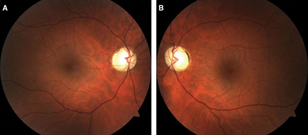
At the 5‐month outpatient review, the patient reported vision being “less washed out” than previously. BCVA had improved in the right eye to 6/24; left eye was counting fingers. Fundus examination revealed pale optic discs bilaterally (Box 1), and optical coherence tomography (OCT) imaging showed significant atrophy of bilateral optic discs (Box 2). These findings were consistent with optic neuropathy after severe methanol poisoning.
Discussion
Significant methanol exposure most commonly occurs through ingestion of alcoholic beverages either inappropriately fortified with methanol or containing excessive methanol as a consequence of inappropriate ethanol fermentation. The optic nerve is vulnerable to formic acid (a metabolite of methanol), and optic nerve atrophy may result from papillomacular bundle damage following mitochondrial injury.1 This atrophy can be appreciated more readily with an OCT scan than a clinical fundus examination, as OCT reveals the subtle loss of ganglion cells in the retinal nerve fibre layer (Box 2). Permanent visual impairment may result when severe methanol toxicity concentrations and systemic metabolic acidosis are present — 25–33% of acute poisonings were found to experience visual sequelae.1,2 However, there is little published evidence to guide effective treatment in the recovery of methanol poisoning‐related visual loss specifically. The management of our patient was based on established guidelines1 to primarily correct metabolic acidosis and inhibit formic acid metabolism. Given the severe visual loss, we also drew from a methanol poisoning case report where a patient treated with oral prednisolone alongside thiamine fully recovered severely impaired vision to 6/6 bilaterally.3 In another interventional case study, six patients with sudden visual loss from methanol poisoning were treated with high dose methylprednisolone for 4 days, followed by 10 days of oral prednisolone4 with subsequent statistically significant improvement in the mean BCVA. Another case study combined steroids with vitamins and erythropoietin to restore vision in two patients who were blinded after methanol poisoning.5
Our patient had severe toxic optic nerve injury from methanol poisoning, but some visual recovery was made following treatment with ethanol as per guidelines (used as a competitive alcohol dehydrogenase inhibitor), haemodialysis and intravenous bicarbonate, alongside adjunctive thiamine and corticosteroids. Treatment of methanol‐induced optic neuropathy with corticosteroids is controversial, as the rarity of methanol toxicity means that there is no high level evidence to support their use. Early treatment with corticosteroids may lead to immunosuppression and increase the likelihood of secondary sepsis in these critically unwell patients. Moreover, methanol poisoning is uncommon in temperate regions, and a randomised controlled trial to investigate correlation between corticosteroid treatment and visual recovery would be difficult.
Visual loss from methanol‐induced optic neuropathy represents a management challenge for clinicians and a debilitating consequence for affected patients.
Lessons from practice
-
Acute methanol poisoning can damage the optic nerve (through its associated metabolic byproducts), resulting in severe and permanent visual loss.
-
There is little published evidence to guide effective treatment in the recovery of visual loss; however, patients should be managed as per established guidelines to correct metabolic acidosis and inhibit formic acid metabolism.
-
Some visual recovery after methanol‐induced optic atrophy may be achieved in patients treated with adjunctive corticosteroids, in addition to systemic treatment for methanol poisoning as per guidelines.
Box 2 – Optical coherence tomography (OCT) imaging showing significant atrophy of bilateral optic discs over time*
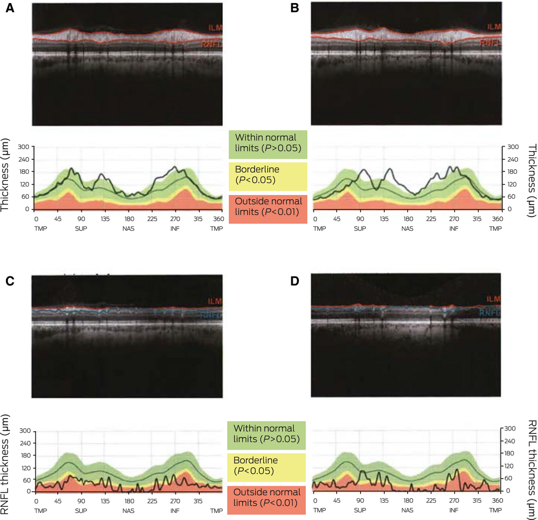
RNFL = retinal nerve fibre layer. * OCT images of both optic nerves showing the thickness of the RNFL and corresponding graphical representation. The bold black line in each graph represents the patient's optic nerve. A. Right eye RNFL at 2 weeks after hospital discharge, with optic nerve thickness largely within normal limits. B. Left eye RNFL at 2 weeks after hospital discharge, with optic nerve thickness largely within normal limits. C. Right eye RNFL showing significant atrophy at 5 months after hospital discharge. D. Left eye RNFL showing significant atrophy at 5 months after hospital discharge.
Competing interests
No relevant disclosures.
References
- Barceloux DG, Bond GR, Krenzelok EP, et al. American Academy of Clinical Toxicology practice guidelines on the treatment of methanol poisoning. J Toxicol Clin Toxicol 2002; 40: 415–446.
- Zakharov S, Pelclova D, Diblik P, et al. Long‐term visual damage after acute methanol poisonings: longitudinal cross‐sectional study in 50 patients. Clin Toxicol (Phila) 2015; 53: 884–892.
- Rotenstreich Y, Assia EI, Kesler A. Late treatment of methanol blindness. Br J Ophthalmol 1997; 81: 416–417.
- Abrishami M, Khalifeh M, Shoayb M, Abrishami M. Therapeutic effects of high‐dose intravenous prednisolone in methanol‐induced toxic optic neuropathy. J Ocul Pharmacol Ther 2011; 27: 261–263.
- Pakravan M, Sanjari N. Erythropoietin treatment for methanol optic neuropathy. J Neuroophthalmol 2012; 32: 325–328.
Provenance: Not commissioned; externally peer reviewed.
Motor Neuron Disease Mortality Trends in Australia From 1986 to 2023: A Population-Based Study
Carol M. Y. Lee, Rupendra N. Shrestha, Julian Gold, Mark Stevenson, Kelly L. Williams, Lyndal Henden, Sandrine K. K. Chan Moi Fat, Zoe Zussa, Amanda Wright, Martin Jude, Mark A. S. Laidlaw, Willem J. R. Fokkink, Dominic B. Rowe
Mild traumatic brain injury and concussion and persisting post‐concussion symptoms: new guidelines to support evidence‐based assessment and management in Australia and Aotearoa New Zealand
Karen M Barlow, Jennie L Ponsford, Alice Theodom, Gill Cowen, Gavin A Davis, Vicki Anderson, Franz E Babl, David Cole, Jennifer Cullen, Stuart R Dalziel, Melinda Fitzgerald, Howard Flavell, Caroline Yates, Rebecca Kimble, John H Olver, Rhonda Orr, Mark Ralfe, Michael Rose, Nick Rushworth, Julia Treleaven, Gary Browne, Nathan Delang, Sarah Harris, Gary Mitchell, Sean Tweedy
The prevalence of and variation in indicators of the quality and safety of long term aged care in Australia, 2019: a cross‐sectional population‐based study
Tesfahun C Eshetie, Gillian E Caughey, Catherine Lang, Olivia Ryan, Renuka Visvanathan, Craig Whitehead, Keith Evans, Janet K Sluggett, Jyoti Khadka, Carolyn Dawkins, Helena Williams, Miranda Starke, Sara Blunt, Anne Liddell, Megan Corlis, Anna Sheppeard, Penelope Lello, Marilyn Thien, Steven L Wesselingh, Maria C Inacio
Health and economic benefits of improving pre‐hospital identification of stroke in Australian women: a modelling study
Thomas Gadsden, Lei Si, Emily R Atkins, Cheryl Carcel, Xia Wang, Stephen Jan, Mark Woodward, Laura E Downey
Symptom burden, quality of life, and diagnostic journey of people with postural orthostatic tachycardia syndrome, Australia, 2021–24: a descriptive patient registry data study
Marie‐Claire Seeley, Gemma Wilson, Eric Ong, Amy Langdon, Jonathan Chieng, Danielle Bailey, Kristina Comacchio, Amanda Page, Dennis H Lau, Celine Gallagher
The intersection of rurality and dementia prevalence in Australia for Aboriginal and Torres Strait Islander and non‐Indigenous peoples
Antonia J Clarke, Marwan Ahmed, Judith M Katzenellenbogen, John Towney (Wiradjuri), Anna H Balabanski, Adrienne Withall (Dharawal Yuin), Kylie Radford, Amy Brodtmann