




Volume 209 - Issue 2
Thyroid nodules: diagnosis and management
Authors: Rosemary Wong, Stephen G Farrell and Mathis Grossmann
Med J Aust 2018; 209 (2): 92-98. || doi: 10.5694/mja17.01204
Published online: 16 July 2018
Published online: 16 July 2018
Consideration of the clinical context, expert evaluation of thyroid sonography, careful selection of nodules for fine-needle aspiration, and standardised cytology reporting are important components in thyroid nodules assessment
Summary
- Thyroid nodules are common. Their importance lies in the need to assess thyroid function, degree of and future risk of mass effect, and exclude thyroid cancer, which occurs in 7–15% of thyroid nodules.
- There are four key components to thyroid nodule assessment: clinical history and examination, serum thyroid stimulating hormone (TSH) measurement, ultrasound and, if indicated, fine-needle aspiration (FNA).
- If the serum TSH is suppressed, a thyroid scan with 99Tc can distinguish between a solitary hot nodule, a toxic multinodular goitre or, less commonly, thyroiditis or Graves’ disease within a coexisting nodular thyroid. Scintigraphically cold nodules are evaluated in the same way as in the setting of normal or elevated serum TSH levels.
- Thyroid ultrasonography should be performed only for palpable goitre and thyroid nodules and by specialists with expertise in thyroid sonography.
- Routine thyroid cancer screening is not recommended, except in high risk individuals, as the detection of early thyroid cancer has not been shown to improve survival.
- FNA may be performed for nodules ≥ 1.0 cm depending on clinical and sonographic risk factors for thyroid cancer.
- FNA specimens should be read by an experienced cytopathologist and be reported according to the Bethesda Classification System.
- Molecular analysis of indeterminate FNA samples has potential to better discriminate benign from malignant nodules and thus guide management.
- Surgery is indicated for FNA findings of malignancy or indeterminate cytology when there is a high risk clinical context. Surgery may also be indicated for suspicion of malignancy; larger nodules, especially with symptoms of mass effect; and in some patients with thyrotoxicosis.
Thyroid nodules present a common clinical problem. Palpable nodules are present in about 5% of the population, and are noted incidentally on ultrasound in up to 70% of people, with increased prevalence in older patients.1,2 Thyroid nodules detected incidentally during positron emission tomography scanning have a 35% probability of being malignant.3 The importance of thyroid nodules lies in the need to assess for thyroid function, local neck symptoms and malignancy.
This review is based on original research articles, authoritative reviews and societal guidelines published in peer-reviewed journals indexed in the PubMed database from 1970 to February 2016, using the search terms “thyroid nodule”, “thyroid cancer”, “thyroid ultrasound” and “thyroid fine-needle aspiration (FNA)”.
Despite the fact that thyroid nodules are very common, there is a paucity of randomised controlled clinical trial data to guide clinical decision making, in part due to the usually good prognosis of thyroid cancer. Instead, recommendations in societal guidelines such as those from the American Thyroid Association (ATA) are largely based on observational studies and expert opinion.4
The majority of thyroid nodules derive from thyroid follicular cells. Benign follicular nodules, either solitary or as part of a multinodular goitre, are the most common mass lesions. Thyroid cancer occurs in 7–15% of thyroid nodules. Factors that increase the risk of malignancy are shown in Box 1.4 Papillary and follicular (including Hürthle) thyroid cancer represent about 85% and 12% of thyroid cancers, respectively. Rarer cancers are medullary thyroid cancer (2%) and anaplastic thyroid cancer (< 1%). Overall, thyroid cancer carries an excellent prognosis with a 5-year overall survival rate of 96.1%, and 98.2% for those patients who have survived one year after diagnosis.5 This good prognosis is largely driven by papillary thyroid carcinoma, with less favourable 5-year survival rates for follicular thyroid carcinoma and other thyroid cancers.6
Thyroid nodules may also coexist in conditions such as subacute thyroiditis, chronic lymphocytic thyroiditis and Graves’ disease. Conditions such as infiltrative disorders, lymphoma, a metastatic tumour (renal and lung are the most common), lipoma or paraganglioma can also result in a thyroid nodule.7 Thyroid nodules can grow irrespective of whether they are benign or malignant. While growth of benign nodules is accepted, the rate of growth of thyroid nodules may help to differentiate benign from malignant nodules. A recent prospective, 21-month cohort study of 126 malignant nodules and 1363 benign nodules ≥ 1.0 cm showed that malignant nodules grew (defined as a > 20% increase in two or more nodule dimensions) in 25.4% of cases, compared with 14.2% of benign nodules (P < 0.001).8 In a prospective study of 992 patients who had benign FNA followed for 5 years, 15% had an increase in nodule size (mean change in largest diameter, 4.9 mm), and 19% had a decrease in nodule size. During follow-up, thyroid cancer was identified in only five (0.3%) nodules, of which only two had increased in size.9
The role for thyroid cancer screening in the general population is debated.10 The United States Preventive Services Task Force recommends against thyroid cancer screening in asymptomatic adults, except for high risk individuals, such as those with a history of radiation exposure in childhood or adolescence or with inherited genetic syndromes associated with thyroid cancer.11 Therefore, thyroid ultrasound should generally only be performed in patients with a palpable goitre, thyroid nodule or cervical lymphadenopathy.
The main challenge in managing thyroid nodules is to identify those that are malignant, while avoiding inappropriate excess use of thyroid sonography, FNA and surgery. Achieving this balance should be considered in the context of recent evidence from the US, the United Kingdom and Australia, suggesting that the increased incidence of thyroid cancer over the past three decades is not purely due to overdiagnosis but also to a true increase in its incidence.12-14 Reasons for this greater incidence may include increased exposure to potentially modifiable factors, including obesity and environmental influences other than the known effect of ionising radiation, such as chemical exposures.15
Approach to thyroid nodules
History and physical examination
Neck examination including palpation of the thyroid and cervical lymph nodes should be part of the routine physical examination. In patients with thyroid nodules, history and physical examination should include assessing risk factors for malignancy (Box 1); for example, exposure to therapeutic irradiation or ionising irradiation in childhood or adolescence markedly increases the risk for thyroid cancer.16-18
Most typically, thyroid cancer presents as a painless nodule. The patient may not be aware of its growth pattern. Recent rapid growth of a thyroid nodule may indicate aggressive cancer and can be associated with pain. This scenario needs to be differentiated from subacute nodular thyroiditis, which is commonly associated with systemic features. Sudden enlargement, especially if accompanied by sudden onset of pain, suggests haemorrhage into a nodule. This is rarely associated with malignancy. Hoarseness suggests infiltration of the recurrent laryngeal nerve by thyroid cancer. Features of dysphagia; neck discomfort in certain positions, such as recumbency at night; and, rarely, dyspnoea or wheeze, may be the result of mass effect by a large goitre.1 Symptoms of hyperthyroidism or hypothyroidism should be sought.
Physical examination of the neck involves characterisation of the thyroid nodules for size, location and texture. Evidence of nodule firmness, fixation, dysphonia and cervical lymphadenopathy suggest malignancy. More commonly however, thyroid cancer does not manifest overtly malignant clinical features.1
Autoimmune thyroiditis may present as bilateral, firm, sometimes tender goitre with or without nodularity. A hallmark of subacute thyroiditis is a unilateral or bilateral firm tender thyroid that may be accompanied by fever.19 If a goitre is present or the history suggests a mass effect, check for tracheal deviation and percuss for subclavicular dullness, although these signs are not well validated. A predominantly intrathoracic goitre may be difficult to detect, even if large; the Pemberton manoeuvre should be performed to assess for thoracic inlet obstruction, which is often an indication for thyroidectomy.20 Signs of hyperthyroidism and hypothyroidism should be evaluated.
Laboratory tests
Serum thyroid stimulating hormone (TSH) should be measured in all patients with a thyroid nodule (Box 2). Most individuals will have a normal TSH level; a low or suppressed TSH level may suggest a hyperfunctioning nodule or a toxic goitre, and free thyroxine (fT4) and/or free triiodothyronine (fT3) levels should be measured. Uncommonly, Graves’ disease can occur in the context of a multinodular goitre. A persistently suppressed TSH with normal fT4 and fT3 levels defines subclinical hyperthyroidism — a condition associated with an increased risk of atrial fibrillation and bone loss, particularly in postmenopausal women,4 and of developing overt thyrotoxicosis, especially when exposed to excess iodine, such as iodinated contrast.4
Hashimoto thyroiditis, which can present with a transient hyperthyroid phase, is the usual cause of elevated TSH or hypothyroidism. TSH elevation or TSH within the upper normal range has been reported to be associated with an increased risk of malignancy within a thyroid nodule.21,22
Measurement of TSH receptor antibody or thyroperoxidase antibody is not indicated unless autoimmune thyroid disease is suspected.2 Routine serum calcitonin measurement is not recommended, except for family members with multiple endocrine neoplasia syndrome type 2 and patients with suspicious imaging and cytology not consistent with papillary thyroid carcinoma.4,23
Imaging studies
All patients with palpable thyroid nodules or nodules detected by other imaging modalities should have a thyroid ultrasound performed by a specialist with expertise in thyroid sonography. A thyroid ultrasound (Box 3) allows documentation of thyroid size, location and characteristics of individual nodules and commonly detects additional nodules not apparent on physical examination. Nodular characteristics suspicious for malignancy include being solid or predominantly solid, taller than wide, hypoechogenicity, irregular margins, microcalcifications, absent halo and increased vascularity.24-26 Nodules are very likely to be benign if they are purely cystic, have typical colloid echoes of ring down or comet tail artefact or are spongiform (multicystic components occupy > 50% of nodule volume).25,27 Based on these characteristics, the ATA4 classifies nodules into risk categories to aid in selection of nodules for FNA (Box 4). Some Australian radiology providers have begun to adopt the American College of Radiology Thyroid Imaging, Reporting and Data System.28
In contrast to neck ultrasound that allows structural assessment, a radionuclide (technetium-99, 99Tc) thyroid scan provides functional assessment of a nodule. It should only be performed when TSH is suppressed in order to diagnose a hyperfunctioning (“hot”) nodule or a toxic multinodular goitre, where nodules will appear “hot”, “warm” or “cold”; this type of scan should not be performed if the TSH level is normal or elevated. Comparison with the patient’s thyroid ultrasound indicates which nodules are hot. Hot nodules usually do not require FNA, due to negligible risk of malignancy.4 Occasionally, however, an FNA may be warranted for large hot nodules with suspicious ultrasound characteristics.
When a multinodular goitre is present with compressive symptoms, a non-contrast computed tomography (CT) thyroid scan is useful to assess the degree of retrosternal extension, tracheal deviation and calibre of the tracheal lumen. In cases of known thyroid cancer, neck and chest CT scan may also be indicated for better pre-operative assessment of extent of spread and involvement of cervical lymph nodes. The use of intravenous contrast is contentious. On the one hand, intravenous contrast provides better structural resolution and can be important for a well planned operation that is critical for optimal long term prognosis. On the other hand, it will delay the timing of radioactive iodine therapy after thyroidectomy for about 2 months, a delay that has not been associated with less favourable outcomes.29,30
Fine-needle aspiration
FNA provides a cytological assessment of thyroid nodules (Box 2), and its main purpose is to reduce the risk of unnecessary surgery and to facilitate single rather than multiple operations for papillary and medullary thyroid carcinoma. The 2015 ATA guidelines recommend the use of FNA for nodules ≥ 1.5 cm, or for nodules ≥ 1.0 cm if they have high or intermediate risk sonographic characteristics.4 There is no evidence that routine investigation of suspicious nodules < 1.0 cm improves outcomes.31 Suspicious lymph nodes should undergo FNA for cytology and needle washings should be taken for thyroglobulin measurement. A normal lymph node does not contain thyroglobulin, and a measurable concentration is suspicious of metastatic thyroid cancer.32 In the case of very low risk sonographic features, such as spongiform or purely cystic appearance, FNA may be limited to nodules sized ≥ 2.0 cm; alternatively, very low risk nodules could be monitored for clinical or sonographic change. Monitoring may also be appropriate for patients with limited life expectancy or unacceptably high surgical risk. If a multinodular goitre is present, each nodule should be assessed for FNA on the basis of the above criteria. In most cases, either no nodule warrants FNA or only few of the many nodules may warrant FNA. Where no FNA is indicated, a repeat thyroid ultrasound may be considered in 12–24 months.
Thyroid cytopathology should be reported according to the Bethesda Classification System (Box 5).33 Benign cytology is found in about 70% of all FNAs, indeterminate cytology (ie, follicular lesion/atypia of undetermined significance [FLUS/AUS] and follicular neoplasm/suspicious for follicular neoplasm [FN/SFN]) in 10–15%, and non-diagnostic or unsatisfactory smears in about 15%.34
The rate of non-diagnostic or unsatisfactory results may be reduced by appropriate selection of nodules that warrant FNA, performing FNA under ultrasound guidance, undertaking two to five needle passes, targeting the solid components of a cystic nodule, immediate check of the material to ensure adequate sampling (obtaining at least six groups of cells, each with at least ten well preserved thyroid epithelial cells), and evaluation by an experienced thyroid cytopathologist.35 Specimen inadequacy, a larger nodule size (≥ 3.0 cm) and inexpertise of the cytopathologist contribute to the false-negative rate of a thyroid FNA, estimated to be about 5–10%.36-38
Indeterminate cytology (FLUS/AUS and FN/SFN) presents a challenging problem. Follicular carcinoma is diagnosed by invasion of the nodular capsule and/or vascular invasion, and not by the cellular morphology. Thus, FNA of follicular lesions can only assess the degree of cellularity and is only an approximation or surrogate for the risk of malignancy. Within follicular lesions, cellular atypia does not correlate well with follicular carcinoma but may confer an increased risk of follicular variant of papillary carcinoma. The risk of malignancy in cases of such indeterminate cytology is 5–30% (Box 5).4 Possible diagnostic approaches for indeterminate cytology include, depending on the clinical context, careful follow-up with repeat ultrasonography and FNA at 6–12 months, or a diagnostic lobectomy.
Molecular analysis of FNA material — not yet widely available in Australia — is likely to improve nodule selection for surgery by evaluation either for the absence or presence of mutations associated with thyroid carcinoma.39,40 The Afirma Gene Expression Classifier (Veracyte, California)41 analyses mRNA expression of 167 genes, providing a high negative predictive value of 94–95% in nodules with indeterminate cytology, making it a useful rule-out test for malignancy, obviating the need for immediate surgery. ThyGenX (Interpace Diagnostics, New Jersey) uses next generation sequencing to identify alterations across eight thyroid cancer-associated genes, together with RNA translocation fusion markers with a negative predictive value of 94% and a positive predictive value of 74%.42 ThyroSeq v2 (CBLPath, New York)43,44 uses next generation sequencing to analyse a larger array of gene mutations and RNA fusion proteins than ThyGenX and may offer better negative and positive predictive values, although further studies are required. While promising, it is not known whether the performance characteristics of these molecular tools developed at tertiary specialist centres will be the same if used during routine clinical care, thus further validation is needed — molecular testing, and its place in clinical practice, is evolving.
Management of thyroid nodules
A solitary hot nodule or a toxic multinodular goitre with a persistently suppressed TSH should usually be treated with radioactive iodine (131I). If a preceding course of antithyroid medication is necessary, 131I should be given while the TSH is still suppressed, as this protects non-autonomous thyroid tissue from 131I uptake and reduces the risk of hypothyroidism. Surgery may be preferred for large toxic lesions, for those individuals with suspicious imaging characteristics and for young patients.
A total thyroidectomy is usually indicated for FNA cytology that is either diagnostic of or suspicious for malignancy. It may also be considered for indeterminate FNA cytology (FLUS/AUS and FN/SFN) in the setting of high risk clinical factors, or if molecular markers are predictive for malignancy.
Total thyroidectomy has been the usual management in Australia for nearly all thyroid cancer. The latest ATA guidelines allow for consideration of hemithyroidectomy for low risk thyroid carcinoma up to 4.0 cm. However, given the absence of definitive evidence to support this recommendation, the guidelines state that “the treatment team may choose total thyroidectomy to enable [radioactive iodine] therapy or to enhance follow-up based upon disease features and/or patient preferences”.4 While hemithyroidectomy for low risk thyroid cancers < 1.0 cm is recommended in the ATA guidelines, active surveillance of these small cancers may be considered. Of 1465 Japanese patients with papillary thyroid carcinoma < 1.0 cm followed prospectively for 10 years, < 10% progressed and there were no deaths from papillary thyroid carcinoma.45
Minimally invasive follicular thyroid carcinoma < 4.0 cm may also be managed by hemithyroidectomy — this is widely accepted practice. In general, follicular thyroid carcinoma can only be diagnosed after definitive pathology on the diagnostic hemithyroidectomy specimen. The decision as to whether the pathology warrants proceeding to completion thyroidectomy is assessed on a case-by-case basis, preferentially by a thyroid cancer multidisciplinary team and in discussion with the patient.4
Compartment-oriented lymph node dissection is necessary for involved nodes, usually in papillary thyroid carcinoma and medullary thyroid carcinoma but occasionally for follicular thyroid carcinoma and Hürthle cell carcinoma. Prophylactic lymph node dissection remains controversial.
Surgical clearance of cancer remains the best option when possible, and it may entail partial excision of contiguous structures when invaded and in the absence of distant metastases. Radioactive iodine should not be relied on to treat gross residual cancer.4
A diagnostic lobectomy may be warranted when there is diagnostic uncertainty or patient preference. A conservative approach is reasonable if there is poor surgical risk or short life expectancy. If a nodule with an indeterminate FNA is to be monitored, a repeat thyroid ultrasound should be performed in 6–12 months, and a repeat FNA is recommended if there is a 50% increase in nodule volume or ≥ 20% increase in at least two nodule dimensions of at least ≥ 2 mm (Box 2).4 When the patient is younger and the nodule is larger or growing, it may be practical to consider surgery for an apparently benign nodule rather than continue close observation and rebiopsy, particularly if future removal seems highly probable.
Another area of controversy relates to whether surgery should be offered for nodules > 4.0 cm that return a benign FNA. A retrospective cohort analysis of 7348 nodules, of which 927 (13%) were cancerous, showed that while 10.5% of nodules 1.0–1.9 cm were cancerous, 15% of nodules > 2.0 cm were cancerous, a size beyond which cancer risk was unchanged, and there was no increased risk of false-negative aspirates.46 A prospective study of 382 nodules > 4.0 cm reported a thyroid cancer rate of 22% and a false-negative cytology rate of 10.4%; moreover, the absence of suspicious ultrasound features did not reliably exclude malignancy.47 In our opinion, in consultation with the patient, nodules > 4.0 cm should be considered for surgical removal.
There is still uncertainty as to the appropriate frequency of follow-up of nodules with benign FNA findings when the risk of malignancy is 0–3% (Box 4). However, it is reasonable to repeat the thyroid ultrasound after 12–24 months (Box 2). If the nodule has grown by the dimensions given above, FNA should be repeated. If the cytology is once again benign, it may not be necessary to have further ultrasounds unless there are sonographically suspicious features or there is clinical change on nodule palpation.34,48 Thyroxine suppression therapy to slow nodule growth is not recommended as it has not been shown to be effective and is associated with adverse effects.4
Pregnancy
Evaluation of thyroid nodules discovered during pregnancy is similar to the non-pregnant state, as there is no evidence that thyroid cancer behaves more aggressively during pregnancy.49,50 However, thyroid scanning with 99Tc is contraindicated during pregnancy. FNA is indicated on the basis of sonographic nodule characteristics. When papillary thyroid cancer is diagnosed early in gestation, and it grows in dimensions as given above or there is suspicious lymphadenopathy, surgery should be considered in the second trimester. However, if papillary thyroid carcinoma remains stable by mid-gestation, or if it is first diagnosed in the second half of gestation, surgery may be safely delayed until postpartum and thyroxine may be used during pregnancy to maintain serum TSH between 0.3 and 2.0 mU/L.4,51 If post-surgical 131I treatment is indicated, this should be deferred for at least 6 weeks to 3 months after cessation of breastfeeding. The latter will more reliably ensure that lactation-associated increase in sodium iodide symporter activity has normalised.52
Conclusion
The challenge in assessing and managing thyroid nodules lies in finding the right balance between, on the one hand, overinvestigation and overtreatment, and on the other hand, the risk of missing clinically significant thyroid cancer. Consideration of the clinical context, judicious use and expert assessment of thyroid sonography, careful selection of nodules for FNA based on clinical and sonographic risk factors, and standardised cytology reporting of an adequate sample by an expert cytopathologist are all important components of an optimised assessment.
If cytology is diagnostic of, or suspicious for, malignancy, surgery is indicated. Indeterminate FNA findings present a challenge and options include close monitoring or diagnostic lobectomy. In addition, molecular analysis has the potential to aid management decisions. Moreover, the majority of nodules with confirmed benign FNA results do not generally require ongoing close follow-up. The increased incidence of thyroid cancer warrants further study as to the possible contributing and modifiable factors.
Box 1 – Factors predicting increased nodular risk of malignancy
|
|
|||||||||||||||
|
History |
|
||||||||||||||
|
Examination |
|
||||||||||||||
|
Investigations |
|
||||||||||||||
|
|
|||||||||||||||
|
PET = positron emission tomography. TSH = thyroid stimulating hormone. |
|||||||||||||||
Box 2 – Approach to thyroid nodules*
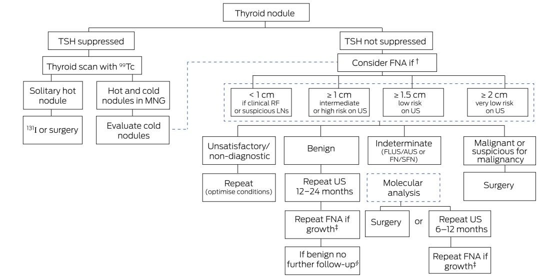
131I = radioactive iodine. FLUS/AUS = follicular lesion/atypia of undetermined significance. FN/SFN = follicular neoplasm/suspicious for follicular neoplasm. FNA = fine needle aspiration. LNs = lymph nodes. MNG = multinodular goitre. RF = risk factor. TSH = thyroid stimulating hormone. US = ultrasound. * A flow chart for the evaluation and management of thyroid nodules, based on thyroid stimulating hormone, thyroid scan with 99Tc, FNA and US characteristics, is presented. † If no indication for FNA, repeat neck ultrasound in 12–24 months. ‡ ≥ 50% increase in nodule volume or ≥ 20% increase in at least two dimensions. § Unless suspicious US features or clinical change in the nodule.
Box 3 – Illustrative ultrasound examples of thyroid pathology
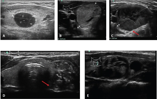
A. Colloid cyst: benign features include a smooth border and bright colloid echoes showing comet tail or ring down artefact. B. Follicular nodule: the differential diagnoses include follicular adenoma, carcinoma or Hürthle cell variant. It is well defined, typically solid or predominantly solid. It is difficult to ascertain if this nodule is benign or malignant unless there are invasive features such as tumour thrombus within a vessel or extrathyroidal spread. C. Suspicious nodule: note the large solitary nodule, more hypoechoic, less well defined margins with some “bulging” (arrow). Absent microcalcification and smoother margin favour follicular over papillary thyroid carcinoma. D. Papillary thyroid cancer: large, solitary, solid, hypoechoic left thyroid nodule with extensive microcalcifications and irregular edge. This lesion was hypervascular (not shown), and abuts the oesophagus and trachea posteriorly (arrow). E. Multinodular goitre: this longitudinal view of the left thyroid lobe shows two of the dominant nodules, both discrete. The larger upper nodule is multicystic, isoechoic in its solid portion and lacks microcalcification. The smaller, lower nodule is predominantly cystic. The risk of malignancy in such multinodular goitres is low.
Box 4 – Thyroid nodule ultrasound characteristics and estimated risk of malignancy
|
Characteristics |
Low risk |
Intermediate risk |
High risk |
||||||||||||
|
|
|||||||||||||||
|
Appearance |
Spongiform/solid |
Solid |
Solid |
||||||||||||
|
Cystic |
Yes |
No |
No |
||||||||||||
|
Echogenicity |
Iso-/hyperechoic |
Iso-/hypoechoic |
Hypoechoic |
||||||||||||
|
Halo |
No |
± |
No (PTC) |
||||||||||||
|
Margin |
Smooth; peripheral “eggshell” calcification |
Smooth |
Irregular; lobulated |
||||||||||||
|
Vascularity |
None |
Peripheral |
Intranodular |
||||||||||||
|
Other |
Comet tail; colloid echoes of ring down |
Macrocalcifications |
Taller than wide; microcalcifications; PET positive‡ |
||||||||||||
|
Malignancy risk |
5–10%* |
10–20% |
70–90% |
||||||||||||
|
Size for FNA |
≥ 1.5 cm† |
≥ 1.0 cm |
≥ 1.0 cm |
||||||||||||
|
|
|||||||||||||||
|
FNA = fine-needle aspiration. FTC = follicular thyroid carcinoma. PET = positron emission tomography. PTC = papillary thyroid carcinoma. * The American Thyroid Association (ATA)4 separates low risk into very low malignancy risk < 3% for spongiform nodules and < 1% malignancy risk for purely cystic nodules. † Very low risk spongiform nodules do not require FNA unless ≥ 2.0 cm; purely cystic nodules, nodules that do not meet the above criteria and nodules < 1.0 cm (with some exceptions) do not require FNA. ‡ PET-positive nodules carry a 35% risk of malignancy. More recently in Australia, some radiology providers have begun to adopt the American College of Radiology Thyroid Imaging, Reporting and Data System (TI-RADS) to risk-stratify nodules and recommend follow-up.28 Both the ATA and TI-RADS reporting systems will take time to become standardised in their usage. |
|||||||||||||||
Box 5 – Bethesda classification for reporting thyroid cytopathology33
|
Diagnostic category |
Predicted risk of malignancy |
||||||||||||||
|
|
|||||||||||||||
|
Benign |
0–3% |
||||||||||||||
|
FLUS/AUS |
5–15% |
||||||||||||||
|
FN/SFN |
15–30% |
||||||||||||||
|
Suspicious for malignancy |
60–75% |
||||||||||||||
|
Malignant |
97–99% |
||||||||||||||
|
Non-diagnostic/unsatisfactory |
1–4% |
||||||||||||||
|
|
|||||||||||||||
|
FLUS/AUS = follicular lesion/atypia of undetermined significance. FN/SFN = follicular neoplasm/suspicious for follicular neoplasm. |
|||||||||||||||
Competing interests
No relevant disclosures.
References
- Burman KD, Wartofsky L. Clinical practice. Thyroid nodules. N Engl J Med 2015; 373: 2347-2356.
- Walsh JP. Managing thyroid disease in general practice. Med J Aust 2016; 205: 179-184.
- Soelberg KK, Bonnema SJ, Brix TH, Hegedüs L. Risk of malignancy in thyroid incidentalomas detected by 18F-fluorodeoxyglucose positron emission tomography: a systematic review. Thyroid 2012; 22: 918-925.
- Haugen BR, Alexander EK, Bible KC, et al. 2015 American Thyroid Association management guidelines for adult patients with thyroid nodules and differentiated thyroid cancer: the American Thyroid Association Guidelines Task Force on thyroid nodules and differentiated thyroid cancer. Thyroid 2016; 26: 1-133.
- Cancer Australia. Thyroid cancer statistics. Commonwealth of Australia; 2014. https://thyroid-cancer.canceraustralia.gov.au/statistics (viewed Nov 2017).
- Grani G, Lamartina L, Durante C, et al. Follicular thyroid cancer and Hürthle cell carcinoma: challenges in diagnosis, treatment, and clinical management. Lancet Diabetes Endocrinol 2018; 6: 500-514.
- Mazzaferri EL. Management of a solitary thyroid nodule. N Engl J Med 1993; 328: 553-559.
- Angell TE, Vyas C, Medici M, et al. Differential growth rates of benign versus malignant thyroid nodules. J Clin Endocrinol Metab 2017; 102: 4642-4647.
- Durante C, Costante G, Lucisano G, et al. The natural history of benign thyroid nodules. JAMA 2015; 313: 926-935.
- Sosa JA, Duh QY, Doherty G. Striving for clarity about the best approach to thyroid cancer screening and treatment: is the pendulum swinging too far? JAMA Surg 2017; 152: 721-722.
- US Preventive Services Task Force, Bibbins-Domingo K, Grossman DC, Curry SJ, et al. Screening for thyroid cancer: US Preventive Services Task Force recommendation statement. JAMA 2017; 317: 1882-1887.
- Chen AY, Jemal A, Ward EM. Increasing incidence of differentiated thyroid cancer in the United States, 1988–2005. Cancer 2009; 115: 3801-3807.
- Pandeya N, McLeod DS, Balasubramaniam K, et al. Increasing thyroid cancer incidence in Queensland, Australia 1982–2008 — true increase or overdiagnosis? Clin Endocrinol (Oxf) 2016; 84: 257-264.
- Lim H, Devesa SS, Sosa JA, et al. Trends in thyroid cancer incidence and mortality in the United States, 1974–2013. JAMA 2017; 317: 1338-1348.
- Kitahara CM, Sosa JA. The changing incidence of thyroid cancer. Nat Rev Endocrinol 2016; 12: 646-653.
- Schneider AB, Ron E, Lubin J, et al. Dose-response relationships for radiation-induced thyroid cancer and thyroid nodules: evidence for the prolonged effects of radiation on the thyroid. J Clin Endocrinol Metab 1993; 77: 362-369.
- Curtis RE, Rowlings PA, Deeg HJ, et al. Solid cancers after bone marrow transplantation. N Engl J Med 1997; 336: 897-904.
- Shibata Y, Yamashita S, Masyakin VB, et al. 15 years after Chernobyl: new evidence of thyroid cancer. Lancet 2001; 358: 1965-1966.
- Samuels MH. Subacute, silent, and postpartum thyroiditis. Med Clin North Am 2012; 96: 223-233.
- Knobel M. Etiopathology, clinical features, and treatment of diffuse and multinodular nontoxic goiters. J Endocrinol Invest 2016; 39: 357-373.
- Boelaert K, Horacek J, Holder RL, et al. Serum thyrotropin concentration as a novel predictor of malignancy in thyroid nodules investigated by fine-needle aspiration. J Clin Endocrinol Metab 2006; 91: 4295-4301.
- Haymart MR, Repplinger DJ, Leverson GE, et al. Higher serum thyroid stimulating hormone level in thyroid nodule patients is associated with greater risks of differentiated thyroid cancer and advanced tumor stage. J Clin Endocrinol Metab 2008; 93: 809-814.
- Wells SA, Asa SL, Dralle H, et al. Revised American Thyroid Association guidelines for the management of medullary thyroid carcinoma. Thyroid 2015; 25: 567-610.
- Hoang JK, Lee WK, Lee M, et al. US Features of thyroid malignancy: pearls and pitfalls. Radiographics 2007; 27: 847-860; discussion 861-865.
- Brito JP, Gionfriddo MR, Al Nofal A, et al. The accuracy of thyroid nodule ultrasound to predict thyroid cancer: systematic review and meta-analysis. J Clin Endocrinol Metab 2014; 99: 1253-1263.
- Remonti LR, Kramer CK, Leitão CB, et al. Thyroid ultrasound features and risk of carcinoma: a systematic review and meta-analysis of observational studies. Thyroid 2015; 25: 538-550.
- Bonavita JA, Mayo J, Babb J, et al. Pattern recognition of benign nodules at ultrasound of the thyroid: which nodules can be left alone? AJR Am J Roentgenol 2009; 193: 207-213.
- Tessler FN, Middleton WD, Grant EG, et al. ACR Thyroid Imaging, Reporting and Data System (TI-RADS): white paper of the ACR TI-RADS Committee. J Am Coll Radiol 2017; 14: 587-595.
- Tsirona S, Vlassopoulou V, Tzanela M, et al. Impact of early vs late postoperative radioiodine remnant ablation on final outcome in patients with low-risk well-differentiated thyroid cancer. Clin Endocrinol (Oxf) 2014; 80: 459-463.
- Suman P, Wang CH, Abadin SS, et al. Timing of radioactive iodine therapy does not impact overall survival in high-risk papillary thyroid carcinoma. Endocr Pract 2016; 22: 822-831.
- Topliss D. Thyroid incidentaloma: the ignorant in pursuit of the impalpable. Clin Endocrinol (Oxf) 2004; 60: 18-20.
- Yap NS, Maher R, Learoyd DL. Any detectable thyroglobulin in lymph node biopsy washouts suggests local recurrence in differentiated thyroid cancer. Endocr Connect 2014; 3: 150-155.
- Cibas ES, Ali SZ, National Cancer Institute Thyroid FNA State of the Science Conference. The Bethesda System for reporting thyroid cytopathology. Am J Clin Pathol 2009; 132: 658-665.
- Tamhane S, Gharib H. Thyroid nodule update on diagnosis and management. Clin Diabetes Endocrinol 2016; 2: 17.
- Baloch ZW, Cibas ES, Clark DP, et al. The National Cancer Institute thyroid fine needle aspiration state of the science conference: a summation. CytoJournal 2008; 5: 6.
- Oertel YC, Miyahara-Felipe L, Mendoza MG, Yu K. Value of repeated fine needle aspirations of the thyroid: an analysis of over ten thousand FNAs. Thyroid 2007; 17: 1061-1066.
- Bongiovanni M, Spitale A, Faquin WC, et al. The Bethesda System for reporting thyroid cytopathology: a meta-analysis. Acta Cytol 2012; 56: 333-339.
- Giles WH, Maclellan RA, Gawande AA, et al. False negative cytology in large thyroid nodules. Ann Surg Oncol 2015; 22: 152-157.
- Mond M, Alexiadis M, Eriksson N, et al. Nuclear receptor expression in human differentiated thyroid tumors. Thyroid 2014; 24: 1000-1011.
- Zhang M, Lin O. Molecular testing of thyroid nodules: a review of current available tests for fine-needle aspiration specimens. Arch Pathol Lab Med 2016; 140: 1338-1344.
- Alexander EK, Kennedy GC, Baloch ZW, et al. Preoperative diagnosis of benign thyroid nodules with indeterminate cytology. N Engl J Med 2012; 367: 705-715.
- Labourier E, Shifrin A, Busseniers AE, et al. Molecular testing for miRNA, mRNA, and DNA on fine-needle aspiration improves the preoperative diagnosis of thyroid nodules with indeterminate cytology. J Clin Endocrinol Metab 2015; 100: 2743-2750.
- Nikiforova MN, Wald AI, Roy S, et al. Targeted next-generation sequencing panel (ThyroSeq) for detection of mutations in thyroid cancer. J Clin Endocrinol Metab 2013; 98: E1852-E1860.
- Nikiforov YE, Carty SE, Chiosea SI, et al. Highly accurate diagnosis of cancer in thyroid nodules with follicular neoplasm/suspicious for a follicular neoplasm cytology by ThyroSeq v2 next-generation sequencing assay. Cancer 2014; 120: 3627-3634.
- Ito Y, Miyauchi A, Oda H. Low-risk papillary microcarcinoma of the thyroid: a review of active surveillance trials. Eur J Surg Oncol 2018; 44: 307-315.
- Kamran SC, Marqusee E, Kim MI, et al. Thyroid nodule size and prediction of cancer. J Clin Endocrinol Metab 2013; 98: 564-570.
- Wharry LI, McCoy KL, Stang MT, et al. Thyroid nodules (≥ 4 cm): can ultrasound and cytology reliably exclude cancer? World J Surg 2014; 38: 614-621.
- Kwak JY, Koo H, Youk JH, et al. Value of US correlation of a thyroid nodule with initially benign cytologic results. Radiology 2010; 254: 292-300.
- Herzon FS, Morris DM, Segal MN, et al. Coexistent thyroid cancer and pregnancy. Arch Otolaryngol Head Neck Surg 1994; 120: 1191-1193.
- Moosa M, Mazzaferri EL. Outcome of differentiated thyroid cancer diagnosed in pregnant women. J Clin Endocrinol Metab 1997; 82: 2862-2866.
- Gharib H, Papini E, Garber JR, et al. American Association of Clinical Endocrinologists, American College of Endocrinology, and Associazione Medici Endocrinologi medical guidelines for clinical practice for the diagnosis and management of thyroid nodules — 2016 update. Endocr Pract 2016; 22: 622-639.
- American Thyroid Association Taskforce on Radioiodine Safety; Sisson JC, Freitas J, McDougall IR, et al. Radiation safety in the treatment of patients with thyroid diseases by radioiodine 131I: practice recommendations of the American Thyroid Association. Thyroid 2011; 21: 335-346.
Provenance: Commissioned; externally peer reviewed.
When ‘Liver Enzymes’ Are Not Hepatic: Late-Onset Pompe Disease
Shauna Madigan, Georgina England, Wayne Rankin
Primary Hyperparathyroidism in Adults: Recent Developments in Diagnosis and Management
Elizabeth Wootton, Sunita M. C. De Sousa, Richard L. Prince, Donald S. A. McLeod, David A. Pattison, Mathis Grossmann
Severe Hypoglycaemia Secondary to Chronic Opioid-Induced Hypothalamic–Pituitary–Adrenal Axis Suppression: An Under-Recognised Phenomenon
Michael Do, Annabelle Hayes, Malgorzata Brzozowska
Rethinking diabetes care for Indigenous Australians: the need for Indigenous‐codesigned and led diabetes models of care
Natalie Nanayakkara, Sharon Atkinson‐Briggs, Alicia J Jenkins, Neale D Cohen
The first Australian evidence‐based guidelines on male infertility
Darren J Katz, Liza O’Donnell, Robert I McLachlan, Tim J Moss, Clare V Boothroyd, Veena Jayadev, Sarah R Catford