




Volume 209 - Issue 10
Hypertrophic lichen planus mistaken for squamous cell carcinoma
Authors: Emily X Shao, Benjamin Carew and James Muir
Med J Aust 2018; 209 (10): 462. || doi: 10.5694/mja18.00577
Published online: 19 November 2018
Published online: 19 November 2018
To the Editor:
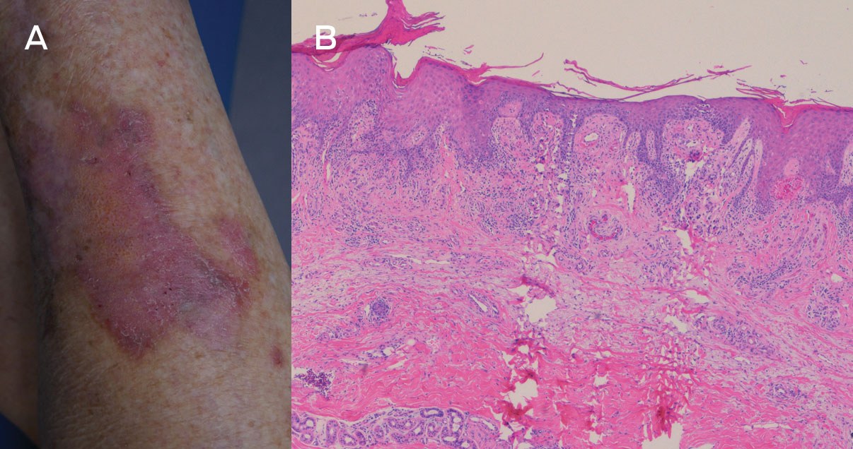
Lichen planus is an autoimmune mucocutaneous inflammatory disorder. Diagnosis is often made clinically and confirmed on biopsy.1 Hypertrophic lichen planus is a distinct subtype characterised by pruritic, hyperkeratotic plaques. Histopathological findings may not have the typical features of lichen planus and can mimic squamous cell carcinoma (SCC).2 Distinguishing between hypertrophic lichen planus and SCC can be difficult for clinicians and pathologists.
In our dermatology practice, we encountered three patients initially diagnosed with SCC, but on review, the cases were consistent with lichen planus. One patient was a 52-year-old woman presenting with asymmetrical, raised and violaceous lesions to her lower legs. She was referred to a skin cancer clinic that performed biopsies of these lesions, which were reported as well differentiated SCC. These lesions were excised, but they were recurrent and were excised again. The second patient was a 54-year-old man who presented with a one-year history of eruptive raised, violaceous lesions to his chest and legs. Biopsies were reported as SCC and multiple lesions were excised by a general surgeon. The third patient was a 77-year-old woman with a 2-year history of pruritic lesions to the lower legs (Box). Biopsies were reported as well differentiated SCC. Each of these patients underwent numerous excisions before being referred to our practice. The patients were reassessed and new biopsies taken, and the clinical picture was discussed with a dermatopathologist. Hypertrophic lichen planus was confirmed as the diagnosis in each of these patients, and they responded well to prednisone, acitretin and topical steroid treatment.
SCC may arise in long-standing hypertrophic lichen planus, but it should be emphasised that cases of supposed SCC with atypical history should not be treated without consideration of the many mimics of SCC, including pseudoepitheliomatous hyperplasia, irritated seborrhoeic keratosis, coral reef granuloma, hypertrophic lupus erythematosus and hypertrophic lichen planus. Clinicians should provide clinical description and a list of potential differentials when referring to a pathologist. Adequate biopsy depth is important, as lichenoid activity may only be present at the tips of the rete ridges, which may be missed on a superficial biopsy.
These cases highlight the difficulties in distinguishing hypertrophic lichen planus from SCC. In the cases we described, correct diagnosis was made after re-evaluation and clinicopathological correlation.
Box – Figure showing violaceous hyperkeratotic patches on the patient’s lower leg, with original biopsies reported as squamous cell carcinoma (A). Histopathology showed a lichenoid inflammatory infiltrate confined to the tips of the rete processes (B)*
* Infiltrate is composed of predominantly lymphocytes with few eosinophils and plasma cells. While these features are typical of hypertrophic lichen planus, superficial shave biopsies may not capture the lichenoid infiltrate at the rete processes.
Competing interests
No relevant disclosures.
Acknowledgements
We thank the pathologists involved in the interpretation of the histology as well as providing the digital images of the histology slides used in this report: Duncan Lambie of IQ Pathology, West End, Queensland, and Louis Pool of Sullivan Nicolaides Pathology, Bowen Hills, Queensland.
References
- Tan E, Malik R, Quirk CJ. Hypertrophic lichen planus mimicking squamous cell carcinoma. Australas J Dermatol 1998; 39: 45-47.
- Weedon D. The lichenoid reaction pattern (“interface dermatitis”). In: Weedon D. Weedon’s skin pathology, 3rd ed. Edinburgh: Churchill Livingstone, 2010; pp 35-70.
From Ambition to Action: Closing Policy, Equity and Implementation Gaps in the Australian Cancer Plan's ‘Enhanced Consumer Experience’
Carla Thamm, Elise Button, Oluwaseyifunmi A. Agbejule, Reegan Knowles, Karen van Gorp, Mark B. Pinkham, Meinir Krishnasamy, Alexandra L. McCarthy, Raymond J. Chan
Sovereignty and Survival: Understanding Lung Cancer Outcomes for Aboriginal and Torres Strait Islander People in Australia, a Case–Control Study
Alice R. T. Bergin, Luc te Marvelde, Roger L. Milne, Benjamin J. Solomon, Kris Ivanova, Peter Savas, Stephen J. Luen, Jay Hamann, Andrea Casey, Nicole Watt, Sue M. Evans, Gail Garvey, Sherene Loi
The Changing Indications for Adult Liver Transplantation in Australia: A Narrative Review
Fadak Mohammadi, Alan J. Wigg, Ken Liu, Simone I. Strasser
Uneven Ground: Survival Differences Among Victorian Lung Cancer Patients by Location of Residence (2011–2023): A Retrospective Cohort Study
Evangeline Samuel, Eldho Paul, Mike Lloyd, Sanuki Tissera, Craig Underhill, Sagun Parakh, Phillip Parente, Inger Olesen, Javier Torres, Katharine See, Gavin M. Wright, David Langton, Thomas John, Matthew Conron, James Bartlett, Nicola Atkin, Nikolajs Zeps, Susan V. Harden, Wasek Faisal, John R. Zalcberg, Rob G. Stirling
Early-Onset Colorectal Cancer With Liver-Only Metastases: A Retrospective Cohort Study Integrating Prospectively Collected Real-World Clinical and Molecular Data From an Australian National Database (2009–2024) to Guide Treatment Planning
Savio G. Barreto, Christos S. Karapetis, Shahid Ullah, Matthew Burge, Susan Caird, Angus Campbell, Azim Jalali, Ross Jennens, Muhammad A. Khattak, Belinda Lee, Stephanie H. Lim, Shehara Mendis, Louise Nott, Timothy J. Price, Jeremy D. Shapiro, Jeanne Tie, Javier Torres, Colin Williams, Rachel Wong, Vanessa Wong, Peter Gibbs
Australian Pathways for Specialist Pain Management and Early Palliative Care for People With Pancreatic Cancer: Developed Using a Community Consensus Approach
Jennifer Philip, Melanie R. Lovell, Kylee Bellingham, Gail Garvey, Gregory B. Crawford, Nicole M. Rankin, Kara Burns, Isabel Young, Vivienne Milch, Dorothy Keefe, Katrina Anderson, James Lawson, Meinir Krishnasamy