




Volume 208 - Issue 2
Abnormal uterine bleeding: managing endometrial dysfunction and leiomyomas
Authors: Annabelle Brennan and Martha Hickey
Med J Aust 2018; 208 (2): 90-95. || doi: 10.5694/mja17.00726
Published online: 5 February 2018
Published online: 5 February 2018
Management of abnormal uterine bleeding should be patient-focused
Summary
- Abnormal uterine bleeding refers to any change in the regularity, frequency, heaviness or length of menstruation. There are several potential causes for bleeding disturbance, the two most common being primary endometrial dysfunction and fibroids. Management of abnormal uterine bleeding involves both medical and surgical options and will largely depend on a patient’s fertility plans.
- The use of levonorgestrel-releasing intrauterine devices for heavy menstrual bleeding is increasing in Australia, and they are considered first-line medical management for women accepting of hormonal therapies. Tranexamic acid, non-steroidal anti-inflammatory drugs, the combined oral contraceptive pill and oral progestins offer alternatives.
- Hysterectomy offers a definitive surgical approach to abnormal uterine bleeding and is associated with high levels of patient satisfaction.
- Women wishing to preserve their fertility, or avoid hysterectomy, may be offered myomectomy. Submucosal fibroids should be removed via hysteroscopy in symptomatic or infertile patients. Intramural and subserosal fibroids may be removed via an open or laparoscopic approach.
- There are several minimally invasive options, including uterine artery embolisation, magnetic resonance-guided focused ultrasound and endometrial ablation, but patients should be aware that there is insufficient evidence to ensure fertility preservation with these procedures and further research is needed.
- Areas for additional research include cost-effectiveness of treatments and quality of life comparisons between management options using patient reported outcome measures to evaluate patient satisfaction.
Abnormal uterine bleeding refers to any change in the regularity, frequency, heaviness or length of menstruation. It may be acute, requiring immediate intervention to prevent further blood loss, or more commonly, a chronic concern, where bleeding disturbance persists for months. The current framework for classifying abnormal uterine bleeding, created by the International Federation of Gynecology and Obstetrics Menstrual Disorders Working Group, outlines the potential causes of bleeding disturbance, including endometrial polyps, adenomyosis, leiomyomas, malignancy, coagulopathies, iatrogenic causes, ovulatory disorders, endometrial dysfunction and various other causes for disturbed menstruation.1
This review provides a clinically focused, evidence-based approach to the medical and surgical management of two of the most common causes of menstrual disturbance: primary endometrial dysfunction and leiomyomas (fibroids). Postmenopausal bleeding and the management of malignancy are beyond the scope of this article.
To evaluate the evidence in this area, we undertook a systematic search using the MEDLINE and Cochrane databases and included the search terms “menorrhagia”, “heavy menstrual bleeding”, “leiomyoma”, “fibroid”, “intrauterine device”, “hysterectomy”, “myomectomy”, “uterine artery embolisation” and “magnetic resonance-guided focused ultrasound”. We limited the results to human studies published in English, and assessed the relevance by title and abstract. Systematic reviews and national and organisational guidelines were of particular interest, and additional results were identified after evaluating the reference lists of the studies included.
Initial diagnosis
Patients may present with abnormal uterine bleeding in general practice or a specialist gynaecological setting, and a thorough but focused history and examination of the patient should include the menstrual and sexual history and potential comorbidities, including coagulopathies and endocrinopathies. In addition, cervical screening should be up to date and sexually transmissible infections and pregnancy should be excluded.
A full blood count is indicated to exclude anaemia, but other biochemical investigations, such as thyroid function and iron studies, should only be ordered when clinically indicated.2,3 Ultrasound imaging will help identify potential structural causes of menstrual disturbance, and current guidelines consider transvaginal ultrasound as first-line investigation and advise performing ultrasound imaging when the physical examination suggests a fibroid uterus, the medical treatment has been unsuccessful or malignancy is suspected.2,3 In addition, endometrial sampling is indicated in women over the age of 40 years, women with abnormal uterine bleeding not responding to medical treatment or women at increased risk of endometrial cancer.2,3 The risk factors for endometrial cancer include obesity, polycystic ovarian syndrome, nulliparity, diabetes and a personal or family history of Lynch syndrome. An outpatient endometrial biopsy may be sufficient for generalised endometrial disturbance. However, ultrasound imaging, with consideration of saline infusion sonography, and hysteroscopic sampling should be arranged for discrete uterine lesions.3
Primary endometrial dysfunction
Primary endometrial dysfunction refers to endometrial tissue with abnormal haemostatic mechanisms in a woman with normal ovulatory function and in the absence of other causative problems.1 The primary complaint is usually heavy menstrual bleeding, which affects up to one-third of women,3 but may also include changes to cycle length and intermenstrual bleeding. After excluding structural concerns, medical treatment should be considered first-line management in abnormal uterine bleeding, and the agent used will depend on the patient’s preference, plans for fertility, medical comorbidities and previous treatments. Pharmaceutical options include non-hormonal and hormonal agents, and the choice of treatment should be guided by the need for contraception.
Medical management
Non-hormonal
Tranexamic acid is an antifibrinolytic agent that is effective for the short term management of abnormal uterine bleeding. A recent systematic review showed a mean reduction in menstrual blood loss by up to 56% with daily doses of more than 3 g for 5 days,4 but the available data assessed treatment regimens of less than 6 months duration with no long term follow-up of treatment success or need for subsequent management.
In addition, there are also various non-steroidal anti-inflammatory drugs used in the treatment of abnormal uterine bleeding, which have been shown to be more effective at reducing menstrual blood loss than placebo, but less effective than tranexamic acid and levonorgestrel-releasing intrauterine devices (LNG-IUDs). However, no particular non-steroidal anti-inflammatory drug has been shown to be superior in treating heavy menstrual bleeding.5
Hormonal
Levonorgestrel-releasing intrauterine devices
The guidelines from the Royal College of Obstetricians and Gynaecologists in the United Kingdom recommend LNG-IUDs as a first-line treatment for abnormal uterine bleeding in women who are accepting of hormonal treatment, including nulliparous women.2 LNG-IUDs are increasingly used in Australia to manage heavy menstrual bleeding, and the Australian Commission on Safety and Quality in Health Care is currently developing a clinical care standard to guide management in the Australian context. Box 1 and Box 2 show that the use of IUDs in Australia continues to increase, and while the data do not distinguish LNG-IUDs from others, the increasing number of insertions for heavy menstrual bleeding suggests a more widespread use of LNG-IUDs. In Australia, the number of IUDs inserted for heavy menstrual bleeding has more than quadrupled over the past decade, with 3365 insertions in 2016, increasing from 3% to 5% over this period.6
There is high-level evidence supporting the efficacy of LNG-IUDs for heavy menstrual bleeding, showing a reduction in blood loss of up to 97% and complete amenorrhoea in about 50% of patients over a 5-year treatment period. Moreover, LNG-IUDs are more effective at reducing heavy blood loss compared with oral treatments, including combined oral contraceptives, non-steroidal anti-inflammatory drugs and oral progestogen regimens.7-10 There is currently insufficient evidence to demonstrate significant differences in quality of life or patient satisfaction between these management options, but continuation rates at 12 months appear to be almost 80% for women with heavy menstrual bleeding and 96% for women with leiomyomas, suggesting that this treatment is well tolerated.4
Combined oral contraceptives
Combined oral contraceptives are effective for abnormal uterine bleeding, while offering contraception. Several randomised trials demonstrate their efficacy in reducing menstrual blood loss, which appears to be between 35–69%, with varying formulations.4,8
Oral progestogens
Several systematic reviews indicate that short regimens of luteal phase oral progestogens, typically involving days 16–26 of a cycle, do not reduce menstrual blood loss.4,8,9,11 Longer regimens of 21 days per cycle appear to significantly improve menstrual blood loss; however, medication side effects may limit the long term use of this approach, with one study showing that less than one-quarter of patients persist with treatment beyond 3 months.11,12
Surgical management
Level 1 evidence indicates that surgical management, including hysterectomy and endometrial ablation, offers more effective reduction in bleeding and greater patient satisfaction compared with oral medical management. A systematic review showed that almost 60% of patients receiving oral medical treatment require surgical management in the following 2 years, with almost one in five women undergoing a hysterectomy.13 Nevertheless, endometrial ablation and LNG-IUD are similar in efficacy in the short and long term, with similar rates of patient satisfaction and impact on quality of life.9,13,14 Of the surgical procedures indicated, hysterectomy is more effective for bleeding, but does not differ from endometrial ablation and LNG-IUD in the level of patient satisfaction or quality of life.9,14 Moreover, almost half of women (46%) treated with LNG-IUD for heavy menstrual bleeding will undergo hysterectomy in the following 10 years,15 which does not necessarily indicate treatment failure, but may reflect patient choices regarding fertility at different ages.
Leiomyomas
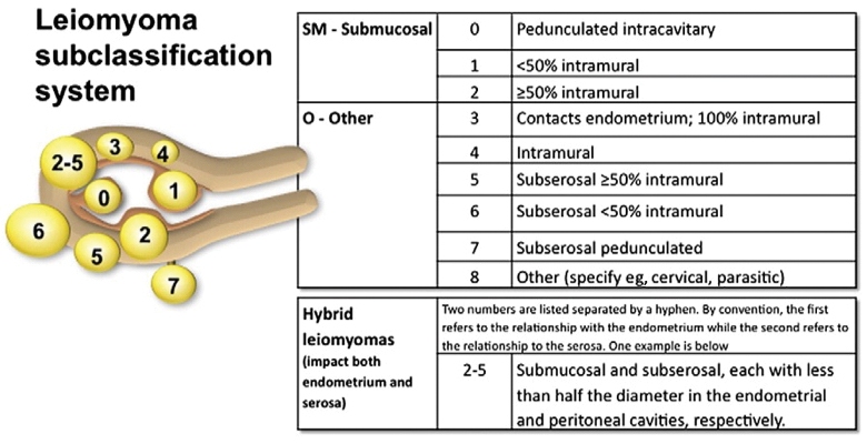
Leiomyomas, commonly referred to as fibroids, are benign tumours of the uterus consisting of smooth muscle cells of the myometrium and may occur in various locations (Box 3). Around 70% of women aged 50 years have uterine fibroids;16 often these patients are asymptomatic, but up to one-third will experience heavy menstrual bleeding associated with fibroids17 — other symptoms may include pelvic pain and pressure. Moreover, in younger women there are also potential implications for fertility and pregnancy, as submucosal leiomyomas may distort the uterine cavity and are associated with an increased risk of miscarriage, placenta praevia, placental abruption, intrauterine growth restriction, pre-term labour, malpresentation and post-partum haemorrhage.18
Ultrasonography is the first-line investigation of leiomyomas, and their abnormal appearance on ultrasound and increasing size or symptoms, particularly in patients who are postmenopausal or in the setting of medical therapy, require further investigation with magnetic resonance imaging (MRI) to exclude malignancy. The rate of leiomyosarcoma in leiomyomas previously considered benign is thought to be less than 1%.19 Asymptomatic fibroids do not require treatment, and there are various approaches to manage symptomatic leiomyomas which will depend on the patient’s preference; the patient’s age and menopause status; the plans for fertility; location, size and number of leiomyomas; and available facilities.
Medical management
Ovarian sex steroids directly influence leiomyoma growth, and oestrogen acts to upregulate the expression of progesterone receptors, increasing the myometrial cells’ sensitivity to progesterone and stimulating growth.20 The medical management of leiomyomas blocks sex steroid effects and the agent chosen will largely depend on the patient’s preference and plans for fertility. Women who wish to conceive or those seeking to avoid hormonal therapies may be offered non-steroidal anti-inflammatories, tranexamic acid or mefenamic acid.2 Women with symptomatic uterine fibroids, who do not currently wish to conceive and are accepting of hormonal therapy, may be offered medical treatment as outlined above. In addition, the selective progesterone receptor modulator ulipristal acetate, which is approved for use in Australia for the management of leiomyomas,21 can be used in a treatment course of 3 months and be repeated up to four times, with a recommended treatment break of at least one menstrual cycle between courses.22 A randomised controlled trial showed significant symptom improvement using ulipristal acetate, with almost 80% of patients experiencing amenorrhoea and a mean reduction in leiomyoma size of 45% after one course.23 However, the symptoms recur when the treatment is stopped; therefore, ulipristal acetate may be more useful for pre-operative optimisation.
Pre-operative medical therapy using gonadotropin-releasing hormone agonists may also be considered. These medications may cause eventual down regulation of intrinsic gonadotropin release and hypo-oestrogenism, which limits leiomyoma growth, and the common side effects include vasomotor symptoms, vulvovaginal atrophy, loss of libido, and mood disturbance.24 The most significant concern in the use of gonadotropin-releasing hormone agonists is the loss of bone density, which may be as high as 6% per 12 months of treatment,25 but may partially recover after treatment cessation.26 However, symptoms rapidly recur when treatment is stopped and, thus, current guidelines advise limiting the use of gonadotropin-releasing hormone agonists to 6 months as pre-operative preparation.2,27-29
Surgical management
Hysterectomy
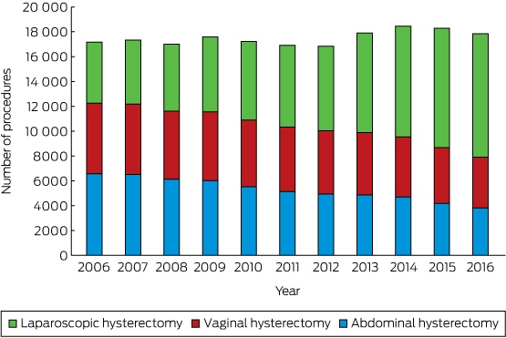
Hysterectomy offers a permanent solution for symptomatic leiomyomas and, as evidence shows, it has high levels of patient satisfaction (up to 90%).30,31 The rates of hysterectomy in Australia have been relatively stable over the past decade, but the proportion of procedures being performed laparoscopically has steadily risen (Box 4).6 Current guidelines recommend hysterectomy as a first-line therapy for patients who have finished childbearing and seek definitive treatment of fibroids or for patients with symptoms uncontrolled by minimally invasive management.2,27,28 It should not necessarily be considered first-line treatment in patients whose sole complaint is heavy menstrual bleeding, as the risks of surgical morbidity associated with hysterectomy may be avoided with a trial of an LNG-IUD.2
Women approaching menopause should be advised that menopausal hormone therapy may increase the size of leiomyomas;32 nevertheless, this therapy should not be withheld from patients with menopausal symptoms who are seeking treatment.27,28,33 In the absence of menopausal hormone therapy, postmenopausal patients with leiomyomas increasing in size or with worsening symptoms should be investigated to exclude the possibility of sarcoma, which may involve an MRI scan.27,28,33
Myomectomy including hysteroscopic resection
Myomectomy is a uterine-sparing procedure recommended for women with symptomatic fibroids who wish to preserve their fertility or avoid hysterectomy. Guidelines from Australia, France and Canada advise that submucosal fibroids should be removed via hysteroscopy in patients who are infertile or otherwise symptomatic,27,34,35 but a recent systematic review indicated that the evidence for fertility benefit is unclear.36 Moreover, the guidelines do not directly address hysteroscopic myomectomy for recurrent pregnancy loss, and while intramural fibroids may affect fertility, there is insufficient evidence to demonstrate improved fertility after this procedure. In addition, subserosal leiomyomas have not been shown to influence fertility and do not need to be removed in patients who are asymptomatic but subfertile.34,37
The surgical approach to myomectomy may be influenced by the leiomyoma location, size and number as well as the practitioner’s surgical experience. Intracavity leiomyomas can be accessed via hysteroscopy in an inpatient or outpatient setting — depending on available services — and subserosal and some intramural leiomyomas may be excised through an open or laparoscopic approach. There is increasing interest in robotic-assisted myomectomy, but there is no evidence of this approach being cost-effective or offering superior clinical outcomes when compared with traditional laparoscopic procedures.38-40 It is important to advise all patients about the risk of leiomyoma recurrence with any uterine-sparing procedure, which may be as high as one-third of patients,41 and there is no evidence suggesting a difference in leiomyoma recurrence between laparoscopic and open approaches.42 The risk of uterine rupture in pregnancy after a previous myomectomy is estimated to be less than 1%, but there is insufficient evidence to assess the difference in risk between the two surgical approaches.27,43,44 Therefore, patients should be counselled about this risk and be informed of the laparoscopic suturing experience of the surgeon.
Minimally invasive management
Uterine artery embolisation
Uterine artery embolisation (UAE) uses interventional radiology to administer embolic material into the uterine circulation. In this procedure, the end arterioles supplying a leiomyoma are blocked, causing necrosis, while vascular anastomoses ensure adequate supply to the normal myometrium. UAE is a uterine-sparing procedure that may be offered to symptomatic patients wanting to retain their uterus, and has been shown to be highly effective at treating leiomyoma symptoms, with up to 90% initial improvement.27,45,46 Moreover, a recent systematic review showed shorter procedure time and hospital stay with reduced blood loss and need for transfusion when compared with both myomectomy and hysterectomy.45
However, UAE is associated with a higher rate of minor complications, most commonly, with post-embolisation syndrome.45 Pedunculated subserosal leiomyomas are also at risk of detachment after embolisation, and similarly, large intracavity leiomyomas may slough off causing pain, heavy bleeding and infection. Therefore, current guidelines advise that endoscopic and laparoscopic services should be available to manage these post-procedural complications if required.27,47,48 Despite initial efficacy, patients are two to four times more likely to require re-intervention after UAE when compared with hysterectomy and myomectomy,27,45 and over a 5-year follow-up after UAE, the rate of hysterectomy for persistent leiomyoma symptoms may be as high as 28%.27 Nonetheless, there is currently no high-level evidence demonstrating a difference in patient satisfaction between UAE, myomectomy and hysterectomy.45
Fertility concerns associated with UAE include non-target embolisation of the ovary, endometrial deterioration from poor perfusion and abnormal placental implantation.49,50 Pregnancy concerns include intrauterine growth restriction, malpresentation, pre-term labour, uterine rupture, post-partum haemorrhage and increased risk of caesarean delivery.46,50,51 While live births have been reported after the procedure, the evidence regarding the impact of UAE on fertility is poor. Low-level data from subgroup analyses of small studies suggest slightly improved outcomes with myomectomy (ie, lower rates of miscarriage and higher rates of live births).45 However, the quality of the evidence is low, with insufficient comparison between the two procedures to assess fertility outcomes, but it appears that UAE does not increase the risk of radiation-associated cancer or radiation-related infertility.46,50
The rate of ovarian failure after UAE may be up to 7% and it most commonly occurs in women aged over 45 years.52 A recent systematic review showed no significant difference in ovarian failure between UAE and hysterectomy or myomectomy, and current guidelines suggest that UAE should only be offered to symptomatic women wishing to preserve their fertility, after a full discussion about the potential impact of this procedure on fertility.28,47,48 Moreover, the guidelines suggest that UAE is contraindicated in patients who do not accept the small risk of hysterectomy associated with procedural complications (estimated to be less than 1%),53 have significant renal disease and are not on dialysis, have an active or recent pelvic infection, or have an uncertain diagnosis of benign leiomyoma.2,27,47,48 There are several other minimally invasive methods used to treat symptomatic leiomyomas (outlined below), but there is no high-level evidence indicating greater efficacy of one procedure over another.
Magnetic resonance-guided focused ultrasound surgery
Magnetic resonance-guided focused ultrasound surgery is a thermoablative procedure using ultrasound waves to cause tissue necrosis in the leiomyoma while using MRI temperature mapping to protect nearby normal tissue. Evidence from systematic reviews shows significant symptom improvement after magnetic resonance-guided focused ultrasound surgery; in the 12 months after the treatment, symptom severity scores measured by the Uterine Fibroid Symptom and Quality of Life scale were halved, but almost one-third of patients required further intervention for treatment failure.54,55
Complications in magnetic resonance-guided focused ultrasound surgery are uncommon and they are generally minor, including skin burns and abdominal wall oedema; other complications such as venous thromboembolism, damage to bowel or bladder and sciatic paraesthesia are rare.56,57 Although pregnancy has been recorded after magnetic resonance-guided focused ultrasound surgery, current guidelines advise that there is insufficient evidence to guide its routine use in women who want to preserve their fertility,2,27,29,35 and the Royal Australian and New Zealand College of Obstetricians and Gynaecologists recommends that patients seeking to preserve fertility should only be offered this procedure in the setting of approved clinical trials.34
Endometrial ablation
Endometrial ablation is an effective management of heavy bleeding associated with submucosal leiomyomas in women who no longer wish to preserve their fertility.58
Myolysis
Myolysis uses a range of techniques, including cryoablation, radiofrequency, microwaves or bipolar energy, to destroy leiomyoma tissue, but current guidelines advise that there is insufficient evidence to recommend its use outside of clinical trials.27,34,35
Gene therapy
Gene therapy involves the use of a vector to administer genetic material into a tissue to interfere with genetic and, subsequently, tissue function. Research continues in this area to broaden our understanding of the genetic mechanisms involved in leiomyoma formation.59
Areas for further research
It is necessary to conduct more research on the long term cost-effectiveness of different surgical procedures for the treatment of fibroids in the Australian context. In addition, it is important to obtain direct quality of life comparisons between management options for abnormal uterine bleeding, highlighting the use of patient-reported outcome measures to evaluate patient satisfaction. Other areas that require further investigation include: comparison between laparoscopic and open myomectomy regarding the integrity of the uterine wound for future pregnancies; the role of tranexamic acid prior to open myomectomy; and a comparison of various minimally invasive procedures to assess the impact on quality of life, cost-effectiveness, symptom improvement and fertility.
Conclusion
Abnormal uterine bleeding is a common condition in premenopausal women and there are several effective medical and surgical treatment options. The decisions about treatment should be patient-focused, evaluate the impact of symptoms on quality of life and function, outline the risks of intervention and appreciate the patient’s plans for fertility.
Box 1 – Number of intrauterine device (IUD) insertions in Australia for the preceding decade6
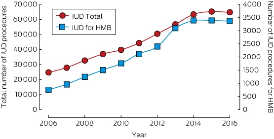
HMB = heavy menstrual bleeding.
Box 2 – Proportion of intrauterine device (IUD) insertions for heavy menstrual bleeding over the preceding decade6
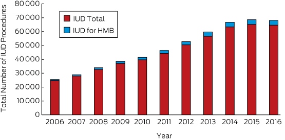
HMB = heavy menstrual bleeding.
Competing interests
No relevant disclosures.
References
- Munro MG, Critchley HOD, Broder MS, et al. FIGO classification system (PALM-COEIN) for causes of abnormal uterine bleeding in nongravid women of reproductive age. Int J Gynaecol Obstet 2011; 113: 3-13.
- National Institute for Health and Care Excellence. Heavy menstrual bleeding: assessment and management. Clinical guideline CG44. London: NICE; 2007. https://www.nice.org.uk/guidance/cg44/chapter/Recommendations#pharmaceutical-treatments-for-hmb (viewed June 2017).
- The Society of Obstetricians and Gynaecologists of Canada. Abnormal uterine bleeding in pre-menopausal women. J Obstet Gynaecol Can 2013; 35(5): S1-S25.
- Bitzer J, Heikinheimo O, Nelson AL, et al. Medical management of heavy menstrual bleeding: a comprehensive review of the literature. Obstet Gynecol Surv 2015; 70: 115-130.
- Lethaby A, Duckitt K, Farquhar C. Non-steroidal anti-inflammatory drugs for heavy menstrual bleeding. Cochrane Database Syst Rev 2013; (1): CD000400.
- Department of Human Services. Medicare item reports. Canberra: Commonwealth of Australia; 2017. http://medicarestatistics.humanservices.gov.au/statistics/mbs_item.jsp (viewed June 2017).
- Nelson A, Teal S. Medical therapies for chronic menorrhagia. Obstet Gynecol Surv 2007; 62: 272-281.
- Matteson K, Rahn D, Wheeler T, et al. Nonsurgical management of heavy menstrual bleeding. Obstet Gynecol 2013; 121: 632-643.
- Lethaby A, Hussain M, Rishworth JR, Rees MC. Progesterone or progestogen-releasing intrauterine systems for heavy menstrual bleeding. Cochrane Database Syst Rev 2015; (4): CD002126.
- Uhm S, Perriera L. Hormonal contraception as treatment for heavy menstrual bleeding. Clin Obstet Gynecol 2014; 57: 694-717.
- Lethaby A, Irvine GA, Cameron IT. Cyclical progestogens for heavy menstrual bleeding. Cochrane Database Syst Rev 2008; (1): CD001016.
- Irvine G, Campbell-Brown M, Lumsden M, et al. Randomised comparative trial of the intrauterine system and norethisterone levonorgestrel for treatment of idiopathic menorrhagia. BJOG 1998; 105: 592-598.
- Marjoribanks J, Lethaby A, Farquhar C. Surgery versus medical therapy for heavy menstrual bleeding. Cochrane Database Syst Rev 2016; (1): CD003855.
- Kaunitz A, Meredith S, Inki P, et al. Levonorgestrel-releasing intrauterine system and endometrial ablation in heavy menstrual bleeding. Obstet Gynecol 2009; 113: 1104-1116.
- Hurskainen R, Teperi J, Rissanen P, et al. Quality of life and cost-effectiveness of levonorgestrel-releasing intrauterine system versus hysterectomy for treatment of menorrhagia: a randomised trial. Lancet 2001; 357: 273-277.
- Baird DD, Dunson DB, Hill MC, et al. High cumulative incidence of uterine leiomyoma in black and white women: ultrasound evidence. Am J Obstet Gynecol 2003; 188: 100-107.
- Drayer SM, Catherino WH. Prevalence, morbidity, and current medical management of uterine leiomyomas. Int J Gynaecol Obstet 2015; 131: 117-122.
- Ezzedine D, Norwitz E. Are women with uterine fibroids at increased risk for adverse pregnancy outcome? Clin Obstet Gynecol 2016; 59: 119-127.
- Brohl AS, Li L, Andikyan V, et al. Age-stratified risk of unexpected uterine sarcoma following surgery for presumed benign leiomyoma. Oncologist 2015; 20: 433-439.
- Moravek M, Yin P, Ono M, et al. Ovarian steroids, stem cells and uterine leiomyoma: therapeutic implications. Hum Reprod Update 2014; 21: 1-12.
- Whitaker LHR, Williams ARW, Critchley HOD. Selective progesterone receptor modulators. Curr Opin Obstet Gynecol 2014; 26: 237-242.
- Australian Government Department of Health, Therapeutic Goods Administration, Australian Public Assessment Reports for Prescription Medicines: Ulipristal acetate. Published 2 November 2016. https://www.tga.gov.au/auspar/auspar-ulipristal-acetate-0 (viewed June 2017).
- Donnez J, Vázquez F, Tomaszewski J, et al. Long-term treatment of uterine fibroids with ulipristal acetate. Fertil Steril 2014; 101: 1565-1573.
- Moroni RM, Vieira CS, Ferriani RA, et al. Pharmacological treatment of uterine fibroids. Ann Med Health Sci Res 2015; 4: 185-192.
- Hornstein MD, Surrey ES, Weisberg GW, et al. Leuprolide acetate depot and hormonal add-back in endometriosis: a 12-month study. Obstet Gynecol 1998; 91: 16-24.
- Surrey ES, Hornstein MD. Prolonged GnRH agonist and add-back therapy for symptomatic endometriosis: long-term follow-up. Obstet Gynecol 2002; 99: 709.
- Marret H, Fritel X, Ouldamer L, et al. Therapeutic management of uterine fibroid tumors: updated French guidelines. Eur J Obstet Gynecol Reprod Biol 2012; 165: 156-164.
- The Society of Obstetricians and Gynaecologists of Canada. The management of uterine leiomyomas. J Obstet Gynaecol Can 2015; 37: 157-178.
- Burke CT, Funaki BS, Ray CH Jr, et al. ACR Appropriatenes criteria on treatment of uterine leiomyomas. J Am Coll Radiol 2011; 8: 228-234.
- Hehenkamp WJ, Volkers NA, Birnie E, et al. Symptomatic uterine fibroids: treatment with uterine artery embolization or hysterectomy — results from the randomized clinical Embolisation Versus Hysterectomy (EMMY) trial. Radiology 2008; 246: 823-832.
- Kjerulff K, Rhodes J, Langenberg P, Harvey L. Patient satisfaction with results of hysterectomy. Am J Obstet Gynecol 2000; 183: 1440-1447.
- Wise LA, Laughlin-Tommaso SK. Epidemiology of uterine fibroids: from menarche to menopause. Clin Obstet Gynecol 2016; 59: 1-24.
- American College of Obstetricians and Gynecologists. ACOG Practice Bulletin: alternatives to hysterectomy in the management of leiomyomas. Obstet Gynecol 2008; 112: 387-400.
- Women’s Health Committee and Australasian CREI Consensus Expert Panel on Trial Evidence Group. Fibroids in infertility: consensus statement of the Australian and New Zealand College of Obstetricians and Gynaecologists and the ACCEPT Group. Sydney: RANZCOG; 2014. https://www.ranzcog.edu.au/RANZCOG_SITE/media/RANZCOG-MEDIA/Women's%20Health/Statement%20and%20guidelines/Clinical%20-%20Gynaecology/Fibroids-in-Infertility-(C-Gyn-27)-Review-November-2014.pdf?ext=.pdf (viewed June 2017).
- The Society of Obstetricians and Gynaecologists of Canada. The management of uterine fibroids in women with otherwise unexplained infertility. J Obstet Gynaecol Can 2015; 37: 277-285.
- Bosteels J, Kasius J, Weyers S, et al. Hysteroscopy for treating subfertility associated with suspected major uterine cavity abnormalities. Cochrane Database Syst Rev 2013; (1): CD009461.
- Brady PA, Stanic AK, Styer AK. Uterine fibroids and subfertility: an update on the role of myomectomy. Curr Opin Obstet Gynecol 2013; 25: 255-259.
- Lewis EI, Garguilo AR. The role of hysteroscopic and robot-assisted laparoscopic myomectomy in the setting of infertility. Clin Obstet Gynecol 2016; 59: 53-65.
- Göçmen A, Şanlıkan F, Uçar MG. Comparison of robotic-assisted laparoscopic myomectomy outcomes with laparoscopic myomectomy. Arch Gynecol Obstet 2013; 287: 91-96.
- Pundir J, Pundir V, Walavalkar R, et al. Robotic-assisted laparoscopic vs abdominal and laparoscopic myomectomy: systematic review and meta- analysis. J Minim Invasive Gynecol 2013; 20: 335-345.
- Rossetti A, Sizzi O, Soranna L, et al. Long-term results of laparoscopic myomectomy: recurrence rate in comparison with abdominal myomectomy. Hum Reprod 2001: 16: 770-774.
- Jin C, Hu Y, Chen X, et al. Laparoscopic versus open myomectomy – a meta-analysis if randomized controlled trials. Eur J Obstet Gynecol Reprod Biol 2009; 145: 14-21.
- Bhave Chittawar P, Franik S, Pouwer AW, Farquhar C. Minimally invasive surgical techniques versus open myomectomy for uterine fibroids. Cochrane Database Syst Rev 2014; (10): CD004638.
- Chittawar PB, Kamath MS. Review of nonsurgical/minimally invasive treatments and open myomectomy for uterine fibroids. Curr Opin Obstet Gynecol 2015; 27: 391-397.
- Gupta JK, Sinha A, Lumsden MA, Hickey M. Uterine artery embolization for symptomatic uterine fibroids. C Cochrane Database Syst Rev 2014; (12): CD005073.
- Silberzweig JE, Powell DK, Matsumoto AH, Spies JB. Management of uterine fibroids: a focus on uterine-sparing interventional techniques. Radiology 2016; 280: 675-692.
- Royal College of Obstetricians and Gynaecologists and the Royal College of Radiologists. Clinical recommendations on the use of uterine artery embolisation (UAE) in the management of fibroids, 3rd ed. London: RCOG and RCR; 2013. https://www.rcog.org.uk/en/guidelines-research-services/guidelines/uterine-artery-embolisation-in-the-management-of-fibroids (viewed June 2017).
- Women’s Health Committee, Royal Australian and New Zealand College of Obstetricians and Gynaecologists. Uterine artery embolisation for the treatment of uterine fibroids. Sydney: RANZCOG: 2014. https://www.ranzcog.edu.au/RANZCOG_SITE/media/RANZCOG-MEDIA/Women's%20Health/Statement%20and%20guidelines/Clinical%20-%20Gynaecology/Uterine-Artery-Embolisation-(C-Gyn-23)-Review-November-2014.pdf?ext=.pdf (viewed June 2017).
- Donnez J, Dolmans MM. Uterine fibroid management: from the present to the future. Hum Reprod Update 2016; 22: 665-686.
- Hickey M, Hammond I. What is the place of uterine artery embolisation in the management of symptomatic uterine fibroids. Aust N Z J Obstet Gynaecol 2008; 48: 360-368.
- Czuczwar P, Stępniak A, Wrona W, et al. The influence of uterine artery embolisation on ovarian reserve, fertility, and pregnancy outcomes – a review of literature. Menopause Rev 2016; 15: 205-209.
- Spies JB, Myers ER, Worthington-Kirsch R, et al. FIBROID Registry Investigators. The FIBROID Registry: symptom and quality-of-life status 1 year after therapy. Obstet Gynecol 2005; 106: 1309-1318.
- Dutton S, Hirst A, McPherson K, et al. A UK multicentre retrospective cohort study comparing hysterectomy and uterine artery embolisation for the treatment of symptomatic uterine fibroids (HOPEFUL study): main results on medium-term safety and efficacy. BJOG 2007; 114: 1340-1351.
- Clark NA, Mumford SL, Segars JH. Reproductive impact of MRI-guided focused ultrasound surgery for fibroids: a systematic review of the evidence. Curr Opin Obstet Gynecol 2014; 26: 151-161.
- Fennessy FM, Tempany CM. A review of magnetic resonance imaging guided focused ultrasound surgery of uterine fibroids. Top Magn Reson Imaging 2006; 17: 173-179.
- Hesley GK, Gorny KR, Henrichson TL, et al. A clinical review of focused ultrasound ablation with magnetic resonance: an option for treating uterine fibroids. Ultrasound Q 2008; 24: 131-139.
- Hindley J, Gedroyc WM, Regan L, et al. MRI guidance of focused ultrasound therapy of uterine fibroids: early results. AJR Am J Roentgenol 2004; 183: 1713-1719.
- Lethaby A, Hickey M, Garry R, et al. Endometrial resection/ablation techniques for heavy menstrual bleeding. Cochrane Database Syst Rev 2009; (4):CD001501.
- Al-Hendy A, Salama S. Gene therapy and uterine leiomyoma: a review. Hum Reprod Update 2006; 12: 385-400.
Linked content
-
MJA InSight: On the rise: use of intrauterine devices for abnormal bleeding
Provenance: Commissioned; externally peer reviewed.
Reducing Nitrous Oxide Emissions Across the Melbourne Biomedical Precinct
Ross Robertson, Andrew Downey, Daryl Williams, Bjorn Makein, Ben Dunne, Tugce Ozturk, Ying Gu, Rebecca McIntyre
Multimorbidity Clusters Among People Aged 65 Years and Over in Australia: A Nationwide Cross-Sectional Data Linkage Study
Weisi Chen, Christine Y. Lu, Sarah N. Hilmer, Alice A. Gibson, Edwin C. K. Tan
When ‘Liver Enzymes’ Are Not Hepatic: Late-Onset Pompe Disease
Shauna Madigan, Georgina England, Wayne Rankin
West Nile virus Kunjin subtype in rural NSW
Emily Gibson, Megan Whitley, Peter Murray, Linda Hueston, Jane Bennett, Raguharan Kathiresu, David N Durrheim
The impact of the Breast Screen NSW transition from film to digital mammography, 2002–2016: a linked population health data analysis
Rachel Farber, Nehmat Houssami, Katy J L Bell