




Medical education
Clinical skills
Volume 207 - Issue 8
Recognising severe cutaneous adverse drug reactions
Author: Paul Chee
Med J Aust 2017; 207 (8): 325-327. || doi: 10.5694/mja17.00522
Published online: 16 October 2017
Published online: 16 October 2017
Severe cutaneous adverse drug reactions are a major cause of morbidity and mortality in Australia and recognition is important
Severe cutaneous adverse drug reactions (SCARs) are a major cause of morbidity and mortality in Australia and recognition is important. Key diagnostic features are present early in the presentation and common drug associations exist. Clinicians should become familiar with these reaction patterns to more effectively manage these dermatological emergencies.
Exanthemic eruptions represent the majority of drug reactions. Erythematous macules and papules begin on the trunk and may generalise, but patients are usually systemically well. Almost any drug can be responsible; common triggers include penicillins and anticonvulsants. Reactions typically appear after 7 to 14 days; a careful history usually uncovers the culprit medication, and cessation results in complete recovery. Like many drug eruptions, reactions are more common with concomitant viral infections, including human immunodeficiency virus.1
SCARs represent type IV delayed hypersensitivity. These T lymphocyte-mediated responses are driven by small molecule drugs acting as haptens presented by human leucocyte antigen class 1 proteins.1 Antiepileptics, antimicrobials and allopurinol are commonly responsible, and these reactions carry significant morbidity and mortality.
Making the correct diagnosis is vital; the causative agent can be identified and ceased immediately and early management initiated. Distinct clinical and morphological patterns of the eruption are clues (Box 1). Key clinical features to consider in suspected drug eruptions are shown in Box 2.
Stevens–Johnson syndrome and toxic epidermal necrolysis
Epidermal cell death and mucosal ulceration are common to Stevens–Johnson syndrome (SJS) and toxic epidermal necrolysis (TEN), classified by the extent of body surface area involved: SJS if < 10%, TEN if 30%, and SJS/TEN overlap if 10–30%. Full thickness epidermal necrosis on biopsy is characteristic.
Drugs are responsible for the majority of cases. In a small number, no clear link is established.2 Mycoplasma pneumonia infection is associated in children.3
Patients are unwell, with flu-like symptoms, fever and eye irritation. Painful skin and early mucosal involvement are red flags and key diagnostic features (Box 3, A). Two or more sites are involved in 80% of cases, including ocular, oral, nasopharyngeal and anogenital mucosa.2 Severe lip crusting reflects oropharyngeal erosion and patients may be unable to swallow. Conjunctival erosions may lead to adhesions and blindness, and require aggressive treatment.
The rash appears dusky red and macular, with irregular targetoid lesions (Box 3, B). Epidermal necrosis evolves within these areas, becoming confluent (Box 3, C). In TEN, large areas of epidermis can sheet off exposing bright red erosions, behaving like an extensive superficial burn (Box 3, D).
Management requires intensive supportive care, ideally in a burns unit, with barrier nursing. Secondary infection is a major risk. Treatment is controversial and includes systemic corticosteroids, cyclosporin, intravenous immunoglobulin and anti-tumour necrosis factor agents.1,2
Drug reaction with eosinophilia and systemic symptoms
Drug reaction with eosinophilia and systemic symptoms (DRESS), also known as drug-induced hypersensitivity syndrome, presents as a maculopapular drug reaction with fever, haematological changes and internal organ involvement. It may resemble a mononucleosis-like syndrome, including arthralgia, lymphadenopathy and pharyngitis.4
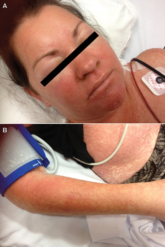
Facial and periorbital erythema and swelling are early red flags (Box 4, A). The rash spreads in a cephalocaudal pattern to become generalised (Box 4, B). Lack of skin or mucosal detachment distinguishes it from SJS/TEN.
Internal involvement predominates. Eosinophilia and atypical lymphocytosis are typical. The liver is most commonly affected (> 50%) and hepatitis may be fulminant.4 Renal, cardiac (myocarditis) and central nervous system inflammation may develop. Thyroiditis and other forms of delayed autoimmunity can manifest 2–3 months later, and the early tests for visceral and thyroid involvement must therefore be repeated.4
A protracted clinical course is typical, with cutaneous and visceral organ involvement lasting weeks or months after drug withdrawal. Flares commonly follow initial improvement.
The suspected medication is ceased immediately. Topical corticosteroids under wet dressings and antihistamines are useful. Prednisolone (1–2 mg/kg/day) is usually given with visceral involvement or severe symptoms, and typically needs to be continued for 50–60 days. Intravenous immunoglobulin and cyclosporin are options.1
There are fewer causative drugs and onset is later. The mortality rate of 2–10% reflects visceral involvement.4
Acute generalised exanthematous pustulosis
Acute generalised exanthematous pustulosis (AGEP) is characterised by early rapid onset, often within 48 hours following drug exposure, usually accompanied by fever.1
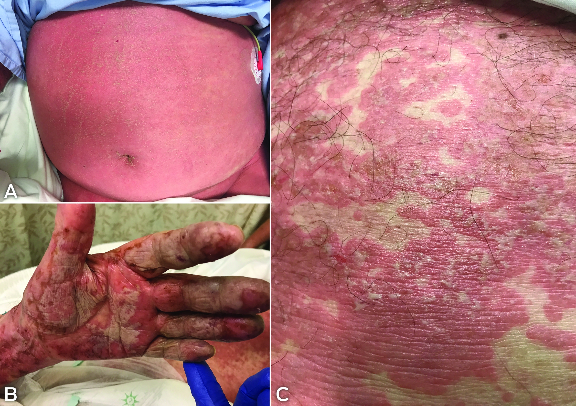
Widespread erythema and oedema is rapidly followed by the appearance of hundreds or thousands of small sterile pustules (Box 5, A). The superficial pustules are the diagnostic key (Box 5, B). These coalesce with superficial epidermal shedding. The rash favours body folds, and palms and soles are involved (Box 5, C).
The dramatic onset and appearance is often mistaken for SJS/TEN, but mucous membrane involvement occurs in a minority of cases, and is typically mild and non-erosive. Cutaneous changes dominate and internal involvement is rare. Facial involvement in AGEP differs from that in DRESS in being pustular rather than exanthematous.
Despite an acute and severe reaction, the prognosis is favourable. It is self-limited, and once the offending drug is withdrawn, the rash resolves with peeling and without scarring. Systemic corticosteroids are given only in severe cases.5
Most cases are drug induced, and re-exposure produces a dramatic recurrence. Viral and bacterial infections may trigger AGEP.5
Summary
In severe adverse drug reactions, clinical signs may evolve sequentially. Awareness of key diagnostic features is necessary for early diagnosis, intervention and referral that may be lifesaving.
Box 1 – Severe cutaneous adverse reactions (SCARs) and associated medications
|
SCAR |
Onset |
Diagnostic keys and red flags |
Most common causative drugs |
||||||||||||
|
|
|||||||||||||||
|
Stevens–Johnson syndrome |
4–35 days |
Early mucosal involvement of multiple sites, especially lips and mouth; atypical targetoid lesions; epidermal loss and erosion |
Allopurinol; carbamazepine; lamotrigine; cotrimoxazole; nevirapine; NSAIDs (oxicam type); phenytoin; phenobarbital |
||||||||||||
|
Drug reaction with eosinophilia and systemic symptoms (drug-induced hypersensitivity syndrome) |
2–6 weeks |
Facial and periorbital oedema; mucosal sparing; blood abnormalities; internal organ involvement; prolonged course; delayed autoimmunity |
Allopurinol; anticonvulsants (phenytoin, carbamazepine, phenobarbital, lamotrigine); sulfonamides; nevirapine; dapsone; minomycin |
||||||||||||
|
Acute generalised exanthematous pustulosis |
Rapid: 1–10 days |
Widespread sterile pustules that coalesce; background erythema and oedema; favours creases; mild mucosal involvement; internal involvement rare |
β-Lactam antibiotics; quinolones; sulfonamides; diltiazem |
||||||||||||
|
|
|||||||||||||||
|
NSAIDs = non-steroidal anti-inflammatory drugs. |
|||||||||||||||
Box 2 – Key features to consider in suspected drug-related eruptions
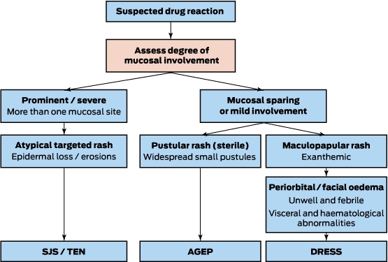
AGEP = acute generalised exanthematous pustulosis. DRESS = drug reaction with eosinophilia and systemic symptoms. SJS = Stevens–Johnson syndrome. TEN = toxic epidermal necrolysis.
Box 3 – Stevens–Johnson syndrome and/or toxic epidermal necrolysis
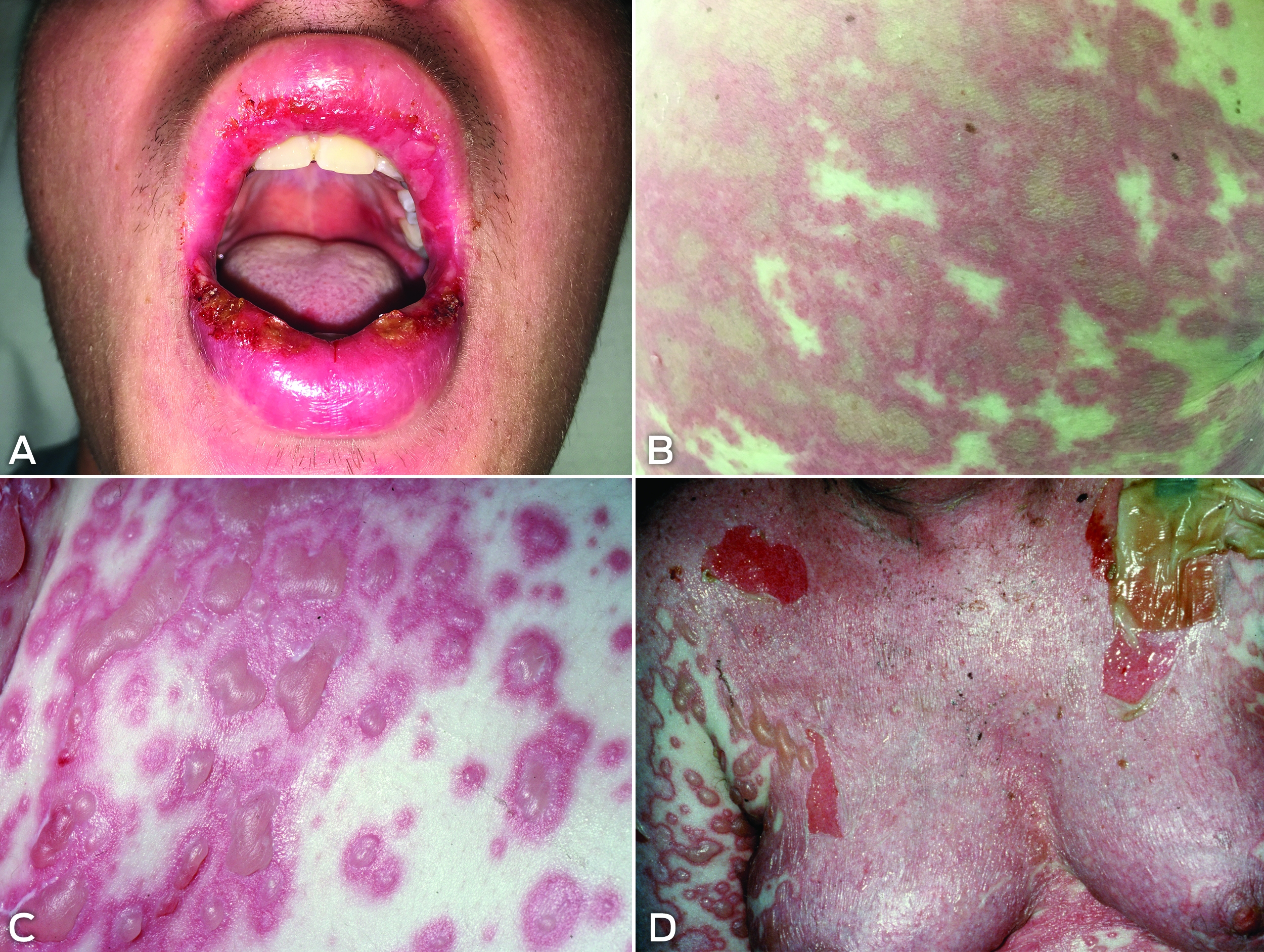
Early signs are lip and mucosal involvement (A). Painful dusky targetoid lesions (B) evolve into epidermal necrosis, blistering (C) and detachment (D).
Competing interests
No relevant disclosures.
References
- Chung WH, Wang CW, Dao RL. Severe cutaneous adverse drug reactions. J Dermatol 2016; 43: 758-766.
- Dibek ME, Guvenir H, Bahceci S, et al. Severe cutaneous adverse drug reactions in pediatric patients: a multicenter study. J Allergy Clin Immunol Pract 2017; 5: 757-763.
- Sassolas B, Haddad C, Mockenhaupt M, et al. ALDEN, an algorithm for assessment of drug causality in Stevens-Johnson Syndrome and toxic epidermal necrolysis: comparison with case-control analysis. Clin Pharmacol Ther 2010; 88: 60-68.
- Kardaun SH, Sekula P, Valeyrie-Allanore L, et al. Drug reaction with eosinophilia and systemic symptoms (DRESS): an original multisystem adverse drug reaction. Results from the prospective RegiSCAR study. Br J Dermatol 2013; 169: 1071-1080.
- Sidoro A, Halevy S, Bavinck JN, et al. Acute generalized exanthematous pustulosis (AGEP) — a clinical reaction pattern. J Cutan Pathol 2001; 28: 113-119.
Provenance: Commissioned; externally peer reviewed.
Intranasal Adrenaline Use for the Management of Insect Venom-Induced Anaphylaxis
Thomas J. Young, Wilhelmina R. F. Johnson, Braydon L. Pennington, Troy Wanandy, Wun Y. Lau, Thanh-Thao A. Le
Gabapentinoid Poisoning Presentations to Victorian Emergency Departments: A 15-Year Trend Analysis
Bishaal Tej Gurung, Amy McNeilage, Angus Skeen, Tina Lam, Joanna F. Dipnall, Jane Hayman, Suzanne Nielsen, Ting Xia
Still Treating Yesterday's Risk? Reconsidering Antiviral Use for Mild-to-Moderate COVID-19 Cases in a Broadly Immune Population
Hadar Mudrik-Zohar, Tim Cutfield, Susan Morpeth, Thomas Hills, Eamon Duffy, Laura J. Edwards, Allen C. Cheng, Steven Y. C. Tong
Birth prevalence, clinical sequelae, and management of congenital cytomegalovirus infections in Australia, 1999–2023: a national prospective study
Ece Egilmezer, Suzy M Teutsch, Carlos Nunez, Stuart T Hamilton, Adam W Bartlett, Pamela Palasanthiran, Elizabeth J Elliott, William D Rawlinson
Concurrent use of hormonal long‐acting reversible contraception by women of reproductive age dispensed teratogenic medications, Australia, 2013–2021
Aaron E Boyce, Tony Caccetta, Jo‐Ann See, Rosemary L Nixon AM
Concurrent use of hormonal long‐acting reversible contraception by women of reproductive age dispensed teratogenic medications, Australia, 2013–2021
Michelle KY Chen, Adrian Lim, Deshan F Sebaratnam