




Volume 207 - Issue 8
Atopic dermatitis: the new frontier
Authors: Victoria R Harris and Alan J Cooper
Med J Aust 2017; 207 (8): 351-356. || doi: 10.5694/mja17.00463
Published online: 16 October 2017
Published online: 16 October 2017
New targeted immunotherapies have had promising results in phase 2 and 3 trials for patients with moderate to severe AD
Summary
- Atopic dermatitis (AD) is the most common inflammatory skin condition in adults and children.
- AD is a chronic disease that has a considerable negative impact on the quality of life of patients and their families.
- Most cases of AD may be effectively treated with topical therapies that are directed at decreasing cutaneous inflammation and alleviating pruritus. These therapies include emollients, antihistamines, topical corticosteroids, topical calcineurin inhibitors and antimicrobial and antiseptic measures; more refractory cases may require additional oral immunosuppression (eg, cyclosporine, azathioprine, methotrexate and mycophenolate).
- Improved understanding of the immune pathogenesis of AD, including the role of T helper cells and the inflammatory pathways involved, has led to breakthrough translational clinical research and treatment.
- New targeted immunotherapies, such as inhibitors of interleukin (IL)-4, IL-13, IL-31, Janus associated kinase and phosphodiesterase, have had promising results from phase 2 and 3 trials for patients with moderate to severe AD.
Atopic dermatitis (AD) is a chronic, pruritic, inflammatory skin disease characterised primarily by a cutaneous barrier deficiency and mediated by cutaneous inflammation. It has a prevalence of 25% in children and of around 7% in adults.1,2 The disease has a significant negative impact on patients’ mental and physical functioning, especially due to its chronic, unremitting time course and associated sleep deprivation from pruritus.3 AD remains a clinical diagnosis with the essential features of pruritus and characteristic eczematous morphology that varies in anatomical distribution with age.4 In addition to a defective cutaneous barrier, the main mediators of cutaneous inflammation in acute AD are type 2 T helper cells (Th2 and Th22).5
The compromised cutaneous barrier in AD also results in colonisation and superinfection, mostly by Staphylococcus aureus and herpes simplex infection. Recent progress in the understanding of the inflammatory pathways involved in AD means that we are on the verge of a revolution in the treatment of severe presentations of this condition.
For this review, we conducted a series of database searches using MEDLINE, PubMed and the Cochrane Central Register of Controlled Trials for meta-analyses, randomised and non-randomised controlled trials, open studies and case series. Additional relevant reviews and articles were isolated from the citations in the reviewed articles. The clinical expertise of the co-authors was used to appraise the selected articles included in this review.
Pathogenesis of atopic dermatitis
Impaired barrier function
The primary event in AD is a cutaneous barrier deficiency.6 Filaggrin (FLG) protein is critically important in the functioning of the outermost cutaneous layer (the stratum corneum). A pivotal study in dermatology was the discovery that loss of function mutations in the FLG gene accounted for the impaired cutaneous barrier function seen in AD.7 The loss of function mutation in the FLG gene has subsequently been associated with more severe AD and early onset of disease in infancy or early childhood.8 Defective epidermal barrier function is not isolated to defective structural proteins and FLG mutations, as patients may develop AD in the absence of FLG mutation.8 It has been proposed that there are also a number of other environmental factors that accumulate to disturb the epidermal barrier, including common irritants (eg, soaps and perfumes) and mechanical factors (eg, chronic scratching).6
Immune response
Another mediator in AD is the observed cutaneous inflammation from increased T cell and circulating cytokine activity. Knowledge of the inflammatory mediators involved in AD has allowed the recent development of targeted biologic therapies for this condition.
Early AD is associated with increased Th2 cells, and skin lesions are associated with the activation of Th2, Th22 and Th17 cytokines.5 A third T helper cell, Th17, which produces interleukin (IL)-17 and IL-22, has also been implicated in acute AD and may have a role in driving Th2 differentiation based on mice model studies.9 In chronic AD, there are additional cellular infiltrates with Th2, Th17 and Th22 activation and a Th1 component.10 The two most important pathways involve Th2 and Th22: Th2 produces IL-4 and IL-13, and Th22 produces IL-22.11 One of the proposed mechanisms to explain the inappropriate inflammatory cascade seen in AD is the prolonged antigen penetration through the defective cutaneous barrier that stimulates the Th2 infiltrate observed in lesional and non-lesional AD.12
Antimicrobial barrier
The antimicrobial barrier of patients with AD is compromised and contributes to observed longstanding S. aureus colonisation, with up to 90% of the lesional skin of a patient with AD being colonised by S. aureus.13 The release of exotoxin by S. aureus has also been shown to induce T cell proliferation and further degrade the already compromised epidermal barrier.14 Therefore, the eradication of biofilms, antimicrobial measures and correction of barrier abnormality with the use of regular emollients should help both the allergen-induced and inflammation cascade observed in AD.12
Environmental factors
It is well documented that the prevalence of AD has increased greatly around the world in areas such as Africa, eastern Asia, western Europe and parts of northern Europe.15 The increase in prevalence of AD raises the possibility that there are multiple potential environmental triggers that lead to flares of AD in predisposed individuals. A recent review has suggested that there is a complex interplay between different environmental factors, including the use of personal care products, exposure to climate, pollution, food and other exogenous factors that may play a role in the pathogenesis of AD.16
Treatment of atopic dermatitis
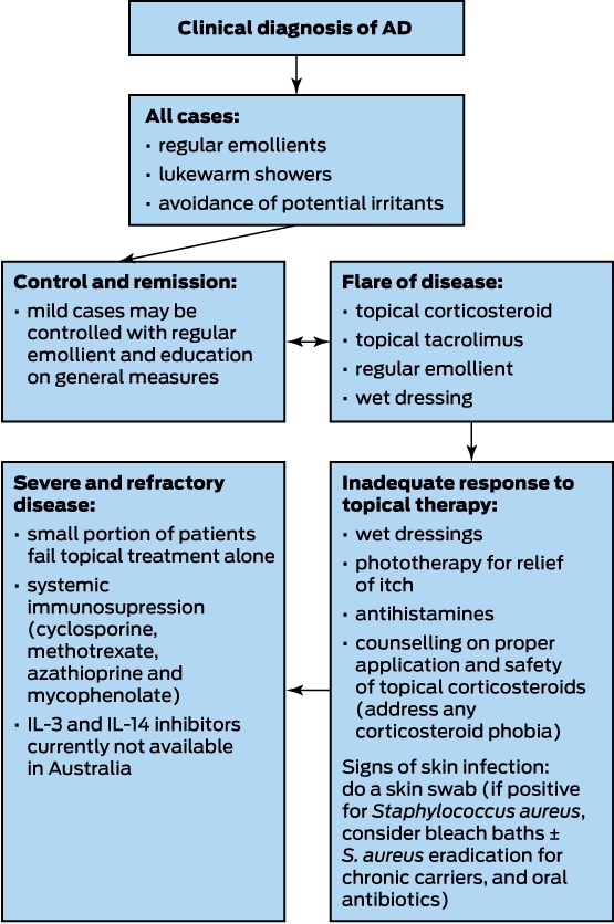
A suggested treatment algorithm is shown in Box 1. All cases may be managed initially with regular emollients and general measures, including avoidance of potential irritants, with specific therapy escalating from topical to oral, according to response, and involvement of a dermatologist when adequate control cannot be maintained.
Topical
Emollients
Counselling on the regular and appropriate use of emollients is essential for the treatment of every case of AD (Box 2). Emollients, such as a combination of emulsifying ointment with 50% water, are available from compounding pharmacists, and they improve the dysfunctional epidermal barrier in AD and help prevent water loss and xerosis. The clinical utility of emollients has been reaffirmed by the strong placebo results seen in recent dupilumab trials, where patients had a compulsory emollient twice daily regimen.17 Recent clinical trials have also shown that the risk of developing AD may be reduced by the early introduction of regular application of a topical emollient within 3 weeks of birth, particularly petrolatum (ie, soft white paraffin) applied at least once daily to neonates at high risk of AD.18
Antiseptics and antibacterial treatments
The increased incidence of S. aureus skin infections is well documented in AD.19 The ongoing threat of antimicrobial resistance highlights the need to further explore the use of antiseptic treatments for prevention of AD flares and to decrease colonisation in patients. This is supported by a recent randomised control trial that found that children with mild clinically infected eczema do not need antibiotic therapy.20 As an alternative to repetitive courses of oral antibiotics for infected AD flares, prophylactic measures may be considered, including the regular use of dilute bleach baths with intermittent intranasal application of mupirocin ointment to decrease S. aureus skin colonisation in cases of chronically infected AD.21 Bleach baths are a safe option and are often used in the paediatric setting as a useful adjunct for patients who have recurrent S. aureus infections. In these cases, patients may be advised to use half a cup of household bleach in a full tub of water, soak for up to 10 minutes and then rinse off. This treatment may be prescribed two to three times per week.
Topical corticosteroids
Due to their local anti-inflammatory effects, topical corticosteroids (TCS) are a first line and very effective treatment for most cases of AD.22 The potency and duration of treatment should be titrated to the response of pruritus and inflammation on a case by case basis. Pruritus is a key symptom for evaluation of treatment success, and tapering should only occur once this symptom has resolved.4 It is important to use corticosteroids with an adjunctive regular emollient to maintain the compromised cutaneous barrier, especially during AD flares. The potency of TCS ranges from mild to moderate, potent and very potent.
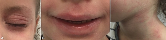
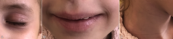
For acute AD flares, it is recommended that TCS be applied to the affected skin once to twice daily, and it may be used less frequently thereafter in a maintenance regimen to prevent future flares.23 Box 3 (A, B and C) shows a child with moderate eczema during an acute flare before treatment. Box 4 (A, B and C) shows marked improvement in the child’s skin 2 weeks later, after the use of moderate potency TCS (ie, methylprednisolone aceponate) on the face twice daily with regular emollient.
A barrier to treatment success in children with AD is the parents’ commonly held belief about the adverse effects of long term use of TCS, which is known as “corticosteroid phobia”.24 This phobia persists in spite of evidence that application of TCS to children does not cause adverse side effects, including cutaneous atrophy and discolouration in moderate to severe cases of AD.25 Moreover, dermatologists generally agree that the benefits of appropriate use of TCS outweigh the theoretical side effects.26
Despite this evidence, the widely held belief that long term corticosteroid use will “thin the skin” is pervasive, and a recent survey-based study of parents found that they are heavily reliant on information about the risk of TCS from non-medical sources, such as the internet, family and friends.27 This risk message is unfortunately often reaffirmed and cited by pharmacists28 and general practitioners29 when dispensing and prescribing TCS. Poor treatment outcomes are unnecessary in most cases and may result from parents’ health beliefs and non-adherence to appropriate TCS use.
Calcineurin inhibitors
Topical calcineurin inhibitors, including tacrolimus ointment and pimecrolimus cream, have shown efficacy in the treatment of AD for their anti-inflammatory and antipruritic effects.30 Topical calcineurin inhibitors are produced by Streptomyces bacteria, and function by blocking the production of cytokines and other inflammatory mediators involved in AD.31 These inhibitors may be used in place of TCS or in areas of increased sensitivity, such as periorbital skin.
Phosphodiesterase type 4 inhibitors
Advances in non-corticosteroid topical treatments include crisaborole 2% ointment (a phosphodiesterase type 4 [PDE4] inhibitor). PDE4 is a key regulator of inflammatory cytokine production in AD through the degradation of cyclic adenosine monophosphate (cAMP).32 PDE4 activity has also found to be increased in the circulating inflammatory cells of patients with AD.33 When topical crisaborole 2% ointment was applied twice daily for 28 days in a study of adults and children, there was reportedly greatly improved disease severity and pruritus in patients with mild to moderate AD.34 Crisaborole 2% ointment was also well tolerated, with the main adverse event being application site irritation reported as burning or stinging. Therefore, this inhibitor may be a useful topical adjunct to TCS in the treatment of mild to moderate AD.
Janus associated kinase inhibitors
Topical tofacitinib is a small molecule Janus associated kinase (JAK) inhibitor. A recent phase 2 study that involved 69 patients with mild to moderate AD showed a significantly greater improvement from baseline for tofacitinib (81.7%) versus vehicle (29.9%).35 There was a significant improvement in itch by Day 2 in this study. Safety and tolerability were similar between the two groups. However, there is concern among dermatologists that the cost of these treatments may be prohibitive.36
Phototherapy
Phototherapy is a useful and effective treatment, particularly for the relief of pruritus. It is usually reserved for second line treatment in conjunction with topical treatments (ie, emollients, steroids and calcineurin inhibitors).4 In addition to symptom relief, phototherapy may potentially decrease colonisation by S. aureus.37 This treatment needs to be introduced on a case by case basis and may not be effective for all patients.
Systemic: cyclosporine, methotrexate, azathioprine and mycophenolate
Cyclosporine is a potent immunosuppressive drug that blocks cytokine production through inhibition of calcineurin and may induce remission in AD when administered orally.38 The efficacy of cyclosporine has been established in cases of severe, refractory AD at doses in the order of 5 mg per kg of bodyweight per day.39 Considerations for the long term use of cyclosporine include the potential side effects of nephrotoxicity and elevation of blood pressure.40
Other systemic treatments in AD with broad T cell targeting mechanisms include methotrexate and azathioprine for their significant anti-inflammatory properties. There is some evidence of clinical benefit with methotrexate in moderate to severe cases of AD at doses ranging from 15 to 25 mg weekly; however, the onset of improvement is slow, with improvement taking up to 3 months to occur.41 The authors’ clinical experience in the paediatric setting is that methotrexate is well tolerated overall and that once weekly dosing is also more practical for children. It is also important that patients are counselled that therapeutic benefit may not be seen for up to 3 months and that patients should be maintained on adjunctive topical treatments during this period.
Azathioprine has also been shown in a randomised control trial to be efficacious in the treatment of severe AD at a dose of 2–5 mg per kg of bodyweight per day. However, it remains second line therapy in severe refractory cases due to its significant side effect profile, including liver and myelotoxicity, and is poorly tolerated due to gastrointestinal disturbances.42
Mycophenolate is another systemic treatment option for a subset of patients with severe refractory cases of AD that cannot be adequately controlled with topical treatments. Mycophenolate may be used at a dosage of 2 g per day, but it is less often used as treatment because it has a slower onset than cyclosporine. In clinical practice, the delayed onset means that patients are often non-compliant after a short period due to perception of inefficacy of treatment. Mycophenolate may be used in children; however, consideration of the side effect profile is important, including gastrointestinal and haematological abnormalities.4
An Australian review on systemic treatments — azathioprine, methotrexate and cyclosporine — used in the paediatric setting recently affirmed the safety of these treatments and a relaxation on the frequency of haematological monitoring during the use of these agents.43 It was also found, in a retrospective study that spanned over 15 years, that there was no significant correlation between observed haematological abnormalities and side effects. Therefore, these systemic agents are useful for treatment of severe cases of AD and may be safely prescribed with adequate monitoring.43
Targeted immune therapy
For the subset of patients who have severe, refractory disease and have inadequate control with existing systemic immunosuppression, there may be hope in the form of new treatment with targeted immune therapy (Box 5). At present, dupilumab is one of the most tested pharmaceutical options of the immune therapies available for adult AD and, while it has been approved by the Food and Drug Administration in the United States, it is still awaiting the Therapeutic Goods Administration approval in Australia for use in adults. In addition to dupilumab, there are a number of other targeted immune therapies under investigation based on the current understanding of the pathogenesis of AD.
IL-4 and IL-13 inhibitor (dupilumab)
Dupilumab is a fully human monoclonal antibody that is directed against the IL-4 receptor α (IL-4Rα) subunit that inhibits both IL-4 and IL-13 signalling. It has been shown convincingly to reverse the inflammation of AD and to correct the skin barrier function defect. A recent trial of dupilumab versus placebo in adult patients with moderate to severe AD with eosinophilia has shown marked reductions in signs, symptoms and Th2-associated biomarker levels.48 Dupilumab has also shown a good safety profile, with side effects balanced between dupilumab and placebo groups, with the placebo groups having higher incidence of skin infections.
The results of these early studies have subsequently been confirmed in a further randomised, placebo-controlled, dose-ranging study in adult patients with moderate to severe AD.17 It is of note that the most recent trial, but not earlier trials, reported that, in patients given dupilumab, there was an increased rate of herpes viral infections of 8% versus 2% in the placebo group.50 Therefore, there is a need for further long term trials of dupilumab, including head-to-head studies with pre-existing systemic agents (eg, cyclosporine) to enable clinicians to make informed decisions on treatment stratification. In addition, this research needs to be extended to the paediatric population, where the disease is most prevalent. A study of dupilumab use in children and adolescents is currently in progress (ClinicalTrials.gov identifier: NCT02407756).
Lebrikizumab (IL-13 inhibitor)
Lebrikizumab is another targeted immune therapy that has undergone phase 2 trials in moderate to severe AD (ClinicalTrials.gov identifier: NCT02340234). It is a cytokine modulator that binds specifically to IL-13, blocking the binding of IL-13 to IL-4Rα and neutralising its inflammatory activity, and has had promising results in the treatment of other atopic diseases, including asthma.51
Ustekinumab (IL-12 and IL-23)
Ustekinumab is a human monoclonal antibody that targets the p40 subunit shared by IL-12 and IL-23. Ustekinumab has proven efficacy for treatment of chronic plaque psoriasis and is currently available in Australia for adults with the disease.52 However, there is a paucity of data available on its efficacy in the treatment of AD. A small case series of three patients with severe AD found a reduction in epidermal hyperplasia and downregulation of Th2 and Th22 expression based on polymerase chain reaction analysis of lesional skin biopsies, and found improvement in disease severity in all three patients.53 However, the long term efficacy of ustekinumab remains questionable following a case report of a patient who flared 5 months into treatment, and a second case where a woman with concurrent psoriasis and eczema failed to have clearance of eczema despite resolution of psoriasis while on treatment.54
Nemolizumab (IL-31 inhibitor)
The receptor for IL-31 is located on keratinocytes and monocytes and it has been proposed that it may be involved in the pathways for pruritus characteristic of AD.49 IL-31 has also been identified as a marker of disease severity in AD, following a case–control study that found serum IL-31 levels were significantly higher in children with severe disease as opposed to children with moderate or low severity AD.46 A recent phase 2 study of nemolizumab for AD found it significantly improved pruritus at a dose of 0.5 mg/kg.55 Nevertheless, the adverse event profile included exacerbation of atopic dermatitis, nasopharyngitis, upper respiratory tract infection, peripheral oedema and increased creatine kinase levels.55
Omalizumab (IgE inhibitor)
Omalizumab is a humanized IgG1 monoclonal antibody that binds IgE, thereby, inhibiting mast cell or basophil activation. It has been tested in a pilot study of adults with severe AD and elevated IgE and had underwhelming results with only two out of ten patients showing significant improvement in eczema severity score.44 This observation is consistent with current understanding of the Th2 axis and its pathogenic role in IgE class-switching without it having a major role in the observed inflammation.45 While IgE has historically been cited as a major mediator in AD, the inflammatory cascade is now better understood and this may not be the case.
JAK inhibitors (tofacitinib)
The JAK-STAT (signal transducer and activator of transcription) pathway is a signalling mechanism for multiple cytokines and growth factors and it has been shown to mediate IL-4, which is a key cytokine in Th2 differentiation observed in AD.47 An oral JAK inhibitor (ABT-494) is currently undergoing phase 2 trials for moderate to severe AD. A small case series involving six patients with moderate to severe AD who were treated with oral tofacitinib citrate for 8–29 weeks reported improvement in AD and particularly in pruritus and sleep.47 Therapeutic use of oral tofacitinib is already approved for use in rheumatoid arthritis.56 Use of tofacitinib in AD is currently limited by the side effect profile.
PDE4 inhibitors (apremilast)
Apremilast is a PDE inhibitor that acts by binding to the catalytic site of PDE4, blocking cAMP degradation resulting in accumulation of cAMP and inhibition of production of tumour necrosis factor α, interferon γ, IL-2, IL-12p70, leukotriene B4 and several chemokines. It has undergone phase 2 trials in refractory AD with underwhelming results.57
Conclusion
The mainstay of management of AD remains restoration of the disrupted epithelial barrier. The gold standard treatment is regular emollient and topical steroid treatment in all cases of AD. The treatment of severe AD is on the verge of a revolution, following recent breakthroughs in the understanding of the inflammatory pathways of the disease — especially with the recent success reported from clinical trials of dupilumab and its role in Th2 pathways of inflammation in AD. The recent successful data for use of dupilumab in AD is encouraging, given the chronicity of this considerable burden of disease for patients who are severely affected. Dermatologists have historically relied on TCS, with great resistance faced by poorly informed parents and the pervasive corticosteroid phobia. In addition, systemic therapies used in moderate to severe cases of AD have had similar drawbacks due to side effect profiles and toxicity with long term use. Therefore, new advances in translational research in targeted biologic AD therapy may revolutionise the treatment of AD in the next decade.
Box 2 – Comparison of cost for topical and systemic treatment currently available for atopic dermatitis
|
Treatment |
Mode |
Frequency |
Cost |
||||||||||||
|
Quantity |
Price |
||||||||||||||
|
|
|||||||||||||||
|
Emulsifying ointment |
Topical |
Twice daily |
100 g jar |
$40* |
|||||||||||
|
Steroids |
Topical |
Twice daily |
15 g tube (methylprednisolone aceponate) |
$17.13 |
|||||||||||
|
Calcineurin inhibitors (tacrolimus) |
Topical |
Twice daily |
100 g (0.03% tacrolimus) |
$120 |
|||||||||||
|
JAK inhibitors |
Topical |
Daily |
100 g jar |
$400 |
|||||||||||
|
Methotrexate |
Systemic |
Weekly |
5-tablet pack, 5 mg/2 mL |
$28.79 |
|||||||||||
|
Mycophenolate |
Systemic |
Daily |
Three 100-tablet packs, 250 mg |
$176.41 |
|||||||||||
|
Cyclosporine |
Systemic |
Daily |
Two 30-tablet pack, 25 mg |
$80.37 |
|||||||||||
|
Azathioprine |
Systemic |
Daily |
100-tablet pack 50 mg |
$31.27 |
|||||||||||
|
|
|||||||||||||||
|
JAK = Janus associated kinase. PBS = Pharmaceutical Benefits Scheme. Costs based on prices MIMSOnline (www.mimsonline.com.au), accessed on 29 August 2017, and subject to change. * Only available from a compounding pharmacy. |
|||||||||||||||
Box 3 – Four-year-old patient with moderate atopic dermatitis: facial erythema, cheilitis and lichenification of periorbital skin
Box 4 – Four-year-old patient showing marked improvement of atopic dermatitis after 2 weeks of treatment with daily application of methylprednisolone aceponate ointment and emollient
Box 5 – Summary of targeted biologics currently under investigation for treatment of moderate to severe atopic dermatitis
|
Inhibition/target |
Oral |
Parenteral |
Topical |
||||||||||||
|
|
|||||||||||||||
|
Tofacitinib |
– |
Tofacitinib |
|||||||||||||
|
IgE46 |
– |
Omalizumab |
– |
||||||||||||
|
PDE447 |
Apremilast |
– |
Crisaborole 2% |
||||||||||||
|
IL-4/IL-1315 |
– |
Dupilumab |
– |
||||||||||||
|
IL-1348 |
– |
Lebrikizumab |
– |
||||||||||||
|
IL-3149 |
– |
Nemolizumab |
– |
||||||||||||
|
|
|||||||||||||||
|
IL = interleukin. JAK = Janus associated kinase. PDE4 = phosphodiesterase type 4. |
|||||||||||||||
Competing interests
No relevant disclosures.
References
- Hanifin JM, Reed ML. A population-based survey of eczema prevalence in the United States. Dermatitis 2007; 18: 82-91.
- Harrop J, Chinn S, Verlato G, et al. Eczema, atopy and allergen exposure in adults: a population-based study. Clin Exp Allergy 2007; 37: 526-535.
- Simpson E, Thomas B, Eckert L, et al. Patient burden of moderate to severe atopic dermatitis (AD): insights from a phase 2b clinical trial of dupilumab in adults. J Am Acad Dermatol 2016; 74: 491-498.
- Wollenberg A, Orange A, Deleuran M, et al. ETFAD/EADV Eczema task force 2015 position paper on diagnosis and treatment of atopic dermatitis in adult and paediatric patients. J Eur Acad Dermatol Venereol 2016; 30: 729-747.
- Gittler JK, Shemer A, Suarez-Farinnas M, et al. Progressive activation of T(H)2/T(H)22 cytokines and selective epidermal proteins characterises acute and chronic dermatitis. J Allergy Clin Immunol 2012; 130: 1344-1354.
- Weidinger S, Novak N. Atopic dermatitis. Lancet 2016; 387: 1109-1122.
- Palmer C, Irvine AD, Terron-Kwiatkowski A. Common loss-of-function variants of the epidermal barrier protein filaggrin are a major predisposing factor for atopic dermatitis. Nat Genet 2006; 38: 441-446.
- Rupnik H, Rijavec M, Korosec P. Filaggrin loss-of-function mutations are not associated with atopic dermatitis that develops in late childhood or adulthood. Br J Dermatol 2015; 172: 455-462.
- Narkajima S, Kitoh A, Egawa G, et al. IL17A as an inducer for Th2 immune responses in murine atopic dermatitis models. J Invest Dermatol 2014; 134: 122-130.
- Malajian D, Guttman-Yassky E. New pathogenic and therapeutic paradigms in atopic dermatitis. Cytokine 2015; 73: 311-318.
- Hamilton JD, Ungar B, Guttman-Yassky E. Drug evaluation review: dupilumab in atopic dermatitis. Immunotherapy 2015; 7: 1043-1058.
- Lias P, Wakefield J. Therapeutic implications of a barrier-based pathogenesis of atopic dermatitis. Clin Rev Allergy Immunol 2011; 41: 282-295.
- Biedermann T. Dissecting the role of infections in atopic dermatitis. Acta Derm Venereol 2006; 86: 99-109.
- Niebuhr M, et al. Staphylococcal exotoxins are strong inducers of IL22: a potential role in atopic dermatitis. J Allergy Clin Immunol 2010; 126: 1176-1183 e4
- Deckers IA, McLean S, Linssen S, et al. Investigating international time trends in the incidence and prevalence of atopic eczema 1990–2010: a systematic review of epidemiological studies. PLoS One 2012; 7: e39803.
- Kantor R, Silverberg JI. Environmental risk factors and their role in the management of atopic dermatitis. Expert Rev Clin Immunol 2017; 13: 15-26.
- Thaci D, Simpson EL, Beck LA, et al. Efficacy and safety of dupilumab in adults with moderate-to-severe atopic dermatitis inadequately controlled by topical treatments: a randomised, placebo-controlled, dose-ranging phase 2b trial. Lancet 2016; 387: 40-52.
- Simpson E, Chalmers J, Hanifin J. Emollient enhancement of the skin barrier from birth offers effective atopic dermatitis prevention. J Allergy Clin Immunol 2014; 134: 818-823.
- Ong PY, Ohtake T, Brandt C, et al. Endogenous antimicrobial peptides and skin infections in atopic dermatitis. N Engl J Med 2002; 347: 1151-1160.
- Francis N, Ridd M, Thomas-Jones E, et al. Oral and topical antibiotics for clinically infected eczema in children: a pragmatic randomized controlled trial in ambulatory care. Ann Fam Med 2017; 15: 124-130.
- Huang J, Abrams M, Tlougan B, et al. Treatment of Staphylococcus aureus colonization in atopic dermatitis decreases disease severity. Paediatrics 2009; 123: e808-e814.
- Eichenfield LF, Wynnis TL, Berger TG. Guidelines for the care and management of atopic dermatitis. J Am Acad Dermatol 2014; 71: 116-132.
- Schmitt J, von Koletzki L, Svensson A, Apfelbacher C. Efficacy and tolerability of proactive treatment with topical corticosteroids and calcineurin inhibitors for atopic eczema: systematic review and meta-analysis of randomized controlled trials. Br J Dermatol 2011; 164: 415-428.
- Smith SD, Stephens AM, Werren JC, Fischer GO. Treatment failure in atopic dermatitis as a result of parental health belief. Med J Aust 2013; 199: 467-469.
- Hong E, Smith SD, Fischer G. Evaluation of the atrophogenic potential of topical corticosteroids in paediatric dermatology patients. Pediatr Dermatol 2011; 28: 393-396.
- Mooney E, Rademaker M, Dailey R, et al. Adverse effects of topical corticosteroids in paediatric eczema: Australasian consensus statement. Australas J Dermatol 2015; 56: 241-251.
- Smith SD, Farrugia LL, Harris V, et al. Evaluation of the influence of family and friends, and the Internet on patient perceptions of long-term topical corticosteroid use. J Dermatolog Treat 2017; doi: 10.1080/09546634.2017.1306017 [Epub ahead of print].
- Smith S, Hong E, Fearns S, et al. Corticosteroid phobia and other confounders in the treatment of childhood atopic dermatitis explored using parent focus groups. Australas J Dermatol 2010: 51: 168-174.
- Smith S, Harris V, Lee A, et al. General practitioners’ knowledge about use of topical corticosteroids in paediatric atopic dermatitis in Australia. Aust Fam Physician 2017; 46: 335-340.
- Eichenfield LF, Lucky AW, Boguniewicz M, et al. Safety and efficacy of pimecrolimus (ASM 981) cream 1% in the treatment of mild and moderate atopic dermatitis in children and adolescents. J Am Acad Dermatol 2002; 46: 495-504.
- Breuer K, Werfel T, Kapp A. Safety and efficacy of topical calcineurin inhibitors in the treatment of childhood atopic dermatitis. Am J Clin Dermatol 2005; 6: 65-77.
- Hanifin JM. Phosphodiesterase and immune dysfunction in atopic dermatitis. J Dermatol Sci 1990; 1: 1-6.
- Hanifin JM, Chan SC, Cheng JB, et al. Type 4 phosphodiesterase inhibitors have clinical and in vitro anti-inflammatory effects in atopic dermatitis. J Invest Dermatol 1996; 107: 51-56.
- Paller A, Tom W, Lebwohl M, et al. Efficacy and safety of crisaborole ointment, a novel, nonsteroidal phosphodiesterase 4 (PDE4) inhibitor for the topical treatment of atopic dermatitis (AD) in children and adults. J Am Acad Dermatol 2016; 75: 494-503.
- Bissonnette R, Papp KA, Poulin Y. Topical tofacitinib for atopic dermatitis: a phase IIa randomized trial. Br J Dermatol 2016; 175: 902-911.
- Rubel D. Biologics beyond psoriasis [unpublished presentation]. 50th Annual Scientific Meeting of the Australasian College of Dermatologist; Sydney (Australia), 6-9 May 2017.
- Silva SH, Guedes ACM, Gontijo B, et al. Influence of narrow-band UVB phototherapy on cutaneous microbiota of children with atopic dermatitis. J Eur Acad Dermatol Venereol 2006; 20: 1114-1120.
- Meagher LJ, Wines N, Cooper A. Atopic dermatitis: review of immunopathogenesis and advances in immunosuppressive therapy. Australas J Dermatol 2002; 43: 247-254.
- Salek MS, Finlay AY, Luscombe DK. Cyclosporin greatly improves the quality of life of adults with severe atopic dermatitis. A randomized, double-blind, placebo-controlled trial. Br J Dermatol 1993; 129: 422-430.
- Granlund H, Erkko P, Remitz A, et al. Comparison of cyclosporin and UVAB phototherapy for intermittent one-year treatment of atopic dermatitis. Acta Derm Venereol 2001; 81: 22-27.
- Goujon C, Bérard F, Dahel K. Methotrexate for the treatment of adult atopic dermatitis. Eur J Dermatol 2006; 16: 155-158.
- Meggitt SJ, Reynolds NJ. Azathioprine for atopic dermatitis. Clin Exp Dermatol 2001; 26: 369-375.
- Yee J, Orchard D. Monitoring requirements for oral azathioprine, methotrexate and cyclosporine in a paediatric dermatology clinic and literature review. Australas J Dermatol 2016; doi: 10.1111/ajd.12526 [Epub ahead of print].
- Kim DH, Park KY, Kim BY, et al. Anti-immunoglobulin E in the treatment of refractory atopic dermatitis. Clin Exp Dermatol 2013; 38: 496-500.
- Noda S, Krueger J, Guttman-Yassky E. The translational revolution and use of biologics in patients with inflammatory skin diseases. J Allergy Clin Immunol 2015; 135: 324-336.
- Ezzat MHM, Hasan ZE, Shaheen KYA. Serum measurement of inteleukin-31 (IL-31) in paediatric atopic dermatitis: elevated levels correlate with severity scoring. J Eur Acad Dermatol Venereol 2011; 25: 334-339.
- Levy L, Urban J, King B, et al. Treatment of recalcitrant atopic dermatitis with the oral Janus Kinase Inhibitor tofacinib citrate. J Am Acad Dermatol 2015; 73: 395-399.
- Beck L, Thaci D, Hamilton JD, et al. Dupilumab treatment in adults with moderate to severe atopic dermatitis. N Engl J Med 2014; 371: 130-139.
- Neis MM, Peters B, Dreuw A, et al. Enhanced expression levels of IL-31 correlate with IL-4 and IL-13 in atopic and allergic contact dermatitis. J Allergy Clin Immunol 2006; 118: 930-937.
- Thaci D, Simpson E, Bieber T, et al. Efficacy and safety of dupilumab in adults with moderate-to-severe atopic dermatitis inadequately controlled by topical treatments: a randomised, placebo-controlled, dose-ranging phase 2b trial. Lancet 2016; 387: 40-52.
- Corren J, Lemanske RF, Hanania NA, et al. Lebrikizumab treatment in adults with asthma. N Engl J Med 2011; 365: 1088-1098.
- Lebwohl M, Strober B, Menter A. Phase 3 studies comparing brodalumab with ustekinumab in psoriasis. N Engl J Med 2015; 373: 1318-1328.
- Weiss D, Schaschinger M, Ristl R, et al. Ustekinumab treatment in severe atopic dermatitis: down-regulation of T-helper 2/22 expression. J Am Acad Dermatol 2017; 76: 91-97e3.
- Samorano LP, Hanfin JM, Simpson El, et al. Inadequate response to ustekinumab in atopic dermatitis – a report of two patients. J Eur Acad Dermatol Venereol 2016; 30: 522-523.
- Ruzicka T, Hanifin JM, Furue M, et al. Anti-interleukin-31 receptor A antibody for atopic dermatitis. N Engl J Med 2017; 376: 826-835.
- Fleischmann R, Kremer J, Cush J. Placebo-controlled trial of tofacitinib monotherapy in rheumatoid arthritis. N Engl J Med 2012; 367: 495-507.
- Volf E, Au S, Dumont N, et al. A phase 2, open-label, investigator-initiated study to evaluate the safety and efficacy of apremilast in subjects with recalcitrant allergic contact or atopic dermatitis. J Drugs Dermatol 2012; 11: 341.
Linked content
-
MJA InSight: Coming revolution in atopic dermatitis
-
MJA Podcast: Professor Alan Cooper
Provenance: Commissioned; externally peer reviewed.
Rhabdomyolysis After COVID-19
Navya Kataria, Abel Devadass, Lana Sundac, Laura De Souza, Nabeel Sheikh, Samantha Ng, Scott Campbell, Yishen Wang, Ross S. Francis
Technological advances in the search for a CURE for asthma
Sara Quon, Timothy M Johanson, Ridhima Wadhwa, Alen Faiz, Anthony Flynn, Gary P Anderson, Amanda J Cox, Nicholas P West, Michael P Menden, Rhys S Allan
The CURE Asthma roadmap
Gary P Anderson, Anthony Flynn, Phil G Bardin, John D Blakey, Shyamali C Dharmage, Paul Foster, Peter G Gibson, Adam Jaffe, Alan James, Christine R Jenkins, Sundram Sivamalai, Peter D Sly, Guy B Marks, Vanessa M McDonald, Judy Wetttenhall
The first live term birth following uterus transplantation in Australia
Rebecca Deans, Brigitte Gerstl, Antonia W Shand, Sarah Lyons, Aaron Budden, Helen L Barrett, Grant Luxton, Mangalee Fernando, Kenneth Yong, Karen Keung, Kaushalya Arulpragasam, Henry Pleass, King Man Wan, Eva Kehag, Jana‐Emily Pittman, Mianna Lotz, Maria Fenn, Erin Nesbitt‐Hawes, Lily Byun, Katrina Tang, Mats Brannstrom, Jason Abbott
The public health impacts of mining in Australia
Javier Cortes‐Ramirez, Ruby N Michael, Leisa‐Maree Toms, Melissa Haswell
Lingual Raynaud phenomenon
Michael Taggart, Mina John