




Medical education
Clinical skills
Volume 207 - Issue 4
Digital rectal examination: indications and technique
Author: Christopher S Pokorny
Med J Aust 2017; 207 (4): 147-148. || doi: 10.5694/mja17.00373
Published online: 21 August 2017
Published online: 21 August 2017
DRE can be embarrassing for the patient and, at times, for the doctor
Digital rectal examination (DRE) is an important component of the physical examination. It is essential when someone presents with rectal bleeding, acute abdominopelvic pain (to check for pelvic peritoneal irritation) or other symptoms suggestive of anorectal or prostatic pathology (Box 1). Indeed, in days gone by, some physicians lived by the maxim: “if you don’t put your finger in, you put your foot in it” (attributed to Hamilton Bailey, English surgeon, 1894–1961).
DRE can be embarrassing for the patient and, at times, for the doctor. It requires a caring approach, and an explanation of both what to expect and the potential benefits. Verbal consent should always be obtained before the examination. Cultural sensitivities may also need to be considered.
Complications from DRE are rare, although vasovagal syncope may occasionally occur. Caution must be exercised in the presence of prostatitis and prostatic abscesses so as not to cause bacteraemia through aggressive palpation. Moreover, DRE is best avoided in anyone who is myelosuppressed, owing to the risk of infection.
Many conditions can be diagnosed by DRE, including rectal masses suggestive of cancer, haemorrhoids and, in men, prostate abnormalities. About 25% of colorectal cancers occur in the rectum and up to half can be palpated, but accuracy depends on training, experience, examination technique and the length of the examining finger.2 Obvious haemorrhoids or other benign conditions should not be assumed to be the cause of rectal bleeding until more sinister pathologies have been excluded.
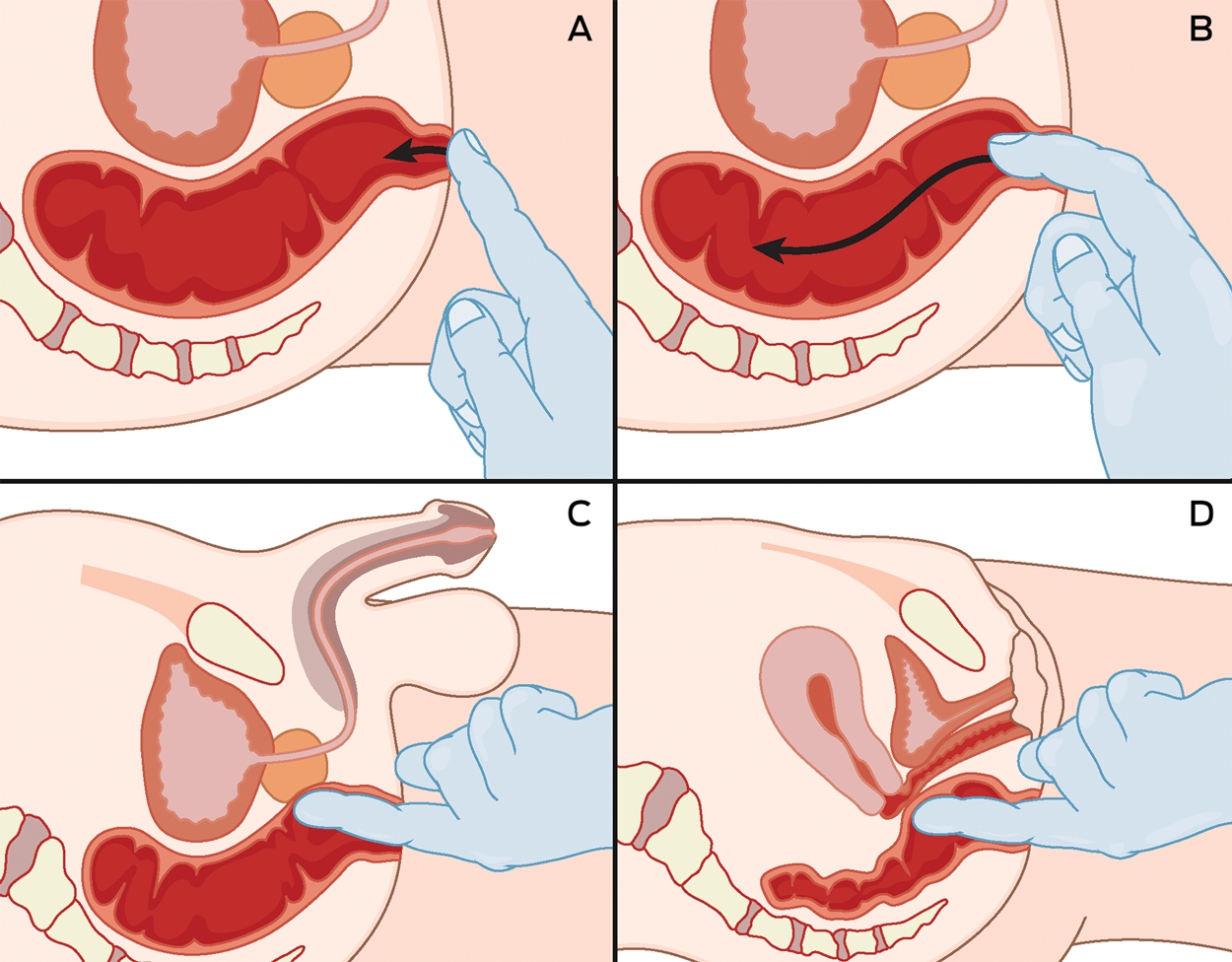
The examination procedure is shown in Box 2. The physician should explain the procedure to the patient and that it may be uncomfortable. It should not be painful, but the presence of pain may suggest pathology. If indicated, have a chaperone present, particularly with patients of the opposite sex.
Position the patient on an examining couch or table in the left lateral position and ask them to remove their lower clothing and draw their knees up to their chest. Ensure that adequate light is present and that the patient is comfortable and not likely to fall off the examination table or couch (a potential cause of litigation).
After parting the buttocks, carefully examine the perianal area, particularly for abscesses, skin conditions such as dermatitis and psoriasis, fissures (a clue is the presence of a sentinel pile or skin tag), fistulae and external haemorrhoids (Box 3). Ask the patient to bear down to assess for rectal prolapse.3 Next place the gloved index finger, with ample water-soluble lubricant, on the posterior part of the anal verge (Box 2, A) and rest for a few seconds to allow the external sphincter to relax. Then rotate the examining finger very slowly in a clockwise direction, similar to a corkscrew, passing from the anus into the rectum, with the finger aimed towards the umbilicus, given that the anal canal runs from the anal verge to the anorectal junction in this direction. Once the anorectal junction is reached, direct the examining finger 90 degrees posteriorly towards the sacrum and advance, as the rectum follows the sacral concavity (Box 2, B). Watch the patient for signs of pain such as grimacing.
Anal sphincter tone can be crudely measured by asking the patient to squeeze the examining finger. Anal sphincter defects (particularly anteriorly) may also be evident. Severe pain is suggestive of a fissure even if not readily visible and, in such instances, the examination should be aborted. Suspected pelvic floor dysfunction may be evaluated with additional special tests.3 Carefully palpate the rectal mucosa anteriorly, posteriorly and laterally for masses (soft, hard, irregular or smooth) and prostatic abnormalities in men (Box 2, C), as well as ovarian and uterine abnormalities in women (Box 2, D). Estimate the size of any palpable nodules or masses. In women, the cervix can often be felt and give the false impression of a mass. Foreign bodies at times may also be encountered. Pelvic peritoneal irritation secondary to acute appendicitis and pelvic inflammatory disease can also be assessed.
On withdrawing the examining finger, look on the glove for visible blood as well as faecal consistency and colour (eg, pale, melaena). Faeces on the examining glove can also be tested for the presence of occult blood using an immunochemical method. The sensitivity and specificity for detecting colorectal neoplasia (carcinomas and colonic adenomas > 1 cm) has been reported as being 60% and 95%, respectively.4 If indicated, proctoscopy and/or rigid sigmoidoscopy may also be carried out at this time with disposable instruments. After completion, clean the perianal area with a tissue to remove any leftover lubricant and faeces.
DRE can be limited by the body habitus of the patient, as well as by the length of the examining finger. For example, it may not be possible to penetrate deep into the rectum, given that it is 12–15 cm in length, resulting in proximal abnormalities being missed. In such instances, when symptoms are suggestive of colorectal pathology, the patient should be referred for flexible sigmoidoscopy or colonoscopy. With regard to detecting prostate cancer, the sensitivity of DRE has been reported at 59%, with a specificity of 94%.5 DRE is rarely indicated in children and, when absolutely necessary, it may be best to use the little, rather than index finger, depending on the child’s age.
DRE is an often neglected but important part of the physical examination and should be performed whenever symptoms suggest anorectal or prostatic pathology. The procedure, including its potential benefits, needs to be carefully explained to patients.
Box 1 – Indications for digital rectal examination
- Bleeding from the rectum
- Mucus discharge from the rectum
- Change in bowel habit
- Faecal urgency
- Obstructed/difficult defecation1
- Faecal incontinence
- Anorectal pain (but avoid digital examination when an obvious fissure is seen on inspection)
- Prostatic symptoms: eg, nocturia, hesitancy, poor stream, difficulty starting urination, dribbling after urination
- Cauda equina syndrome (anal tone)
Box 3 – Information obtained from digital rectal examination
|
Inspection |
Palpation |
||||||||||||||
|
|
|||||||||||||||
|
Skin tags |
Rectal mass |
||||||||||||||
|
External haemorrhoids: protruding or thrombosed |
Pelvic peritoneal irritation |
||||||||||||||
|
Anal fissure |
Polyps |
||||||||||||||
|
Fistulae |
Stenosis |
||||||||||||||
|
Abscesses |
Prostatomegaly |
||||||||||||||
|
Skin conditions: eg, eczema, dermatitis, psoriasis |
Prostate mass |
||||||||||||||
|
Anal warts |
Foreign bodies |
||||||||||||||
|
Anal cancer |
Anal tone |
||||||||||||||
|
Pilonidal sinuses |
Melaena |
||||||||||||||
|
Rectal prolapse |
Steatorrhoea |
||||||||||||||
|
|
Extrarectal pathology: eg, uterine and ovarian masses |
||||||||||||||
|
|
|||||||||||||||
|
|
|||||||||||||||
Competing interests
No relevant disclosures.
References
- Tantiphlachiva K, Rao P, Attaluri A, et al. Digital rectal examination is a useful tool for identifying patients with dyssynergia. Clin Gastroenterol Hepatol 2010; 11: 955-960.
- Wong RK, Drossman DA, Bharucha AE, et al. The digital rectal examination: a multicentre survey of physicians’ and students’ perceptions and practice patterns. Am J Gastroenterol 2012; 107: 1157-1163.
- Talley NJ. How to do and interpret a rectal examination in gastroenterology. Am J Gastroenterol 2008; 103: 820-822.
- Hoepffner N, Shastri YM, Hanisch E, et al. Comparative evaluation of a new bedside faecal occult blood test in a prospective multicentre study. Aliment Pharmacol Ther 2006; 23: 145-154.
- Hoogendam A, Buntinx F, de Vet HC. The diagnostic value of digital rectal examination in primary care screening for prostate cancer: a meta-analysis. Fam Pract 1999; 16: 621.
Provenance: Commissioned; externally peer reviewed.
Early-Onset Colorectal Cancer With Liver-Only Metastases: A Retrospective Cohort Study Integrating Prospectively Collected Real-World Clinical and Molecular Data From an Australian National Database (2009–2024) to Guide Treatment Planning
Savio G. Barreto, Christos S. Karapetis, Shahid Ullah, Matthew Burge, Susan Caird, Angus Campbell, Azim Jalali, Ross Jennens, Muhammad A. Khattak, Belinda Lee, Stephanie H. Lim, Shehara Mendis, Louise Nott, Timothy J. Price, Jeremy D. Shapiro, Jeanne Tie, Javier Torres, Colin Williams, Rachel Wong, Vanessa Wong, Peter Gibbs
Australian Pathways for Specialist Pain Management and Early Palliative Care for People With Pancreatic Cancer: Developed Using a Community Consensus Approach
Jennifer Philip, Melanie R. Lovell, Kylee Bellingham, Gail Garvey, Gregory B. Crawford, Nicole M. Rankin, Kara Burns, Isabel Young, Vivienne Milch, Dorothy Keefe, Katrina Anderson, James Lawson, Meinir Krishnasamy
Cause‐specific mortality among Queensland people with cirrhosis, by cirrhosis aetiology and decompensation status, 2007–22: a retrospective cohort study
Vikas Bhasker, Jessica R Fong, Paul J Clark, Gunter F Hartel, Richard Skoien, James O’Beirne, Elizabeth E Powell, Patricia C Valery
Clinical practice guidelines for hepatocellular carcinoma surveillance for people at high risk in Australia: summary of recommendations
Jacob George, Nicole L Allard, Stuart K Roberts, Leon A Adams, Jane Davies, Behzad Hajarizadeh, Jennifer H MacLachlan, Suzanne E Mahady, Rosalie Altus, Catherine Brown, David C Fry, Belinda Greenwood‐Smith, Natali Smud, Patricia C Valery, Nafisa Yussf, Kate Broun, Denise Campbell, Karen Canfell, Chelsea Carle Harrison, Victoria Freeman, Paul Grogan, Catherine Holliday, Suzanne Hughes, Anna Kelly, Cathelijne Kemenade, Claire Latumahina, Amanda McAtamney, Megan Varlow, Joachim Worthington, Susan Yuill, Eleonora Feletto
Assessment of metabolic dysfunction‐associated fatty liver disease in primary care: a consensus statement summary
Leon A Adams, William W Kemp, Kate R Muller, Elizabeth E Powell, Stuart K Roberts, Luis Calzadilla Bertot, Stephanie Best, Gary Deed, Jon D Emery, Samantha L Hocking, Graham R Jones, John S Lubel, Sinead Sheils, Stephen M Twigg, Gerald F Watts, Jacob George
Treatment outcomes for people with hepatitis C referred to tertiary care in Victoria, 2021–22: a retrospective observational study
Elly Layton, Nicole Matthews, Brendan Quinn, Nasra Higgins, Gabrielle Lindeman, Mielle Abbott, Jennifer MacLachlan, Elizabeth Birbilis, Margaret E Hellard, Joseph Doyle, Benjamin C Cowie, Mark Stoové