




Volume 207 - Issue 3
Three-dimensional printing in medicine
Authors: Jasamine Coles-Black, Ian Chao and Jason Chuen
Med J Aust 2017; 207 (3): 102-103. || doi: 10.5694/mja16.01073
Published online: 7 August 2017
Published online: 7 August 2017
Three-dimensional printing could revolutionise the way we practise medicine
Three-dimensional printing could revolutionise the way we practise medicine
Three-dimensional (3D) printing allows digital 3D models to be converted into physical ones with unprecedented accessibility. It has been hailed as a “disruptive technology” — a term used to describe advances that could change the status quo. The technology involves building 3D structures by depositing material layer upon layer to form a 3D object. This process is achieved via several ways, most commonly via fused deposition modelling (FDM), selective laser sintering and stereolithography.
The most accessible 3D printing technique is FDM, which costs from $300 upwards. It involves the extrusion of heat-softened plastic filaments via a movable printer head to form the 3D structure. The printers are no larger than standard paper printers. The “ink” used is cheap polymer filaments — with a cost of $30 per kilogram roll — such as the thermoplastics acrylonitrile butadiene styrene and polylactic acid, which result in hard plastic models. In addition, newer materials have entered the market, including thermoplastic polyurethane, which is used to produce flexible models. FDM is, therefore, the most accessible 3D printing technology for medical applications.
Selective laser sintering involves sintering powders, such as metals and ceramics, layer by layer. These machines cost tens of thousands of dollars, and the print mediums are also expensive, thus limiting its use to larger institutions. Its applications include printing patient-specific titanium devices.
Another method is stereolithography, which uses ultraviolet lasers on a vat of ultraviolet curable photopolymer, solidifying specified parts on the liquid’s surface to form models (Box 1). Biodegradability is the limiting factor of these models; however, this property has been exploited to create scaffolds for tissue engineering.
Generating three-dimensional models from medical images
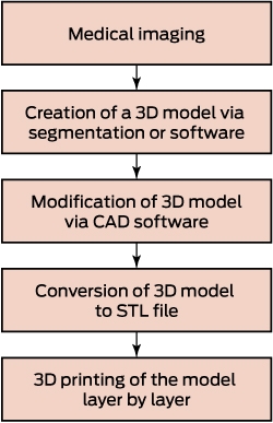
Three-dimensional printed models are readily achieved from computed tomography scans and magnetic resonance imaging. Segmentation software converts imaging into digital models in standard triangle language format, which can then be 3D printed. In addition, 3D models can also be modified or generated for printing de novo using computer-aided design software; for instance, in our laboratory, we have successfully applied a workflow as an adjunct to clinical care using a variety of 3D printers (Box 2).
Applications for three-dimensional printing
Interventional planning
Three-dimensional printed models have been used to assist surgical plans, allowing for surgical rehearsal, and thus reducing operating time and trauma. Orthopaedic case reports have shown a reduction in surgical time, time under anaesthesia, intra-operative blood loss, and better reduction of complex fractures.1 For example, in a case report of recurrent anterior shoulder instability, conventional imaging was inadequate. Hence, a glenohumeral joint model was 3D printed from patient imaging2 to plan an arthroscopic Bankart repair and remplissage. The 3D model was commissioned at just $200, which is a small amount in orthopaedics. Similar reports are published in cardiac surgery3 and plastic surgery.4
Customised prostheses and implantable components
Individualised prostheses and orthoses can be tailor-made to the individual patient, avoiding the need for off-the-shelf prostheses to be modified and resulting in a better fit both mechanically and aesthetically. A study investigating 3D printed calcium phosphate implants for the reconstruction of cranial and maxillofacial defects found the process practical and uncomplicated,5 with the individualised implants showing a high degree of dimensional accuracy, adequate for producing craniofacial structures. Unlike conventional pre-fabricated titanium implants — which are not patient specific, are costly, cannot be integrated into the patient’s own bone tissue, and could potentiate infection — these implants degrade in vivo and are replaced by bone over time. Moreover, an advantage cited in the study is that the implants could be created in 4 hours.
Training and education
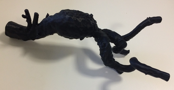
Another area of interest is the fabrication of realistic, inexpensive simulation models, as there is currently a dearth of realistic models in the field (Box 3).6 An example includes temporal bone models printed in acrylonitrile butadiene styrene filament,7 which expert surgeons considered an appropriate and accurate representation of a human temporal bone. Traditional simulation models can cost several hundred dollars, and cadaveric specimens are difficult to access for practitioners working outside of major universities, along with ethical, cultural, contamination and storage concerns associated with cadavers.8
In addition, life-sized 3D printed models have also been used to improve patient understanding of fetal malformations9 and for patient education for people undergoing partial nephrectomy.10
Pharmacology
A 3D printed polypill with separate release profiles for three different drugs has been developed for patients with diabetes who have hypertension.11 This allows complex medication regimes to be combined into a single tablet tailored to the requirements of each patient. 3D printed tablets are imminent, with the United States Food and Drug Administration recently approving a 3D printed levetiracetam pill for seizures.12
Bioprinting and tissue engineering
Regenerative medicine is the single largest application of 3D printing in the literature. Bioprinting involves the combination of living cells, biocompatible materials, and biochemical and physical substances in order to create tissue-like analogues. 3D printed scaffolds can be seeded with cells, but tissues can also be 3D printed using actual cells. The field, although older than other medical applications of 3D printing, has been slow to translate into clinical practice. The three most promising tissues at present include skin, cartilage and liver.13
Barriers remaining in three-dimensional printing for medical applications
Despite the promise 3D printing has shown in the medical literature, clinicians often lack the technical skills required to produce these models.
There are also few approved biocompatible materials to print patient-specific implants.14 Machines that print metal or extracellular matrices are still more expensive than those that print polymers. However, the patent for selective laser sintering metal 3D printers has recently expired and, as has occurred with FDM printers, the costs are likely to reduce.
Apart from humanitarian exceptions targeting rare diseases, there are still no regulatory standards addressing how personalised 3D printed medical devices should be manufactured.
Conclusion
Three-dimensional printing is growing in medical disciplines where novel approaches to complex anatomical relationships have aided therapeutic interventions. An increasing number of institutions are recognising its disruptive potential, establishing institutional hubs and pursuing collaborations with industry leaders.
It is imperative to conduct further studies into the present obstacles and limitations that may impede the adoption of 3D printing by the wider medical community. Nevertheless, current applications of 3D printing in the medical field are promising and, in the future, they may be the norm; but only time will tell if this new technology will truly disrupt the way we approach medical care.
Box 1 – A three-dimensional printed aortic valve and coronary arteries printed on a stereolithography printer*
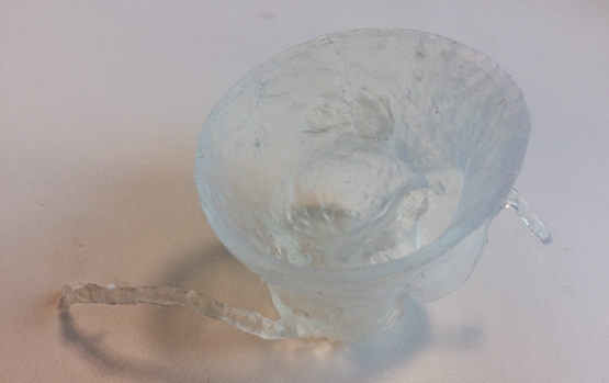
* The model was used in the pre-operative planning of a David procedure.
Competing interests
No relevant disclosures.
Acknowledgements
We thank the 3D Medical Printing Laboratory at Austin Health, Victoria, for use of their images. We also thank the Austin Medical Research Foundation and the Harold and Cora Brennen Benevolent Trust for funding this work.
References
- Giovinco NA, Dunn SP, Dowling L, et al. A novel combination of printed 3-dimensional anatomic templates and computer-assisted surgical simulation for virtual preoperative planning in Charcot foot reconstruction. J Foot Ankle Surg 2012; 51: 387-393.
- Sheth U, Theodoropoulos J, Abouali J. Use of 3-dimensional printing for preoperative planning in the treatment of recurrent anterior shoulder instability. Arthrosc Tech 2015; 4: e311-e316.
- Hossien A, Nithiarasu P, Cheriex E, et al. A multidimensional dynamic quantification tool for the mitral valve. Interact Cardiovasc Thorac Surg 2015; 21: 481-487.
- Chae MP, Lin F, Spychal RT, et al. 3D-printed haptic “reverse” models for preoperative planning in soft tissue reconstruction: a case report. Microsurgery 2014; 35: 148-153.
- Klammert U, Gbureck U, Vorndran E, et al. 3D powder printed calcium phosphate implants for reconstruction of cranial and maxillofacial defects. J Craniomaxillofac Surg 2010; 38: 565-570.
- Tai BL, Rooney D, Stephenson F, et al. Development of a 3D-printed external ventricular drain placement simulator: technical note. J Neurosurg 2015; 123: 1070-1076.
- Mowry SE, Jammal H, Myer C, et al. A novel temporal bone simulation model using 3D printing techniques. Otol Neurotol 2015; 36: 1562-1565.
- McMenamin PG, Quayle MR, McHenry CR, Adams JW. The production of anatomical teaching resources using three-dimensional (3D) printing technology. Anat Sci Educ 2014; 7: 479-486.
- Werner H, Rolo LC, Araujo Júnior E, Dos Santos J. Manufacturing models of fetal malformations built from 3-dimensional ultrasound, magnetic resonance imaging, and computed tomography scan data. Ultrasound Q 2014; 30: 69-75.
- Bernhard J, Isotani S, Matsugasumi T, et al. Personalized 3D printed model of kidney and tumor anatomy: a useful tool for patient education. World Urol 2015; 34: 337-345.
- Khaled SA, Burley JC, Alexander MR, et al. 3D printing of tablets containing multiple drugs with defined release profiles. Int J Pharm 2015; 643-650.
- Kurzrock R, Stewart DJ. Click chemistry, 3D-printing, and omics: the future of drug development. Oncotarget 2016; 7: 2155-2158.
- Berthiaume F, Maguire TJ, Yarmush ML. Tissue engineering and regenerative medicine: history, progress, and challenges. Annu Rev Chem Biomol Eng 2011; 2: 403-430.
- Cheng GZ, San Jose Estepar R, Folch E, et al. Three-dimensional printing and 3D slicer: powerful tools in understanding and treating structural lung disease. Chest 2016; 149: 1136-1142.
Provenance: Not commissioned; externally peer reviewed.
Rapid Growth in Diagnostic CT Knee Imaging in Australia: Implications for Imaging Stewardship
Hanwen Chen, Rachelle Buchbinder, Sean Docking, Ian A Harris
Disease‐specific survival for people with chronic myeloid leukaemia, South Australia, 1980–2020: a retrospective cohort study
Kerri Beckmann, Brendon J Kearney, Naranie Shanmuganathan, David Yeung, Tim Hughes
Technological advances in the search for a CURE for asthma
Sara Quon, Timothy M Johanson, Ridhima Wadhwa, Alen Faiz, Anthony Flynn, Gary P Anderson, Amanda J Cox, Nicholas P West, Michael P Menden, Rhys S Allan
Who gets asthma, and why?
Denby J Evans, Peter D Sly, Paul Foster, Chantal Donovan
The first Australian evidence‐based guidelines on male infertility
Darren J Katz, Liza O’Donnell, Robert I McLachlan, Tim J Moss, Clare V Boothroyd, Veena Jayadev, Sarah R Catford
Sex differences in management and outcomes of people with ST‐elevation myocardial infarction, New South Wales, 2011–2020: a retrospective cohort study
Samia Kazi, Simone Marschner, Haeri Min, Desi Quintans, James JH Chong, Ehsan Khan, David B Brieger, Clara K Chow