




Medical education
Clinical skills
Volume 205 - Issue 1
Ankles jerk — yes, but how?
Author: David B Williams
Med J Aust 2016; 205 (1): 14-15. || doi: 10.5694/mja16.00355
Published online: 4 July 2016
Published online: 4 July 2016
Eliciting an ankle jerk requires the skills of an experienced bell-ringer, an expert golfer and a mechanical engineer
Eliciting an ankle jerk is not a trivial task. It requires the skills of an experienced bell-ringer, an expert golfer and a mechanical engineer. The experience of the examiner and the positioning of the patient are two factors contributing to the accuracy of reported findings.1,2 Accurate findings can provide invaluable clinical information.
When an ankle jerk is elicited, the response can be seen, felt and heard. When the response is present, the tendon produces an undamped oscillation heard as a “boing”, and when it is absent, the damped oscillation is heard as a dull “thud”.
Percussion on the Achilles tendon activates muscle spindles — specialised sensory receptors within the muscle that respond to stretch (lengthening) and vibration. Action potentials move centripetally via large diameter, myelinated, fast conducting fibres, and trigger responses in the spinal cord from α motor neurones that mediate muscle contraction. The magnitude and rapidity of the reflex response is modulated by both autonomic sympathetic tone and descending (upper motor neurone) inhibition.3
Reflex activation is faster and stronger when sympathetic nervous system tone is high. Patients who are anxious, thyrotoxic or withdrawing from alcohol will have stronger, brisker responses. Similarly, if descending inhibition from upper motor neurones is reduced by, for example, stroke, myelopathy or Jendrassik (reinforcement) manoeuvre, there will be stronger, brisker responses.
An intact ankle jerk response confirms normal function in the large diameter, myelinated, sensory fibres, in spinal cord integration at the L5-S1 level, and in the α motor neurones projecting to skeletal muscle. Symmetrical reduction or loss of ankle jerks occurs early in the course of large fibre sensory neuropathy (eg, in diabetes). Asymmetrical loss of the ankle jerk is a common sign of radiculopathy but may also occur with rarer conditions such as plexopathy. Symmetrical exaggeration of ankle jerks may simply reflect increased sympathetic nervous system activity. Whether symmetrically brisk responses are pathological or not depends on the “company they keep”. If the exaggerated ankle jerks are accompanied by clonus, spasticity, spread of reflexes, or an extensor plantar response, they will be judged to be pathologically exaggerated. Asymmetrical exaggeration of one ankle jerk indicates the presence of an upper motor neurone abnormality (either ipsilateral or contralateral, depending on how rostral the lesion is).
Absent ankle jerks are often due to faulty technique. If the tendon is too slack, it will not vibrate when struck, and if it is too taut, the response will be diminished or inevident. If the head of the reflex hammer strikes adjacent bone or connective tissue as well as the tendon, any response may be attenuated or lost. This is the bell-ringing skill. If the head of the hammer is pushed against the tendon, rather than allowed to swing freely against it and bounce off unimpeded, the response may be decreased or absent. This is the skill of the experienced golfer. Patient positioning can affect tendon length, sympathetic tone and the magnitude of descending inhibition. This is the mechanical engineering skill.
Unfortunately, most medical students have only a hazy notion of the required skill and are often taught the patient positioning that is least likely to offer early success. There are at least five different positions that can be used to elicit ankle jerks, each with advantages and disadvantages. The positions are illustrated, and their relative merits described, in Figures 1 - 5. Additional dynamic factors are discussed in the video at mja.com.au.
Figure 1 – Patient kneeling backwards in a chair
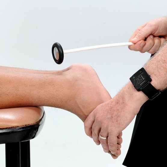
In this position, the lower segment muscles are relaxed and the tendon is near optimal stretch. However, the examiner needs to be aware that gripping the back of the chair is a default Jendrassik manoeuvre, exaggerating the response. This is desirable if the ankle jerk is thought to be absent. This method is reported to have the lowest rate of false-negative results.
Figure 2 – Patient with legs dangling over the edge of the bed
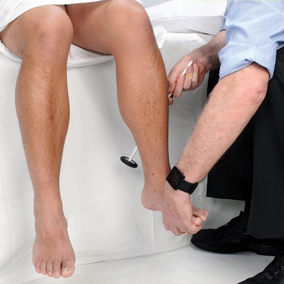
In this position, the examiner must gently position the ankle in the mid-position without the patient “helping” by contracting the ankle dorsiflexors and preventing the reflex plantar flexion.
Figure 3 – Patient seated in a chair with the feet beneath the knees
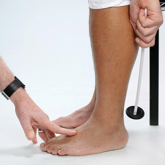
This position almost automatically produces lower limb relaxation and optimal tendon stretch. However, it requires an examiner to “grovel” a little in front of the patient.
Figure 4 – Patient supine in bed with legs relaxed
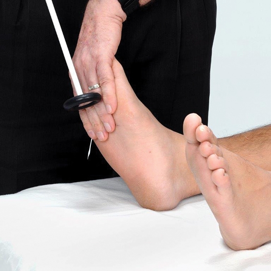
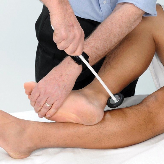
The technique illustrated here requires the examiner to strike his or her hand while it rests on the sole of the foot near the metatarsal heads. Patients will often become tense when first touched, and the examiner has to avoid excessive dorsiflexion at the ankle with this technique. It is said to be a more reproducible technique for general physicians, but it might also produce more false-negative results.
Figure 5 – Patient supine with hip abducted and externally rotated, knee flexed and ankle dorsiflexed
This traditional positioning is easily the most problematic. Patients for whom this position is uncomfortable, unseemly or simply unfamiliar will experience excess muscle tension. In addition, if the patient is unable to hold the position unaided, the examiner is tempted to stabilise the limb by strongly dorsiflexing the foot at the ankle, thereby lengthening the tendon excessively and reducing the chance of obtaining any response. The effect is complete if the examiner, failing to elicit a response, exhorts the patient to relax. Feeling implicitly blamed, the patient inevitably becomes more tense.
Competing interests
No relevant disclosures.
Series editors
Balakrishnan (Kichu) R Nair
Simon O’Connor
Series editors
Balakrishnan (Kichu) R Nair
Simon O’Connor
Acknowledgements
I thank future colleague Joshua Diamoy for his assistance in creating the photographs and video.
References
- O’Keeffe ST, Smith T, Valacio R, et al. A comparison of two techniques for ankle jerk assessment in elderly subjects. Lancet 1994; 344: 319-320.
- Oluwole OSA, Odebode TO, Komolafe MO. Effect of lower limb position on ankle jerk assessment. J Neurol Neurosurg Psychiatry 2001; 70: 266-267.
- Burke D, Gandevia SC, McKeon B. The afferent volleys responsible for spinal proprioceptive reflexes in man. J Physiol 1983; 339: 535-552.
- O’Brien MD. Ankle jerk assessment. Lancet 1995; 345: 331.
Provenance: Commissioned; externally peer reviewed.
Shifting focus to adolescent wellbeing and inclusive participation in the digital age
Allyson R Todd, Elena Wang, Stephanie R Partridge
The clinical presentation, investigation, and management of women diagnosed with endometriosis in Australian general practices, 2011–2021: an open cohort study
Danielle Mazza, Kailash Thapaliya, Sharinne B Crawford, Alissia Hui, Maryam Moradi, Luke E Grzeskowiak
The impact of the Breast Screen NSW transition from film to digital mammography, 2002–2016: a linked population health data analysis
Rachel Farber, Nehmat Houssami, Katy J L Bell
Non‐technical errors associated with deaths in surgical care, Australia, 2012–2019, by surgical specialty (Australian and New Zealand Audit of Surgical Mortality): a retrospective cohort study
Jesse Ey, Victoria Kollias, Octavia Lee, Kelly Hou, Matheesha Herath, John B North, Ellie Treloar, Suzanne Edwards, Martin Bruening, Adam J Wells, Guy J Maddern
The CURE Asthma roadmap
Gary P Anderson, Anthony Flynn, Phil G Bardin, John D Blakey, Shyamali C Dharmage, Paul Foster, Peter G Gibson, Adam Jaffe, Alan James, Christine R Jenkins, Sundram Sivamalai, Peter D Sly, Guy B Marks, Vanessa M McDonald, Judy Wetttenhall
Evidence from Australian cohort studies about asthma trajectories and transitions across the life course: a narrative review
Caroline Lodge, Xin Dai, Ingrid A. Laing, Michael P Menden, Anthony Flynn, Gary P Anderson, Sarath Ranganathan, Shyamali C Dharmage