




Volume 203 - Issue 5
Tattoo-associated mycobacterial infections: an emerging public health issue
Authors: John W Frew and Roland TD Nguyen
Med J Aust 2015; 203 (5): 223. || doi: 10.5694/mja15.00098
Published online: 7 September 2015
Published online: 7 September 2015
Mycobacterial infection is an important consideration in patients with widespread papular eruption in recent tattoos
Three men aged 21–24 years presented to our dermatology clinic with a 2-week history of pruritic erythematosquamous papules coalescing into plaques within areas of recent tattooing. The tattoos were done in Thailand 4 weeks before presentation (Box, A–E). The lesions were concentrated in areas of black shading and overlapping colours, and did not involve non-tattooed skin. All patients were afebrile, systemically well with no palpable lymphadenopathy. Investigations, including a full blood count, biochemistry and inflammatory markers, returned results within the normal range. Serological tests for HIV, hepatitis B, hepatitis C and syphilis were negative. Skin biopsies were performed on all three patients. Histopathology showed a suppurative granulomatous reaction with lymphohistiocytic infiltrate in the upper and mid dermis (Box, F–H). Modified Ziehl–Neelsen staining was negative for acid-fast bacilli. However, cultures showed Mycobacterium mucogenicum in Patients 1 and 2, and M. fortuitum in Patient 3. Empiric antibiotic therapy was commenced with oral clarithromycin 500 mg twice a day for 4 weeks. Patients 1 and 2 required 7 days of intravenous amikacin 750 mg daily and cefoxitin 2 g four times a day for failure to respond based on tissue culture and sensitivities.
The surge in interest in tattoo and body art over the past decade has also led to a surge in tattoo-related complications. While the risk of blood-borne disease and secondary bacterial infection is well known, infection with other organisms has received little publicity. However, it remains a significant public health risk,1 with outbreaks of tattoo-associated mycobacterial infection documented in the United States, France and Germany. Common causative organisms include M. chelonae, M. fortuitum and M. abscessus. Infections with Mycobacterium tuberculosis2 and M. leprae3 have also been reported. Mycobacteria are ubiquitous in the environment and species such as M. chelonae are commonly found in water supplies. However, they typically only cause clinical disease in the immunosuppressed host, or when high concentrations of the organism are introduced via surgery, trauma or tattooing. Sources of mycobacteria in tattooing include tattoo inks,4 with the chemical composition of differing pigments possibly promoting or suppressing organism growth.5 Purple inks (containing manganese) may have the potential for inhibiting organism growth, similar to the action of potassium permanganate used in dermatological practice. The dilution of inks with non-sterile water to produce gradations of colour can also introduce mycobacteria. The clinical distribution of papules in the reported cases demonstrates large numbers of papules coalescing at sites of shading and the borders between colour overlaps. This distribution coincides with areas of high puncture density to give desired colour gradations.
Mycobacterial infection is an important consideration in patients with widespread papular eruption in recent tattoos. Diagnosis can only be made on skin biopsy with tissue culture, and definitive antibiotic therapy should be directed by antimicrobial sensitivities.
Box
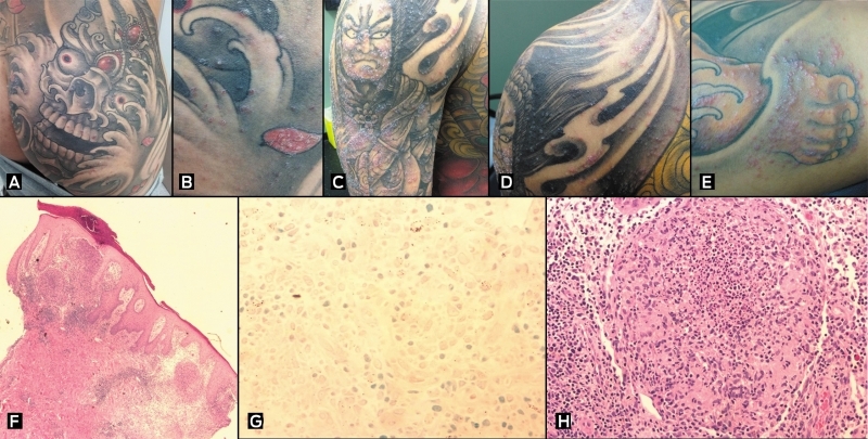
Erythematous scaled papules on presentation, of Patient 1 (A, B); Patient 2 (C, D); and Patient 3 (E). Representative histopathology from Patient 1 demonstrating multiple granulomas in the upper to mid dermis (F, magnification x 4), with a negative Ziehl–Neelsen stain (G, magnification x 80) and suppurative granuloma formation (H, magnification x 40).
Competing interests
No relevant disclosures.
Acknowledgements
We thank Dr C Henderson, Department of Anatomical Pathology, Liverpool Hospital, Sydney, for his contribution to this article.
References
- Shinohara MM, Nguyen J, Gardner J, et al. The histopathologic spectrum of decorative tattoo complications. J Cutan Pathol 2012; 39: 1110-1118.
- Wong HW, Tay YK, Sim CS. Papular eruption on a tattoo: a case of primary inoculation tuberculosis. Australas J Dermatol 2005; 46: 84-87.
- Ghorpade A. Inoculation (tattoo) leprosy: a report of 31 cases. J Eur Acad Dermatol Venereol 2002; 16: 494-499.
- Kennedy BS, Bedard B, Younge M, et al. Outbreak of Mycobacterium chelonae infection associated with tattoo ink. New Engl J Med 2012; 367: 1020-1024.
- Høgsberg T, Saunte DM Frimodt-Møller N, Serup J. Microbial status and product labelling of 58 original tattoo inks. J Eur Acad Dermatol Venereol 2013; 27: 73-80.
Diagnosing Acute Kava Dermopathy: A Case Report of a Characteristic Cutaneous Eruption
Ali Abid, Nicholas Allen, Abeer Hagelamin, Christopher Henderson, Artiene Tatian
Localised Herpes Simplex Following Midline Laparotomy
Jessica S. Bulluss, Paul Chee, Matthew J. Verheyden
No silver lining with health misinformation: argyria caused by intentional silver consumption
Luke Collins, Logesh Palanikumar, Stephen Bacchi
Lingual Raynaud phenomenon
Michael Taggart, Mina John
Melasma in the male: a less well recognised entity
Tim Aung, Rowland Noakes
Age group‐specific changes in keratinocyte cancer treatment rates in Australia, 2012–2021: a retrospective cohort study based on MBS claims data
Catherine M Olsen, Nirmala Pandeya, Rachel E Neale, David C Whiteman