




Volume 201 - Issue 8
Sentinel lymph node biopsy for melanoma: an important risk-stratification tool
Authors: David E Gyorki and Michael A Henderson
Med J Aust 2014; 201 (8): 442-444. || doi: 10.5694/mja14.00645
Published online: 20 October 2014
Published online: 20 October 2014
Biopsy should be routinely considered for intermediate thickness melanoma
This test should be routinely considered for patients with intermediate thickness melanoma
Most patients with a new diagnosis of melanoma present with localised disease without clinical evidence of metastasis and are cured by surgical resection. However, a small proportion will harbour occult metastatic disease in the regional lymph nodes. The presence of nodal involvement is the most significant prognostic factor in patients without clinical evidence of distant metastasis.1 The most sensitive test to identify these patients at presentation is sentinel lymph node biopsy (SLNB).1,2 The sentinel lymph node is the first draining lymph node from the area of skin where the primary tumour arose. The technique was first described over 20 years ago.3 Careful histological examination of the sentinel lymph node by a combination of routine histological and immunohistochemistry tests can accurately identify small-volume metastatic disease.
On the basis of initial reports, SLNB has rapidly become the standard of care in major melanoma centres around the world for patients with melanoma greater than 1 mm in thickness (stage T2 and above; Box 1) and incorporated into the American Joint Committee on Cancer staging system.1 Considerable controversy about the routine use of sentinel node biopsy has focused on the lack of a survival benefit from the procedure.4,5 In this review, we seek to put the rationale for SLNB into context in light of both advances in the management of patients with advanced melanoma and the recent publication of the final report of the Multicenter Selective Lymphadenectomy Trial (MSLT-I).2
The era before sentinel lymph node biopsy
Before the introduction of SLNB, a number of randomised controlled trials had compared elective lymph node dissection (ELND) with nodal observation; none of these trials showed an overall survival benefit for ELND.6 The Intergroup study was the most recent of these trials, and incorporated lymphoscintigraphy to identify the draining nodal basin. This study identified a number of subgroups which appeared to benefit from ELND, including patients with melanoma of Breslow thickness between 1 and 2 mm and patients aged less than 60 years.7 SLNB offered the benefit over ELND of identifying those patients who might benefit from early lymph node dissection while minimising the morbidity to patients without nodal involvement.
Prognostic impact of sentinel lymph node biopsy
The MSLT-I study randomly allocated patients to wide local excision (WLE) with SLNB, and immediate lymphadenectomy for nodal metastases, versus WLE and nodal observation with lymphadenectomy only at nodal relapse (Box 2). The primary end point of the study was disease-specific survival (DSS). The study finished accruing patients in 2002, and the final analysis was recently published.2
The MSLT-I study confirmed that lymph node status is the strongest predictor of DSS for patients with intermediate thickness melanoma (hazard ratio, 2.4; Box 3). Note that intermediate thickness in the MSLT-I study was Breslow thickness 1.2–3.5 mm; this was the thickness of melanomas in the primary analysis. There were too few patients (and events) with thin melanoma (Breslow thickness, < 1.2 mm), so no analysis of this subgroup has been provided to date. Other non-randomised studies have demonstrated the prognostic impact of SLNB in certain patients with thin melanoma (0.75–1.0 mm),9 although SLNB in this group of patients remains controversial.
Therapeutic benefit
The major area of controversy about SLNB is its therapeutic impact on DSS. MSLT-I did not show a significant difference in DSS between the two arms. The rate of sentinel lymph node positivity in the SLNB arm was only 16%, and this is the only population who might benefit from the procedure. MSLT-I was therefore likely underpowered to demonstrate an effect on DSS for the entire group.
A planned post-hoc analysis of node-positive patients was performed, comparing the outcomes of patients with nodal disease diagnosed at SLNB (biopsy-based detection) with the outcomes of those presenting with nodal relapse in the observation group. For patients with intermediate thickness (1.2–3.5 mm) melanoma with nodal involvement, this analysis showed a significant improvement in DSS, with 62.5% 10-year survival in the SLNB arm compared with 41.5% in the observation arm (P = 0.006). This difference remained significant even when patients with a false-negative sentinel node were included in the analysis (intention to treat, P = 0.04). For patients with thick (> 3.5 mm) melanoma, there was no survival benefit from early detection of nodal involvement, which likely reflects the high rate of distant metastatic disease in this less common, high-risk subgroup.
The validity of this post-hoc analysis has been questioned, as critics have suggested that SLNB identifies subclinical micrometastatic disease which is not destined to progress to palpable lymphadenopathy.10 Unlike other tumour types, such as breast cancer and differentiated thyroid cancer, where it is clear that micrometastatic disease can lie dormant without clinical significance, the results of MSLT-I and other non-randomised studies suggest that virtually all patients with microscopic disease will inevitably progress to clinical lymphadenopathy (10-year node-positive rate [mean ± SE], 21.9% ± 1.5% in the SLNB group compared with 19.5% ± 1.9% in the observation group).2 Furthermore, the mean number of lymph nodes harbouring metastatic melanoma in the SLNB arm was 1.4 compared with 3.3 for patients undergoing lymphadenectomy at nodal relapse in the observation arm (P < 0.001), suggesting progression during the period of observation.
Risks of sentinel lymph node biopsy
The major disadvantages of SLNB include cost and morbidity. For some patients, the use of SLNB converts their treatment from one deliverable under local anaesthesia to one requiring a general anaesthetic. Furthermore, SLNB requires preoperative lymphoscintigraphy. However, taking these factors into account, a cost-effectiveness analysis of SLNB in the Australian health care setting showed a significant improvement in quality-adjusted life-years with only a marginal increase in costs.11
Surgical complications from SLNB are seen in 5%–10% of patients, and are mostly minor in severity.8,12 The most common of these are early complications, including seroma and infection, and resolve over time. This is in stark contrast with lymphadenectomy where complication rates are significantly higher, particularly the serious long-term morbidity of lymphoedema.
Some have argued that ultrasound could replace SLNB as a non-invasive means of identifying lymph nodes harbouring metastatic disease.10 However, ultrasound is an operator-dependent technique and published results are variable.13,14 While ultrasound may select patients for immediate lymph node dissection, SLNB in these patients allows a thorough pathological assessment of nodal burden. The volume of disease, as well as the location of the metastatic deposit within the sentinel lymph node, provide independent prognostic information with regard to risk of further lymph node involvement as well as DSS.15,16
What to do with the information
The risk of a positive SLNB is associated with increasing Breslow thickness, presence of ulceration and mitotic count. The current clinical practice guidelines for managing melanoma in Australia and New Zealand recommend that “Patients with a melanoma greater than 1.0 mm in thickness be given the opportunity to discuss sentinel lymph node biopsy to provide staging and prognostic information”.17 At our institution, SLNB is recommended for patients with an expected rate of nodal involvement of greater than 5%. This includes all patients with melanoma greater than 1.0 mm thickness and patients with melanoma between 0.75 mm and 1.0 mm with ulceration or an increased mitotic count. For patients with significant comorbid conditions, the impact of the additional prognostic information provided by SLNB needs to be carefully weighed up against the added morbidity associated with the procedure.
The current standard of care for patients with a positive result on SLNB is completion lymph node dissection (CLND). The question of immediate CLND versus observation for patients with a positive SLNB result will be answered by the MSLT-II trial which will report in the next few years. In the interim, the risk of further lymph node involvement beyond the sentinel lymph node can be estimated from various characteristics of the sentinel lymph node including the size and the location of the tumour deposit within the lymph node.15,16,18 Patients with small deposits, particularly if less than 0.1 mm in size, have a very low risk of further lymph node involvement; this needs to be weighed up against the morbidity of the lymph node dissection as well as the risk of metastatic disease. On the flipside, CLND is associated with lower complication rates than delayed lymphadenectomy at the time of clinical progression,19 so if the risk of further nodal involvement is significant, early intervention may be preferable. Thus, patients with a positive SLNB result require a nuanced discussion in a melanoma multidisciplinary meeting where the individualised pros and cons of CLND are considered.
The past 5 years have seen a dramatic change in the landscape of systemic therapy options for patients with metastatic melanoma. As these therapeutic agents filter into the adjuvant setting, one of the most important roles of SLNB is to select patients for inclusion in these trials. Currently, adjuvant trials for clinically localised melanoma (many of which are recruiting in Australia) are confined to the higher risk subsets of patients, particularly those with a positive SLNB result. Future developments in molecular oncology may identify markers to supersede SLNB. However, currently, SLNB is the best stratification tool available. We strongly recommend that patients with a positive SLNB result should be offered inclusion in these trials with a hope of seeing these new therapeutic agents reach the clinic for routine adjuvant use.
Conclusion
Sentinel lymph node biopsy is the most important prognostic test for patients with intermediate thickness melanoma. It identifies patients at high risk of relapse and allows them to be offered involvement in trials of adjuvant therapy. Data from the MSLT-I study suggest that, for patients with intermediate thickness melanoma with spread to regional lymph nodes, biopsy-based detection of nodal disease using SLNB is associated with a significant improvement in DSS. For these reasons a discussion about SLNB should be standard of care for patients with melanoma greater than 1.0 mm thick.17
1 Primary melanoma staging and corresponding Breslow thicknesses
Melanoma T stage | Breslow thickness* | Description | |||||||||||||
T1 | < 1.0 mm | Thin melanoma | |||||||||||||
T2 | 1.0–2.0 mm | Intermediate thickness melanoma | |||||||||||||
T3 | 2.0–4.0 mm | Intermediate thickness melanoma | |||||||||||||
T4 | > 4.0 mm | Thick melanoma | |||||||||||||
* Tumour thickness measured between the upper layer of the epidermis and the deepest point of tumour penetration. | |||||||||||||||
2 Design for the Multicenter Selective Lymphadenectomy Trial (MSLT-I)
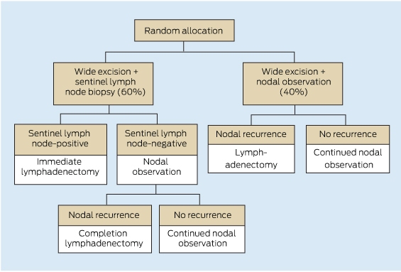
Stratification was by Breslow thickness (1.2–1.8 mm v 1.8–3.5 mm) and primary tumour site (limb v other site).8
3 Predictors of disease-specific survival2
Prognostic indicator | Hazard ratio | P | |||||||||||||
Sentinel node status (positive v negative result) | 2.40 | < 0.001 | |||||||||||||
Ulceration (present v absent) | 1.79 | 0.002 | |||||||||||||
Breslow thickness* (per 1 mm increase) | 1.59 | < 0.001 | |||||||||||||
Sex (male v female) | 1.22 | 0.32 | |||||||||||||
Clark level† (IV or V v III) | 1.07 | 0.73 | |||||||||||||
* Tumour thickness measured between the upper layer of the epidermis and the deepest point of the tumour; see Box 1. † Measure of tumour penetration into the layers of the skin, defined as follows: Level l, confined to the epidermis; Level II, invasion of the papillary dermis; Level III, filling of the papillary dermis, but no extension in to the reticular dermis; Level IV, invasion of the reticular dermis; Level V, invasion of subcutaneous tissue. | |||||||||||||||
Competing interests
No relevant disclosures.
References
- Balch CM, Gershenwald JE, Soong SJ, et al. Final version of 2009 AJCC melanoma staging and classification. J Clin Oncol 2009; 27: 6199-6206. _ENREF_1
- Morton DL, Thompson JF, Cochran AJ, et al. Final trial report of sentinel-node biopsy versus nodal observation in melanoma. N Engl J Med 2014; 370: 599-609. _ENREF_2
- Morton DL, Wen DR, Wong JH, et al. Technical details of intraoperative lymphatic mapping for early stage melanoma. Arch Surg 1992; 127: 392-399. _ENREF_3
- Thomas JM. Where is the evidence base for benefits of sentinel node biopsy in melanoma? BMJ 2013; 346: f675. _ENREF_4
- Torjesen I. Sentinel node biopsy for melanoma: unnecessary treatment? BMJ 2013; 346: e8645. _ENREF_5
- Lens MB, Dawes M, Goodacre T, Newton-Bishop JA. Elective lymph node dissection in patients with melanoma: systematic review and meta-analysis of randomized controlled trials. Arch Surg 2002; 137: 458-461. _ENREF_6
- Balch CM, Soong SJ, Bartolucci AA, et al. Efficacy of an elective regional lymph node dissection of 1 to 4 mm thick melanomas for patients 60 years of age and younger. Ann Surg 1996; 224: 255-263; discussion 63-66. _ENREF_7
- Morton DL, Thompson JF, Cochran AJ, et al. Sentinel-node biopsy or nodal observation in melanoma. N Engl J Med 2006; 355: 1307-1317. _ENREF_11
- Han D, Zager JS, Shyr Y, et al. Clinicopathologic predictors of sentinel lymph node metastasis in thin melanoma. J Clin Oncol 2013; 31: 4387-4393. _ENREF_8
- Thomas JM. Prognostic false-positivity of the sentinel node in melanoma. Nat Clin Pract Oncol 2008; 5: 18-23. _ENREF_9
- Morton RL, Howard K, Thompson JF. The cost-effectiveness of sentinel node biopsy in patients with intermediate thickness primary cutaneous melanoma. Ann Surg Oncol 2009; 16: 929-940. _ENREF_10
- Wrightson WR, Wong SL, Edwards MJ, et al. Complications associated with sentinel lymph node biopsy for melanoma. Ann Surg Oncol 2003; 10: 676-680. _ENREF_12
- Sanki A, Uren RF, Moncrieff M, et al. Targeted high-resolution ultrasound is not an effective substitute for sentinel lymph node biopsy in patients with primary cutaneous melanoma. J Clin Oncol 2009; 27: 5614-5619. _ENREF_13
- Voit C, Van Akkooi AC, Schäfer-Hesterberg G, et al. Ultrasound morphology criteria predict metastatic disease of the sentinel nodes in patients with melanoma. J Clin Oncol 2010; 28: 847-852. _ENREF_14
- Murali R, Desilva C, Thompson JF, Scolyer RA. Non-Sentinel Node Risk Score (N-SNORE): a scoring system for accurately stratifying risk of non-sentinel node positivity in patients with cutaneous melanoma with positive sentinel lymph nodes. J Clin Oncol 2010; 28: 4441-4449. _ENREF_15
- van der Ploeg AP, van Akkooi AC, Rutkowski P, et al. Prognosis in patients with sentinel node-positive melanoma is accurately defined by the combined Rotterdam tumor load and Dewar topography criteria. J Clin Oncol 2011; 29: 2206-2214. _ENREF_16
- Australian Cancer Network Melanoma Guidelines Revision Working Party. Clinical practice guidelines for the management of melanoma in Australia and New Zealand. Sydney and Wellington: Cancer Council Australia, Australian Cancer Network and New Zealand Guidelines Group, 2008. http://www.cancer.org.au/content/pdf/HealthProfessionals/ClinicalGuidelines/ClinicalPracticeGuidelines-ManagementofMelanoma.pdf (accessed Aug 2014).
- van der Ploeg AP, van Akkooi AC, Haydu LE, et al. The prognostic significance of sentinel node tumour burden in melanoma patients: an international, multicenter study of 1539 sentinel node-positive melanoma patients. Eur J Cancer 2014; 50: 111-120. _ENREF_18
- Faries MB, Thompson JF, Cochran A, et al. The impact on morbidity and length of stay of early versus delayed complete lymphadenectomy in melanoma: results of the Multicenter Selective Lymphadenectomy Trial (I). Ann Surg Oncol 2010; 17: 3324-3329. _ENREF_19
Provenance: Not commissioned; not externally peer reviewed.
Diagnosing Acute Kava Dermopathy: A Case Report of a Characteristic Cutaneous Eruption
Ali Abid, Nicholas Allen, Abeer Hagelamin, Christopher Henderson, Artiene Tatian
Localised Herpes Simplex Following Midline Laparotomy
Jessica S. Bulluss, Paul Chee, Matthew J. Verheyden
No silver lining with health misinformation: argyria caused by intentional silver consumption
Luke Collins, Logesh Palanikumar, Stephen Bacchi
Lingual Raynaud phenomenon
Michael Taggart, Mina John
Melasma in the male: a less well recognised entity
Tim Aung, Rowland Noakes
Age group‐specific changes in keratinocyte cancer treatment rates in Australia, 2012–2021: a retrospective cohort study based on MBS claims data
Catherine M Olsen, Nirmala Pandeya, Rachel E Neale, David C Whiteman