




Notable Cases
Volume 197 - Issue 8
Australia’s first liver–intestinal transplant
Authors: Mayur Garg, Robert M Jones, Darius Mirza, Bao Zhong Wang, Michael A Fink, Graham Starkey, Rhys B Vaughan and Adam G Testro
Med J Aust 2012; 197 (8): 463-465. || doi: 10.5694/mja12.10605
Published online: 15 October 2012
Published online: 15 October 2012
With the completion of the first liver-intestinal transplant in Australia in July 2010, the Austin Hospital in Victoria became one of only 35 active centres worldwide to offer this life-saving procedure to patients with irreversible intestinal failure and associated life-threatening complications of parenteral nutrition.Clinical recordA 32-year-old man was initially referred to our centre in 2005 after the progression of multiple complications of long-term parenteral nutrition (PN) for ...
With the completion of the first liver–intestinal transplant in Australia in July 2010, the Austin Hospital in Victoria became one of only 35 active centres worldwide to offer this life-saving procedure to patients with irreversible intestinal failure and associated life-threatening complications of parenteral nutrition.
Clinical record
A 32-year-old man was initially referred to our centre in 2005 after the progression of multiple complications of long-term parenteral nutrition (PN) for short gut syndrome. He had concomitant bilateral hydronephrosis and dilated renal pelvises, suggesting a visceral myopathy. A long-strip rectal biopsy confirmed hypoganglionosis.
The patient had experienced bowel dysfunction since birth, manifest as constipation and recurrent functional large bowel obstruction. By the age of 18 years, he had had multiple operations including a right pyeloplasty, later followed by a nephrectomy, a total colectomy, and multiple small bowel resections and bypasses. After an end-jejunostomy, an estimated 90 cm of the jejunum remained; hence, PN was commenced in 1998.
His subsequent treatment was complicated by prerenal renal dysfunction, multiple central venous access-related thromboses and polymicrobial infections, with isolation of multiple organisms including Staphylococcus aureus and Candida species. In 1999, he developed jaundice, and a liver biopsy showed changes consistent with chronic cholestasis, leading to a diagnosis of intestinal failure-associated liver disease (IFALD). With increasing portal hypertension manifest as oesophageal varices, splenomegaly and pancytopenia, he was referred to our unit for consideration of combined liver-intestinal transplantation, a procedure never previously performed in Australia.
In the months leading up to his transplant, he developed progressive jaundice (total serum bilirubin concentration approaching 300 μmol/L; reference interval [RI], < 20 μmol/L) and hypoalbuminaemia (albumin concentration, 21 g/L; RI, 35–45 g/L).
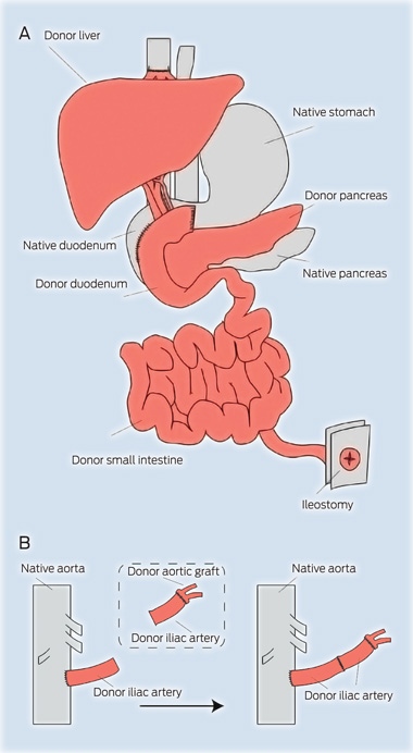
On 2 July 2010, a combined team of 20 surgeons, anaesthetists and theatre staff performed the cadaveric donor liver–intestinal transplant in an operation that lasted 13.5 hours (Box 1).
The ideal donor for a combined liver–intestinal transplant is younger than 50 years, of compatible blood group, and smaller in size than the recipient, with no history of prolonged hypotension or of conditions requiring inotropic therapy.
Before the donor organs were retrieved, selective gut decontamination was performed with an antibacterial and antifungal lavage. The donor operation comprised en-bloc resection of the liver and biliary tree in continuity with the duodenum, pancreas and small intestine. The arterial supply to the gut was taken back to the origin of the coeliac and superior mesenteric arteries, which were dissected on a common “patch” of abdominal aortic wall. Donor iliac arteries were also retrieved. The cold ischaemic time for the donor organs was 6.5 hours.
On the back table, the arterial anastomoses were performed (Box 1B). A donor iliac artery was divided in two. One of these arterial grafts was anastomosed to the donor aortic patch, including the origin of the coeliac axis and superior mesenteric artery. The other donor iliac artery graft was anastomosed end-to-side to the recipient infrarenal aorta (Box 1B).
The recipient operation involved removal of the diseased native liver and residual intestine. The recipient’s duodenum and pancreas remained in situ. An end-to-side portacaval shunt was performed, allowing venous drainage of the native pancreaticoduodenal complex into the systemic circulation. The donor liver, pancreas and small bowel were then implanted en bloc (Box 1A). The organs were reperfused after the end-to-end anastomosis of the two iliac arterial grafts as described above (Box 1B). The donor suprahepatic inferior vena cava was anastomosed to the recipient right, middle and left hepatic veins in a standard “piggyback” fashion. A side-to-side duodeno–duodenal anastomosis was performed, and an end ileostomy was fashioned. A nasojejunal tube was inserted for initial enteral feeding.
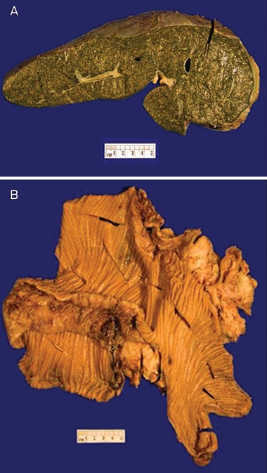
The explanted organs are shown in Box 2. Histopathological examination showed cirrhosis of the liver with severe canalicular cholestasis and hepatolithiasis (Box 2A). The intestinal remnant was 51 cm long and showed good villous adaptation and serosal fibrosis (Box 2A), consistent with previous surgery. Interestingly, there were no specific features of enteropathy, with plentiful ganglion cells and no abnormality of the myenteric plexus.
The patient’s immunosuppression regimen comprised the anti-CD25 antibody basiliximab given on Day 1 and Day 4, with 2 g of intravenous methylprednisolone given at the induction of anaesthesia, and a further 1 g given immediately before reperfusion. Treatment with tacrolimus was started on Day 1, intravenously for the first 10 days, and then enterally, aiming for a trough level of 15–20 ng/mL. Intravenous methylprednisolone was reduced gradually to 20 mg daily until full enteral function was restored on postoperative Day 23, at which stage treatment was started with 20 mg of oral prednisolone daily. The preferred, modern approach of preconditioning using a lymphocyte depleting agent, such as antithymocyte globulin or alemtuzumab (a monoclonal anti-CD52 antibody), was contraindicated in this case because of the severe underlying thrombocytopenia and portal hypertension.
The patient was monitored for rejection using protocol ileal biopsies twice a week for 4 weeks and then weekly, to enable rejection to be detected before the appearance of clinical symptoms or signs. There was evidence of mild histologic rejection on biopsies on Day 73. He was treated with a bolus of intravenous methylprednisolone, and good histological recovery was observed with no clinical sequelae.
Given the susceptibility of the patient to local and systemic infections, he was treated with prophylactic systemic antimicrobial therapy, including the antibacterial agents meropenem, vancomycin and co-trimoxazole; (combined trimethoprim and sulfamethoxazole) the antifungal agents caspofungin, topical nystatin and amphotericin; and the antiviral agent gancyclovir. He was monitored weekly for cytomegalovirus and Epstein–Barr virus.
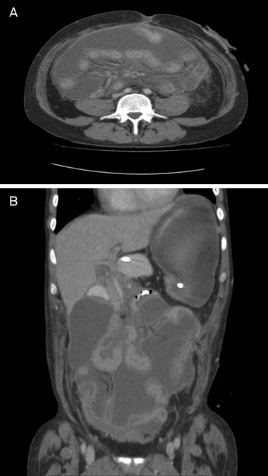
Mycoplasma hominis-infected chylous ascites became evident on Day 27 (Box 3), when the patient experienced generalised abdominal distension and pain. This resolved with intravenous antibiotic therapy and fat-free feeding for 2 weeks. On Day 83, the patient developed sepsis of the peripherally inserted central catheter line. He was treated with intravenous antibiotics, and the line was removed.
Nasojejunal feeding was started on Day 4, and was increased to meet total nutritional requirements by Day 14, when PN was stopped. With subsequent increasing oral intake, nasojejunal feeds were gradually reduced and stopped on Day 26. He was discharged home on postoperative Day 40. By 3 months postoperation, his stomal output had reduced to 1 L/day, his weight was stable, his albumin level had improved to 39 g/L, and his micronutrient levels, including those of vitamin B12, folate, vitamin D, zinc and selenium, had normalised. His iron stores required intravenous supplementation.
He remains clinically well 2 years post-transplant and is being monitored closely on an outpatient basis. His psychological wellbeing and quality of life have improved markedly, and he has the freedom of unlimited oral intake. He is employed full-time for the first time in 14 years, and no longer requires a disability support pension.
Discussion
Over the three decades since the first human intestinal transplant was performed,1 there have been enormous advances in immunosuppression protocols, surgical technique and postoperative care. Intestinal transplantation is now an established and accepted procedure for patients with irreversible intestinal failure with PN-related complications.2 Graft and patient survival rates exceed those expected for patients with complications of home PN. Figures from the 2011 Intestinal transplant registry report show 1-year graft and patient survival rates of more that 80% for transplants performed between 2006 and 2011, and 5-year survival rates of more than 50%.3 Some larger units report improved survival rates of over 90% at 1 year and 70% at 5 years.4 Despite the improvement in short-term survival, intestinal transplantation is currently reserved as a last-resort treatment for patients with irreversible and life-threatening complications of PN, including recurrent central venous thrombosis or catheter-associated sepsis, IFALD, or failure of PN with recurrent episodes of dehydration, electrolyte depletion, or hyperoxaluria-associated renal calculi.5-10
Apart from infection and rejection, long-term morbidity related to immunosuppressive therapy in intestinal transplant recipients includes calcineurin inhibitor-associated kidney disease, metabolic complications such as diabetes, hypertension and osteoporosis, and malignancies (particularly cutaneous and lymphoid).11 Nevertheless, modern preconditioning immunosuppression protocols using aggressive T-cell depleting therapies may enable a marked reduction in tacrolimus requirements in the long term, with some patients completely weaned from all immunosuppression.4 Such advances in protocols will likely improve future outcomes for recipients.
Independence in daily activities and successful occupational rehabilitation is achieved in 92% of patients after 6 months.12 Health and physical functioning of child recipients is similar to those of age-matched controls.13
The cost-effectiveness of intestinal transplantation is well established. The total cost of maintaining PN is estimated to be greater than $150 000 per year, excluding hospitalisations resulting from line complications and liver disease. Intestinal transplantation therefore becomes cost-effective after about 2 years of a functioning graft.13
The prevalence of PN in Australia is currently not known, but extrapolation from European data and suppliers of PN gives an estimated prevalence of 80–100 patients and an incidence of 40–60 patients per year. A small proportion of these patients suffer from PN-associated complications. Hence, we welcome the establishment of a multidisciplinary program in Australia for the management of patients with intestinal failure with the option of rescue intestinal transplantation — a program that is now in place at the Austin Hospital and Royal Children’s Hospital, Victoria. This service is available to all Victorian patients, and extends to patients in other Australian states on a case-by-case basis, within the limitations of geography and funding.
Competing interests
No relevant disclosures.
References
- Kirkman RL. Small bowel transplantation. Transplantation 1984; 37: 429-433. 1
- Abu-Elmagd K, Bond G, Reyes J, Fung J. Intestinal transplantation: a coming of age. Adv Surg 2002; 36: 65-101. i1140555
- Intestinal Transplant Association. Intestinal transplant registry report. Proceedings of the XII International Small Bowel Transplant Symposium; 2011 Sep 15-18; Washington DC. i1140558
- Abu-Elmagd KM, Costa G, Bond GJ, et al. Five hundred intestinal and multivisceral transplantations at a single center: major advances with new challenges. Ann Surg 2009; 250: 567-581. i1140561
- Howard L, Heaphey L, Fleming CR, et al. Four years of North American registry home parenteral nutrition outcome data and their implications for patient management. J Parenter Enteral Nutr 1991; 15: 384-393. i1140563
- Chan S, McCowen KC, Bistrian BR, et al. Incidence, prognosis, and etiology of end-stage liver disease in patients receiving home total parenteral nutrition. Surgery 1999; 126: 28-34. i1140565
- Buchman AL, Iyer K, Fryer J. Parenteral nutrition-associated liver disease and the role for isolated intestine and intestine/liver transplantation. Hepatology 2006; 43: 9-19. i1140567
- Fishbein TM, Matsumoto CS. Intestinal replacement therapy: timing and indications for referral of patients to an intestinal rehabilitation and transplant program. Gastroenterology 2006; 130 (2 Suppl 1): S147-S151. i1140569
- Fishbein TM. Intestinal transplantation. N Engl J Med 2009; 361: 998-1008. i1140571
- Garg M, Jones RM, Vaughan RB, Testro AG. Intestinal transplantation: current status and future directions. J Gastroenterol Hepatol 2011; 26: 1221-1228. i1140573
- Horslen SP. Optimal management of the post-intestinal transplant patient. Gastroenterology 2006; 130 (2 Suppl 1): S163-S169. i1140575
- Abu-Elmagd KM. Intestinal transplantation for short bowel syndrome and gastrointestinal failure: current consensus, rewarding outcomes, and practical guidelines. Gastroenterology 2006; 130 (2 Suppl 1): S132-S137. i1140577
- Sudan D. Cost and quality of life after intestinal transplantation. Gastroenterology 2006; 130 (2 Suppl 1): S158-S162. i1140580
Early-Onset Colorectal Cancer With Liver-Only Metastases: A Retrospective Cohort Study Integrating Prospectively Collected Real-World Clinical and Molecular Data From an Australian National Database (2009–2024) to Guide Treatment Planning
Savio G. Barreto, Christos S. Karapetis, Shahid Ullah, Matthew Burge, Susan Caird, Angus Campbell, Azim Jalali, Ross Jennens, Muhammad A. Khattak, Belinda Lee, Stephanie H. Lim, Shehara Mendis, Louise Nott, Timothy J. Price, Jeremy D. Shapiro, Jeanne Tie, Javier Torres, Colin Williams, Rachel Wong, Vanessa Wong, Peter Gibbs
Australian Pathways for Specialist Pain Management and Early Palliative Care for People With Pancreatic Cancer: Developed Using a Community Consensus Approach
Jennifer Philip, Melanie R. Lovell, Kylee Bellingham, Gail Garvey, Gregory B. Crawford, Nicole M. Rankin, Kara Burns, Isabel Young, Vivienne Milch, Dorothy Keefe, Katrina Anderson, James Lawson, Meinir Krishnasamy
Cause‐specific mortality among Queensland people with cirrhosis, by cirrhosis aetiology and decompensation status, 2007–22: a retrospective cohort study
Vikas Bhasker, Jessica R Fong, Paul J Clark, Gunter F Hartel, Richard Skoien, James O’Beirne, Elizabeth E Powell, Patricia C Valery
Clinical practice guidelines for hepatocellular carcinoma surveillance for people at high risk in Australia: summary of recommendations
Jacob George, Nicole L Allard, Stuart K Roberts, Leon A Adams, Jane Davies, Behzad Hajarizadeh, Jennifer H MacLachlan, Suzanne E Mahady, Rosalie Altus, Catherine Brown, David C Fry, Belinda Greenwood‐Smith, Natali Smud, Patricia C Valery, Nafisa Yussf, Kate Broun, Denise Campbell, Karen Canfell, Chelsea Carle Harrison, Victoria Freeman, Paul Grogan, Catherine Holliday, Suzanne Hughes, Anna Kelly, Cathelijne Kemenade, Claire Latumahina, Amanda McAtamney, Megan Varlow, Joachim Worthington, Susan Yuill, Eleonora Feletto
Assessment of metabolic dysfunction‐associated fatty liver disease in primary care: a consensus statement summary
Leon A Adams, William W Kemp, Kate R Muller, Elizabeth E Powell, Stuart K Roberts, Luis Calzadilla Bertot, Stephanie Best, Gary Deed, Jon D Emery, Samantha L Hocking, Graham R Jones, John S Lubel, Sinead Sheils, Stephen M Twigg, Gerald F Watts, Jacob George
Treatment outcomes for people with hepatitis C referred to tertiary care in Victoria, 2021–22: a retrospective observational study
Elly Layton, Nicole Matthews, Brendan Quinn, Nasra Higgins, Gabrielle Lindeman, Mielle Abbott, Jennifer MacLachlan, Elizabeth Birbilis, Margaret E Hellard, Joseph Doyle, Benjamin C Cowie, Mark Stoové