




Volume 197 - Issue 3
Terson syndrome: the need for fundoscopy in subarachnoid haemorrhage
Authors: Akbar N Ashrafi, Rahul Chakrabarti and John Laidlaw
Med J Aust 2012; 197 (3): 152. || doi: 10.5694/mja12.10642
Published online: 6 August 2012
Published online: 6 August 2012
To the Editor: Patients who survive subarachnoid haemorrhage (SAH) are at significant risk of visual impairment, further complicating their recovery. This may arise as a result of Terson syndrome,1 which is the phenomenon of intraocular haemorrhage associated with any intracranial bleed and raised intracranial pressure. We report a case of missed Terson syndrome at a tertiary referral centre.
A 36-year-old previously well woman presented with a ...
To the Editor: Patients who survive subarachnoid haemorrhage (SAH) are at significant risk of visual impairment, further complicating their recovery. This may arise as a result of Terson syndrome,1 which is the phenomenon of intraocular haemorrhage associated with any intracranial bleed and raised intracranial pressure. We report a case of missed Terson syndrome at a tertiary referral centre.
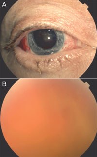
A 36-year-old previously well woman presented with a Glasgow Coma Scale (GCS) score of 7 after a World Federation of Neurological Surgeons (WFNS) Clinical Grade 4 SAH, with equal and reactive pupils and no afferent pupillary defect. She was treated with craniotomy and clipping of a ruptured aneurysm of the right M2 (Sylvian segment of the middle cerebral artery). Despite her poor neurological condition, the patient made slow but gradual progress. It was not until Day 65, when her GCS score was 14, that she described difficulty with her vision. Ophthalmic examination of both eyes revealed visual acuity of counting fingers only and a poor red reflex, and fundoscopy showed an obscured view of the retina due to vitreous haemorrhage (Box). The patient underwent bilateral vitrectomy, and was subsequently transferred for rehabilitation. Review at 6 months showed acuities of 6/36 and 6/24 in the right and left eye, respectively.
The incidence of Terson syndrome in patients who survive SAH is approximately 2%–14%.2,5 It is usually bilateral, and can progress to retinal traction, retinal detachment, and proliferative retinopathy if not treated.3,4 Terson syndrome is associated with increased morbidity compared with SAH without an intraocular bleed.5
Diagnosis of Terson syndrome may be delayed for numerous reasons. In patients with SAH, the need for neurological monitoring often restricts the dilatation of pupils for ophthalmic assessment. Further, patients with higher WFNS grade SAH have greater cognitive impairment, limiting their ability to communicate visual disturbance or comply with testing. Unfortunately, these patients are also at most risk of Terson syndrome. We recommend routine bedside ophthalmoscope examination in all patients with SAH, as soon as the patient’s neurological status is stable.
Visual impairment in patients recovering from SAH has important prognostic and functional implications. The timely diagnosis of Terson syndrome may prevent further morbidity and facilitate recovery and rehabilitation. This case emphasises the need for early ophthalmic screening in all patients with SAH.
Competing interests
No relevant disclosures.
References
- Terson A. De l’hémorrhagie dans le corps vitre au cours de l’hémorrhagie cerebrale [French]. Clin Ophthalmol 1900; 6: 309-312. 0_CIGDCBAF
- Linn FH, Rinkel GJ, Algra A, van Gijn J. Incidence of subarachnoid hemorrhage: role of region, year, and rate of computed tomography: a meta-analysis. Stroke 1996; 27: 625-629. 0_CBBJGDFA
- McCarron MO, Alberts MJ, McCarron P. A systematic review of Terson’s syndrome: frequency and prognosis after subarachnoid haemorrhage. J Neurol Neurosurg Psychiatry 2004; 75: 491-493. 0_CBBDDGID
- Frizzell RT, Kuhn F, Moris R, et al. Screening for ocular hemorrhages in patients with ruptured cerebral aneurysms: a prospective study of 99 patients. Neurosurgery 1997; 4: 529-533; discussion 533-534. 0_CBBHAGBI
- Sung W, Arnaldo B, Sergio C, et al. Terson’s syndrome as a prognostic factor for mortality of spontaneous subarachnoid haemorrhage. Acta Ophthalmol 2011; 89: 544-547. 0_i1142889
Motor Neuron Disease Mortality Trends in Australia From 1986 to 2023: A Population-Based Study
Carol M. Y. Lee, Rupendra N. Shrestha, Julian Gold, Mark Stevenson, Kelly L. Williams, Lyndal Henden, Sandrine K. K. Chan Moi Fat, Zoe Zussa, Amanda Wright, Martin Jude, Mark A. S. Laidlaw, Willem J. R. Fokkink, Dominic B. Rowe
Mild traumatic brain injury and concussion and persisting post‐concussion symptoms: new guidelines to support evidence‐based assessment and management in Australia and Aotearoa New Zealand
Karen M Barlow, Jennie L Ponsford, Alice Theodom, Gill Cowen, Gavin A Davis, Vicki Anderson, Franz E Babl, David Cole, Jennifer Cullen, Stuart R Dalziel, Melinda Fitzgerald, Howard Flavell, Caroline Yates, Rebecca Kimble, John H Olver, Rhonda Orr, Mark Ralfe, Michael Rose, Nick Rushworth, Julia Treleaven, Gary Browne, Nathan Delang, Sarah Harris, Gary Mitchell, Sean Tweedy
The prevalence of and variation in indicators of the quality and safety of long term aged care in Australia, 2019: a cross‐sectional population‐based study
Tesfahun C Eshetie, Gillian E Caughey, Catherine Lang, Olivia Ryan, Renuka Visvanathan, Craig Whitehead, Keith Evans, Janet K Sluggett, Jyoti Khadka, Carolyn Dawkins, Helena Williams, Miranda Starke, Sara Blunt, Anne Liddell, Megan Corlis, Anna Sheppeard, Penelope Lello, Marilyn Thien, Steven L Wesselingh, Maria C Inacio
Health and economic benefits of improving pre‐hospital identification of stroke in Australian women: a modelling study
Thomas Gadsden, Lei Si, Emily R Atkins, Cheryl Carcel, Xia Wang, Stephen Jan, Mark Woodward, Laura E Downey
Symptom burden, quality of life, and diagnostic journey of people with postural orthostatic tachycardia syndrome, Australia, 2021–24: a descriptive patient registry data study
Marie‐Claire Seeley, Gemma Wilson, Eric Ong, Amy Langdon, Jonathan Chieng, Danielle Bailey, Kristina Comacchio, Amanda Page, Dennis H Lau, Celine Gallagher
The intersection of rurality and dementia prevalence in Australia for Aboriginal and Torres Strait Islander and non‐Indigenous peoples
Antonia J Clarke, Marwan Ahmed, Judith M Katzenellenbogen, John Towney (Wiradjuri), Anna H Balabanski, Adrienne Withall (Dharawal Yuin), Kylie Radford, Amy Brodtmann