




Volume 196 - Issue 8
Brain abscess due to Propionibacterium propionicum in Eisenmenger syndrome
Authors: Anthony M T Chau, Lileane L Xu, Jacob M Fairhall, Joga Chaganti and Brendan J McMullan
Med J Aust 2012; 196 (8): 525-526. || doi: 10.5694/mja11.10768
Published online: 7 May 2012
Published online: 7 May 2012
Clinical record
A 33-year-old man with Eisenmenger syndrome due to a congenital ventricular septal defect presented with a 3-week history of headache, blurred vision and expressive dysphasia,
and unintentional weight loss of 10% of his body weight. His medications included sitaxsentan sodium (a sulfonamide endothelin-receptor antagonist, recently withdrawn because of hepatotoxicity) for pulmonary arterial hypertension. He ...
Clinical record
A 33-year-old man with Eisenmenger syndrome due to a congenital ventricular septal defect presented with a 3-week history of headache, blurred vision and expressive dysphasia, and unintentional weight loss of 10% of his body weight. His medications included sitaxsentan sodium (a sulfonamide endothelin-receptor antagonist, recently withdrawn because of hepatotoxicity) for pulmonary arterial hypertension. He had no history of neurosurgery. He was centrally cyanosed and drowsy but afebrile. Results of a neurological examination were otherwise normal.
The patient had no laboratory findings suggestive of infection: white blood cell count was normal at 6.9 × 10⁹/L (reference interval [RI], 4.0–11.0 × 10⁹/L), and C-reactive protein level was normal at 4 mg/L (RI, < 10.0 mg/L). He was polycythaemic with a haematocrit of 0.62 (RI, 0.40–0.54). Magnetic resonance imaging (MRI) revealed a well defined, multiloculated, homogenous, cystic lesion (measuring 58 × 43 mm in the axial plane), consistent with haematogenous spread. Significant surrounding oedema in the right frontal lobe was causing a mass effect, including midline shift, ipsilateral ventricular effacement and subfalcine herniation. The lesion was hypointense on T1-weighted imaging with ring enhancement after gadolinium injection (Figure 1A) and hyperintense on T2-weighted imaging (Figure 1B), consistent with glioma, mucinous metastasis or brain abscess. Diffusion-weighted imaging showed high signal intensity (Figure 1C), favouring the diagnosis of an abscess.
A craniotomy performed under stereotactic guidance confirmed the lesion to be an abscess; 60 mL of purulent fluid was removed and the patient’s symptoms resolved in the immediate postoperative period. Histological analysis of the aspirated material demonstrated reactive gliosis of the abscess wall, and necrotic components and abundant polymorphs in the abscess cavity (Figure 2A). A Gram stain of the abscess fluid revealed filamentous gram-positive bacilli, leading to the suspicion of infection with Nocardia species. The patient was therefore treated with intravenous (IV) sulfamethoxazole, trimethoprim and meropenem, as well as oral phenytoin. However, after 48 hours of incubation, aerobic cultures were negative while anaerobic cultures yielded catalase-negative, beaded filamentous gram-positive bacilli (Figure 2B). The species was not able to be identified with conventional biochemical tests, but results of 16S ribosomal RNA gene sequencing, obtained 3 weeks after isolation of the gram-positive bacilli, were concordant with Propionibacterium propionicum, an extremely rare cause of brain abscess. The patient was then started on IV benzylpenicillin.
Reappearance of neurological symptoms 4 weeks after the craniotomy necessitated drainage of a further 45 mL of fluid. No organisms were seen on Gram stain of this fluid and no growth was detected on aerobic or anaerobic cultures. Therapy was changed to a 5-week course of IV ceftriaxone (for convenience of outpatient daily dosing) and the patient was discharged with full symptom resolution. At 6-month follow-up, MRI revealed no new lesion formation (Figure 1D).
Figure
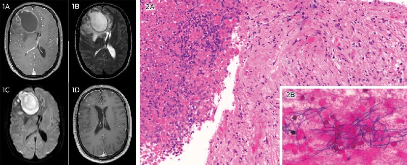
1: Brain MRI scans before and after surgery — T1-weighted imaging after gadolinium injection (1A) and T2-weighted imaging (1B) before surgery, consistent with both cystic or necrotic brain tumour and brain abscess; diffusion-weighted imaging before surgery (1C), favouring diagnosis of abscess; and T1-weighted imaging 6 months after surgery (1D), showing lesion resolution.
2: Samples collected during surgery — haematoxylin and eosin stain of aspirated biopsy sample (2A; original magnification, x 100), with abscess wall on right side (reactive gliosis is visible, with microglia, some neutrophils and proliferating blood vessels) and abscess cavity on left side (necrotic components and abundant polymorphs are visible); and Gram stain of abscess fluid after 48 hours of incubation (2B; original magnification, x 400), demonstrating the beaded filamentous rods of Propionibacterium propionicum.
Brain abscesses are potentially life-threatening intracerebral collections of pus circumscribed by well vascularised capsules. The differentiation of brain abscess from tumour can be difficult on both clinical and radiological grounds. Patients with brain abscess classically present with the triad of fever, headache and focal neurological deficit, but this triad may occur in only 15%–30% of patients.1
Cyanotic heart disease has been implicated in 12.8%–69.4% of published cases of brain abscess.2 In these cases, infection is usually deep-seated, and located at the grey–white matter junction and in the distribution of the middle cerebral artery.3 With the intracardiac right-to-left shunting seen in Eisenmenger syndrome, bacteria bypass the phagocytic component of the pulmonary circulation and enter the cerebral circulation unfiltered.2 Poorly perfused cerebral areas due to chronic hypoxaemia and increased blood viscosity secondary to cyanotic-induced polycythaemia provide fertile seeding conditions for circulating microorganisms.3
Brain abscess in cyanotic heart disease is often attributable to aerobic or anaerobic streptococci.1 We could find only one other report of brain abscess due to Propionibacterium propionicum (under its former name, Arachnia propionica), occurring in a 32-year-old man who also had Eisenmenger syndrome.4 Propionibacterium species are anaerobic gram-positive rods found as normal components of human skin, oral and gastrointestinal flora. It can be difficult to differentiate these organisms from Actinomyces israelii using conventional biochemical methods, but many species can be identified by 16S ribosomal RNA gene sequencing (a tool that is increasingly available to clinical microbiology laboratories).
Diffusion-weighted imaging provides a method of differentiating cerebral abscess from malignancy, by evaluating the diffusion properties of water molecules in the affected tissue.5 Pyogenic abscesses typically exhibit restricted diffusion (increased signal intensity compared with malignant processes) due to increased fluid viscosity from cellular and proteinaceous debris. This phenomenon is about 90% sensitive and specific for brain abscess, as opposed to malignancy, but may occasionally be seen in certain tumours with liquefaction or radiation necrosis.5,6
This case illustrates that in the clinical diagnostic dilemma between brain tumour and abscess, a relatively long history of symptoms, absence of fever, normal levels of inflammatory markers and results of conventional MRI can be unreliable indicators of either aetiology. Abscess diagnosis requires a high index of suspicion in susceptible patients so that appropriate and timely imaging is ordered and necessary intervention is not delayed. As growing numbers of children with cyanotic heart disease survive into adulthood, awareness of its potential complications is becoming increasingly important.
Lessons from practice
Differentiation between brain abscess and brain tumour can be a diagnostic dilemma. Brain abscess may require urgent treatment.
Patients with congenital cyanotic heart disease have an increased risk of brain abscess due to haematogenous spread. In these patients, a high index of suspicion for brain abscess is thus required, even when the classic triad of fever, headache and focal neurological deficit is incomplete.
Diffusion-weighted magnetic resonance imaging is useful for differentiating abscess from malignancy, by demonstrating restricted diffusion of the pus-filled lesion.
Propionibacterium propionicum infection is a rare cause of brain abscess which should be treated with intravenous antibiotics and may require surgery.
Competing interests
No relevant disclosures.
Acknowledgements
We thank Peter Earls (Anatomical Pathology, St Vincent’s Hospital) for his assistance in preparing and interpreting the histopathology slides included in this article.
References
- Erdogan E, Cansever T. Pyogenic brain abscess. Neurosurg Focus 2008; 24: E2. 0_i1140560
- Moorthy RK, Rajshekhar V. Management of brain abscess: an overview. Neurosurg Focus 2008; 24: E3. 0_i1140562
- Takeshita M, Kagawa M, Yato S, et al. Current treatment of brain abscess in patients with congenital cyanotic heart disease. Neurosurgery 1997; 41: 1270-1279. 0_i1140564
- Riley TV, Ott AK. Brain abscess due to Arachnia propionica. Br Med J (Clin Res Ed) 1981; 282: 1035. 0_i1140566
- Chang SC, Lai PH, Chen WL, et al. Diffusion-weighted MRI features of brain abscess and cystic or necrotic brain tumors: comparison with conventional MRI. Clin Imaging 2002; 26: 227-236. 0_i1140568
- Tung GA, Evangelista P, Rogg JM, Duncan JA 3rd. Diffusion-weighted MR imaging of rim-enhancing brain masses: is markedly decreased water diffusion specific for brain abscess? AJR Am J Roentgenol 2001; 177: 709-712. 0_i1140572
Fatty Liver Disease in Australia: A Narrative Review on the Epidemiology, Natural History, Prognostication and Management in People With Metabolic Dysfunction
Karl Vaz, Daniel Clayton-Chubb, William W. Kemp, Stuart K. Roberts, Ammar Majeed
Sex Differences in Management and Outcomes of People With ST-Elevation Myocardial Infarction, New South Wales, 2011–2020: A Retrospective Cohort Study
Adam C. Livori, Jedidiah I. Morton, Dianna J. Magliano, Jonathan E. Shaw
Rethinking Rheumatic Fever Diagnosis: Progress, Pitfalls and the Path Towards a Definitive Test
Anna P. Ralph, Rachel Webb, Tom Parks, Nicole J. Moreland, Asha C. Bowen, Benjamin Jones, Emma Ndagire, Andrea Z. Beaton
Sex Differences in Management and Outcomes of People With ST-Elevation Myocardial Infarction, New South Wales, 2011–2020: A Retrospective Cohort Study
Samia Kazi, Simone Marschner, Clara K. Chow
Distribution of Cardiovascular Disease Risk Based on the Updated 2023 Guideline-Recommended Australian Cardiovascular Disease Risk Algorithm and Comparison With the 2012 Algorithm: An Observational Study
Nina Lazarevic, Meghana Bhat, Grace Joshy, Danielle C. Butler, Mark Woodward, Anushka Patel, Rod T. Jackson, Garry Jennings, Rosemary Wyber, Ellie Paige, Emily Banks
They Brought the Hospital to Me
Hugh N. Burrill