Connect
MJA
MJA

Multisegment jejunojejunal intussusception in gastrojejunostomy

Uei Pua
Med J Aust 2011; 195 (3): 148. || doi: 10.5694/j.1326-5377.2011.tb03244.x
Published online: 1 August 2011

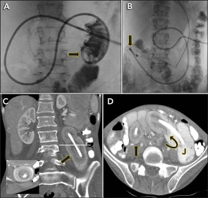
A 27-year-old woman was being treated with gastrojejunostomy feeding for severe anorexia nervosa. The position of the gastrojejunostomy tube was checked by fluoroscopy on the day of insertion (Figure, A, arrow). The next day, the patient presented with abdominal pain and “shortening” of the external portion of the tube. Repeat fluoroscopy showed migration of the tube (Figure, B, arrow). Computed tomography showed two segments of jejunojejunal intussusception (Figure, C, arrow and inset, and D, straight arrow) centred around the tube (Figure, D, curved arrow), with an intervening segment of normal jejunum (Figure, D, label J). The patient’s gastrointestinal tract was intact. The intussusceptions were reduced by applying traction on the tube under fluoroscopic guidance, and the patient resumed tube feeding without recurrence.


Provenance: Not commissioned; externally peer reviewed.

  • Uei Pua

  • Tan Tock Seng Hospital, Singapore, Singapore.


Correspondence: druei@yahoo.com

Author

remove_circle_outline Delete Author
add_circle_outline Add Author

Comment
Do you have any competing interests to declare? *

I/we agree to assign copyright to the Medical Journal of Australia and agree to the Conditions of publication *
I/we agree to the Terms of use of the Medical Journal of Australia *
Email me when people comment on this article

Online responses are no longer available. Please refer to our instructions for authors page for more information.