




Volume 195 - Issue 1
A couplet: a case of anal ulceration and another of inguinal swelling
Authors: Marcus Y Chen, Timothy R H Read, David E Leslie and Melanie Bissessor
Med J Aust 2011; 195 (1): 47-48. || doi: 10.5694/j.1326-5377.2011.tb03191.x
Published online: 4 July 2011
Published online: 4 July 2011
In recent years, lymphogranuloma venereum, which is caused by C. trachomatis serotypes L1–L3, has emerged as a problem among men who have sex with men (MSM). Cases of LGV have been reported among MSM in the United States, United Kingdom, Europe and Australia, and have been overrepresented among HIV-positive MSM.1,2 In contrast to infections seen in developing areas where LGV remains endemic, the more recent reports among MSM in Western countries have been characterised by a predominance of proctitis cases, with fewer genital or inguinal presentations.1-3 Furthermore, nearly all recent cases have involved a clonal strain of the C. trachomatis L2b serovar.4
Genital LGV infections are commonly heralded by transient genital ulceration, followed by lymphatic spread and the development of local lymphadenopathy. A transient genital ulcer reported by Patient 2 had resolved before the development of the bubo.
LGV infections causing ulceration and proctitis are readily detected using commercially available C. trachomatis nucleic acid amplification assays such as PCR or SDA. However, for LGV to be confirmed microbiologically, further genotyping is required to identify C. trachomatis serovars L1–L3, as distinct from the more common non-LGV-associated serovars D–K. This requires awareness on the part of the clinician, as LGV genotyping is only available from reference laboratories in most countries. In a symptomatic patient with a positive C. trachomatis nucleic acid amplification assay result, the clinician must specifically request referral of the original specimen or DNA extract for further testing, potentially delaying the final diagnosis. Newer amplification assays that can directly detect C. trachomatis serovars from clinical specimens are under development, but none are commercially available and they may not be cost-effective for routine diagnostic use in most settings.5
C. trachomatis serological testing can sometimes be useful for diagnosing LGV when there is no obvious lesion for sampling by PCR. However, a positive serological result does not discriminate between LGV and non-LGV chlamydial infections.6 In a recent study among MSM in Melbourne, 7% (21/292) of chlamydial rectal infections were associated with the L2 or L2b serovar.7 The distinction between LGV and non-LGV chlamydial infection of the rectum is potentially important, as a single 1 g dose of azithromycin is commonly used for non-LGV chlamydial infection, while more prolonged treatment — usually a 3-week course of doxycycline — is recommended for LGV.8-10 The extent to which inadequate treatment might be contributing to further transmission of LGV between men is not known.
The diagnosis of LGV should be considered in MSM with proctitis, anogenital ulcers or inguinal buboes, particularly in HIV-positive men. If LGV genotyping is not readily available, an alternative approach would be to treat such men with a 3-week course of doxycycline or to perform a test-of-cure to ensure that the treatment has cured the patient of LGV.
Clinical records
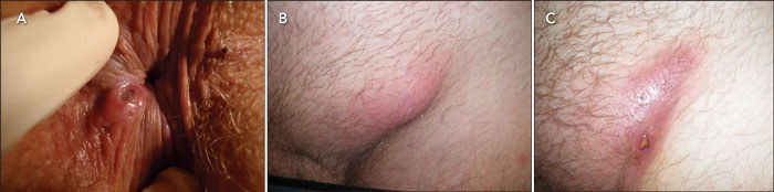
An HIV-positive man (Patient 1) presented with a 4-day history of a painful anal ulcer (Figure, A), which was associated with a small, tender left-sided inguinal lymph node. He was not taking antiretroviral therapy and had a CD4 cell count of 648 cells/μL and an HIV viral load of 25 500 copies/mL.
The man’s male sexual partner (Patient 2), also HIV-positive, presented on the same day with a lump in the left inguinal region (Figure, B). He was taking antiretroviral treatment and had a CD4 cell count of 494 cells/μL, with an undetectable HIV viral load (< 50 copies/mL). The lump had been present for 4 weeks and had enlarged despite treatment with amoxycillin followed by flucloxacillin given at a hospital emergency department. Two weeks before the lump appeared, the patient reported that there had been a sore on the penis but this had resolved before the lump developed. The inguinal lump had overlying erythema and was slightly fluctuant.
Given these men were sexual partners, the diagnosis most likely to explain the presence of an inguinal bubo in one partner and anal ulceration in the other was lymphogranuloma venereum (LGV). Both men were treated with oral doxycycline 100 mg twice daily for 3 weeks.
Patient 1 was reviewed 1 week later, by which time the anal ulcer had begun to resolve and a swab taken from the ulcer at the previous visit had tested positive for Chlamydia trachomatis by strand displacement amplification (SDA) assay and negative for herpes simplex virus and Treponema pallidum by polymerase chain reaction (PCR) assay. In both men, serological tests for syphilis returned negative results, and white cell counts were normal.
Despite the doxycycline therapy, Patient 2’s bubo had enlarged further when examined 3 weeks later, with increased swelling, erythema and fluctuance. A hollow-bore needle was used to aspirate 5 mL of milky pus from the bubo. Light microscopy of the Gram-stained aspirate showed few polymorphs, and no bacteria, parasites or fungi. No acid-fast bacilli were seen on Ziehl–Neelsen staining, and bacterial culture showed no growth. The aspirate tested positive for C. trachomatis and negative for T. pallidum and Neisseria gonorrhoeae by PCR assay. Cytological examination of the aspirate showed acute inflammation, with no organisms identified. Despite the initial aspiration and continued therapy with doxycycline, the bubo reformed and then spontaneously discharged 2 days later, leaving a sinus (Figure, C).
Genotyping of the chlamydia isolates from the anal ulcer and the bubo aspirate confirmed the presence of C. trachomatis serovar L2b in each patient. Both men also tested positive for C. trachomatis IgG and IgA by enzyme immunoassay.
 |
Competing interests
None identified.
Acknowledgements
We thank staff at the Microbiological Diagnostic Unit, University of Melbourne, for their assistance in LGV genotyping.
References
- Ward H, Alexander S, Carder C, et al. The prevalence of lymphogranuloma venereum infection in men who have sex with men: results of a multicentre case finding study. Sex Transm Infect 2009; 85: 173-175. 0_CHDEDCDH
- Nieuwenhuis RF, Ossewaarde JM, Gotz HM, et al. Resurgence of lymphogranuloma venereum in Western Europe: an outbreak of Chlamydia trachomatis serovar L2 proctitis in The Netherlands among men who have sex with men. Clin Infect Dis 2004; 39: 996-1003. 0_CHDGGHFH
- Klausner JD, Kohn R, Kent C. Etiology of clinical proctitis among men who have sex with men. Clin Infect Dis 2004; 38: 300-302. 0_i1095896
- Christerson L, de Vries HJC, de Barbeyrac B, et al. Typing of lymphogranuloma venereum Chlamydia trachomatis strains. Emerg Infect Dis 2010; 16: 1777-1779. 0_i1095898
- Stevens MP, Twin J, Fairley CK, et al. Development and evaluation of an ompA quantitative real-time PCR assay for Chlamydia trachomatis serovar determination. J Clin Microbiol 2010; 48: 2060-2065. 0_i1095900
- van der Snoek EM, Ossewaarde JM, van der Meijden WI, et al. The use of serological titres of IgA and IgG in (early) discrimination between rectal infection with non-lymphogranuloma venereum and lymphogranuloma venereum serovars of Chlamydia trachomatis. Sex Transm Infect 2007; 83: 330-334. 0_i1095902
- Nguyen V, Bissessor M, Lee D, et al. Characteristics of lymphogranuloma venereum (LGV) infection among homosexual men in Melbourne. Australasian Sexual Health Conference; 2010 Oct 18–20; Sydney, Australia. 0_i1095904
- Antibiotic Expert Group. Therapeutic guidelines: antibiotic. Version 14. Genital and sexually transmitted infections: proctitis. Melbourne: Therapeutic Guidelines Limited, 2010: 110. 0_i1095906
- White JA. Manifestations and management of lymphogranuloma venereum. Curr Opin Infect Dis 2009; 22: 57-66. 0_i1095908
- Centers for Disease Control and Prevention. Sexually transmitted diseases treatment guidelines, 2010. MMWR 2010; 59 (No. RR-12): 1-110. http://www.cdc.gov/STD/treatment/2010 (accessed Nov 2010).
Provenance: Not commissioned; externally peer reviewed.
Awareness, Usage and Perceptions of Doxycycline Post-Exposure Prophylaxis (doxyPEP) for Prevention of Sexually Transmitted Infections in Australia: Insights From a National Cross-Sectional Survey
Catriona S. Bradshaw, Dash Heath-Paynter, Benjamin Riley, Daniel Grace, Fabian Y. S. Kong, Eric P. F. Chow
How Can We Ensure Access to Sexual and Reproductive Health Information for Adolescents in Light of Australia's Social Media Restrictions?
Olena Ivanova, Anisa R. Assifi, Danielle Mazza
Differentiated and simplified oral HIV pre‐exposure prophylaxis (PrEP) models hold the key to virtually eliminating HIV transmission in Australia by 2030
Tyson Arapali, Sarah Warzywoda, Anthony K J Smith, Curtis Chan, Timothy R Broady, Erin Sullivan, Catherine MacPhail, Mohamed A Hammoud, Alexander Dowell‐Day, Benjamin R Bavinton
An autoethnographic critique of a past report of inpatient psychiatric treatment for gender diverse children
Jayne McFadyen, Timothy W Jones, Rowena Koek, Fintan Harte, Brendan Jansen, Megan Galbally, Warren Kealy‐Bateman, Catherine Wall, Quinnehtukqut McLamore, Anja Ravine
Cass Review does not guide care for trans young people
Julia K Moore, Cate Rayner, S Rachel Skinner, Katie Wynne, Blake S Cavve, Brodie Fraser, Uma Ganti, Claire McAllister, Gideon Meyerowitz‐Katz, Tram Nguyen, Anja Ravine, Brian Ross, Darren B Russell, Liz A Saunders, Aris Siafarikas, Ken C Pang
Motivations, barriers and enablers for medical and forensic examiners in New South Wales sexual assault services: a qualitative interview study
Natalie Edmiston, Sam Sperring, Rosalie Power, Samantha Ryan, Kathryn Evans, Jane Ussher, Ellie Freedman