




Volume 193 - Issue 3
Breathing life into histology
Author: Tanya Grassi
Med J Aust 2010; 193 (3): 180. || doi: 10.5694/j.1326-5377.2010.tb03841.x
Published online: 2 August 2010
Published online: 2 August 2010
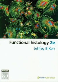
Functional histology. 2nd ed.
Jeffrey B Kerr. Sydney: Mosby Elsevier, 2010 (viii + 512 pp). ISBN 9780729538374.
Histology can be a dry subject. In medical curricula, it has suffered variably from overexposure to underexposure, to the point where graduates may feel perfectly comfortable identifying normal tissues under the light microscope, or squirm uncomfortably when confronted with the same at a multidisciplinary meeting. Finding a balance between pure information and visual references can be difficult and, to paraphrase Elvis, the reader is likely to plead, “a little less longwinded text, a few more pictures please”.
The second edition of Functional histology, by Jeffrey Kerr, strikes a good balance. The book has numerous clear, vivid light micrographs, excellent electron micrographs, and superb drawings and diagrams aimed at describing and explaining the organisation and function of body tissues. Traditionally laid out in systems, the text tells an interesting story for each organ, taking the reader through the structure, microscopic appearance, physiology, and function of each tissue, before delineating common abnormal conditions and clinical features.
Kerr, an Associate Professor of Anatomy and Developmental Biology at Monash University in Melbourne, says he prepared Functional histology for “those with an interest in biological science as applied to the health professions and for the research community”. The book more than accomplishes this goal, being suitable for a range of users from medical students and pathology registrars through to researchers in the biological sciences. It is distinguished from other works in the discipline by the use of concise, plain language. The useful, friendly “tips”, scattered throughout the text, further engage the reader. The addition of topical references to molecular biology keep the content current and this, together with the text’s accessible style, will maintain the interest of those returning to histology for the purposes of research or postgraduate study.
Reducing Nitrous Oxide Emissions Across the Melbourne Biomedical Precinct
Ross Robertson, Andrew Downey, Daryl Williams, Bjorn Makein, Ben Dunne, Tugce Ozturk, Ying Gu, Rebecca McIntyre
Multimorbidity Clusters Among People Aged 65 Years and Over in Australia: A Nationwide Cross-Sectional Data Linkage Study
Weisi Chen, Christine Y. Lu, Sarah N. Hilmer, Alice A. Gibson, Edwin C. K. Tan
When ‘Liver Enzymes’ Are Not Hepatic: Late-Onset Pompe Disease
Shauna Madigan, Georgina England, Wayne Rankin
West Nile virus Kunjin subtype in rural NSW
Emily Gibson, Megan Whitley, Peter Murray, Linda Hueston, Jane Bennett, Raguharan Kathiresu, David N Durrheim
The impact of the Breast Screen NSW transition from film to digital mammography, 2002–2016: a linked population health data analysis
Rachel Farber, Nehmat Houssami, Katy J L Bell