




Volume 192 - Issue 8
Visceral leishmaniasis due to Leishmania donovani in a patient with advanced HIV infection
Authors: Samuel C Hume, Craig A Aboltins, Karin A Thursky, John R Daffy and Peter A Stanley
Med J Aust 2010; 192 (8): 474-475. || doi: 10.5694/j.1326-5377.2010.tb03591.x
Published online: 19 April 2010
Published online: 19 April 2010
An Eritrean-born man observed over an extended period had upper gastrointestinal symptoms, fever, hepatosplenomegaly and pancytopenia in the setting of advanced HIV infection and poor adherence to antiretroviral therapy. Despite thorough investigation, it was not until a repeat gastroscopic examination and gastric biopsy were performed 18 months after initial presentation that Leishmania infection was diagnosed. The species was identified by polymerase chain reaction assay as L. donovani. Physicians managing HIV-infected patients from regions where Leishmania is endemic should consider visceral leishmaniasis, even in patients who have not lived in a Leishmania-endemic region for many years.
Clinical record
A 42-year-old Eritrean-born man who had previously lived in Sudan arrived in Australia in 1995. He had not travelled overseas since that time. He was diagnosed with HIV infection in 1996. He presented in 2005 with a 2-month history of odynophagia, dysphagia and vomiting.
The patient was non-adherent to antiretroviral therapy and his CD4 count was 40 cells/μL (reference range [RR], 410–1590 cells/μL). He had low-grade fever, hepatosplenomegaly and pancytopenia (haemoglobin level, 118 g/L [RR, 130–180 g/L]; white cell count, 3.2 × 10⁹/L [RR, 4.0–11.0 × 10⁹/L]; and platelet count, 111 × 109/L [RR, 150–400 × 109/L]). A computed tomography scan confirmed hepatosplenomegaly, but there was no intra-abdominal or pelvic lymphadenopathy. A bone marrow biopsy showed hypercellular marrow with no granulomas or malignancy. Bone marrow fungal and mycobacterial culture and a polymerase chain reaction (PCR) assay for parvovirus DNA were negative. No abnormality was visible on gastroscopic examination. Histological examination of oesophageal biopsies showed chronic inflammation, but no fungal elements were detected by fungal stains, and fungal cultures were negative. A herpes multiplex PCR assay (capable of detecting herpes simplex virus types 1 and 2, cytomegalovirus and varicella-zoster virus) was negative. No Leishmania amastigotes (the non-flagellate, intracellular form of Leishmania) were identified. Symptoms improved after treatment with omeprazole and a period of improved adherence to antiretroviral therapy.
Odynophagia, dysphagia, weight loss, intermittent fever and mild pancytopenia recurred and continued over the next 18 months. Despite changes to the antiretroviral therapy regimen and efforts to improve adherence, the patient’s CD4 count remained below 50 cells/μL and the HIV viral load was consistently > 750 000 copies/mL.
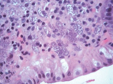
A repeat gastroscopic examination 18 months after initial presentation demonstrated pseudomembrane formation over the upper stomach, and histological examination of a gastric biopsy showed a florid inflammatory cell infiltrate. Within histiocytes, numerous intracellular organisms 1–3 μm in diameter were identified as Leishmania amastigotes (Box). A PCR assay confirmed the presence of L. donovani, leading to a diagnosis of visceral leishmaniasis (VL) in the setting of advanced HIV infection.
Induction treatment was commenced with daily liposomal amphotericin B 4 mg/kg for 1 week, followed by weekly doses for a further 4 weeks. The odynophagia, dysphagia and pancytopenia resolved over the following 4 months. Secondary prophylaxis with monthly doses of liposomal amphotericin B continued, but the patient missed three doses over the next 6 months and symptoms recurred. When repeat endoscopy, biopsy and histology showed that Leishmania was still present, therapy was recommenced and the symptoms resolved. Further unsuccessful attempts were made to optimise the antiretroviral therapy regimen. After 12 months of intermittent adherence to treatment, the patient again developed symptoms. He subsequently returned to East Africa and was lost to follow-up.
Discussion
Leishmaniasis is a protozoal infection caused by species of the genus Leishmania, an intracellular parasite transmitted by sandflies of the genus Phlebotomus. Clinical presentations include visceral, cutaneous and mucocutaneous forms, depending on the infecting species and the strength of the host’s cell-mediated immunity. The Leishmania donovani complex, composed of L. donovani and L. infantum/chagasi, is responsible for most cases of VL. Where co-infection with HIV is present, deteriorating immune function may delay the clinical presentation of VL for many years after the initial infection.1
Although documented in 70 countries, most cases of VL occur in southern Asia (particularly north-eastern India, Nepal, and Bangladesh) and East Africa (mostly Sudan, Ethiopia and Eritrea). In these regions, L. donovani is the predominant species. About 15 000 to 20 000 cases of VL occur annually in Sudan, particularly in the eastern region bordering Ethiopia and Eritrea, and VL–HIV co-infection rates are as high as 29%–40%.2-4 In southern Europe, especially in urban coastal regions, where the less virulent L. infantum is the predominant organism, high rates of VL–HIV co-infection have also been described.5
The usual incubation period of VL is 2–6 months, but a proportion of patients will remain asymptomatic after primary infection, and immunosuppression can lead to reactivation of the disease many years later. The classic presentation of VL includes fever, weight loss, hepatosplenomegaly, pancytopenia and hypergammaglobulinaemia. HIV co-infection does not dramatically alter this presentation.6,7 Gastrointestinal tract involvement is evident in 7%–40% of VL–HIV co-infected patients, and may be accompanied by odynophagia, dysphagia, chronic diarrhoea, malabsorption or abdominal pain.6,8 Most patients with HIV who present with VL are in a state of advanced immunodeficiency: up to two-thirds of patients with concomitant HIV and L. donovani infection have CD4 counts < 200 cells/μL.3
Serological tests for VL are of low sensitivity and thus have limited diagnostic value for VL–HIV co-infected patients.2 Demonstration of amastigotes in tissue or blood is the preferred method of diagnosis. Examination of a bone marrow smear is often the most useful diagnostic test (sensitivity, 67%–94%),2,4,8 and aspirates from other sites can be considered depending on the clinical presentation. A PCR assay for Leishmania DNA may improve the diagnostic yield and allow identification to a species level.
Treatment of VL in patients with underlying HIV infection is associated with lower cure rates, higher rates of drug toxicity, higher relapse rates and greater mortality than treatment of VL in immunocompetent patients.2,9 Optimal treatment regimens have not yet been developed, as there is a paucity of clinical data on patients with VL–HIV co-infection. Treatment options include pentavalent antimonials such as sodium stibogluconate, standard and lipid formulations of amphotericin B, miltefosine, paromomycin and pentamidine.2,9 Although liposomal amphotericin B has not been formally assessed in VL–HIV co-infected patients, it has the highest therapeutic index of all antileishmanial drugs and a superior safety profile, making it the first choice for treatment in resource-rich countries. Although there has been recent interest in miltefosine because of its oral administration, an Ethiopian study showed that miltefosine was less effective than intramuscular sodium stibogluconate for treating HIV-infected patients.10 Maintenance secondary prophylaxis and initiation of effective antiretroviral therapy is required to prevent relapse.9 In a study conducted in a resource-limited setting in Ethiopia, antiretroviral therapy was shown to reduce the risk of VL relapse by about 50%.3
The increasing rate of VL–HIV co-infection in Africa and India is a major concern, but so far, very few cases have been reported in Australia. The case described here highlights the importance of considering the diagnosis of VL in a patient with immunodeficiency and a history of travel to, or residence in, a Leishmania-endemic region; the value of tissue biopsy combined with PCR testing; and the need for ongoing secondary prophylaxis and immune restoration.
Competing interests
None identified.
References
- Visvanathan K, Jones PD, Riordan SM, et al. Delayed reactivation of visceral leishmaniasis complicating HIV infection. Aust N Z J Med 1993; 23: 407. 0_i1091926
- Alvar J, Aparicio P, Aseffa A, et al. The relationship between leishmaniasis and AIDS: the second 10 years. Clin Microbiol Rev 2008; 21: 334-359. 0_i1091928
- ter Horst R, Collin SM, Ritmeijer K, et al. Concordant HIV infection and visceral leishmaniasis in Ethiopia: the influence of antiretroviral treatment and other factors on outcome. Clin Infect Dis 2008; 46: 1702-1709. 0_i1091930
- Lyons S, Veeken H, Long J. Visceral leishmaniasis and HIV in Tigray, Ethiopia. Trop Med Int Health 2003; 8: 733-739. 0_i1091932
- Desjeux P, Alvar J. Leishmania/HIV co-infections: epidemiology in Europe. Ann Trop Med Parasitol 2003; 97 Suppl 1: 3-15. 0_i1091934
- Russo R, Laguna F, Lopez-Velez R, et al. Visceral leishmaniasis in those infected with HIV: clinical aspects and other opportunistic infections. Ann Trop Med Parasitol 2003; 97 Suppl 1: 99-105. 0_i1091936
- Sinha PK, Rabidas VN, Pandey K, et al. Visceral leishmaniasis and HIV coinfection in Bihar, India. J Acquir Immune Defic Syndr 2003; 32: 115-116. 0_i1091938
- Pintado V, Martin-Rabadan P, Rivera ML, et al. Visceral leishmaniasis in human immunodeficiency virus (HIV)-infected and non-HIV-infected patients. A comparative study. Medicine (Baltimore) 2001; 80: 54-73. 0_i1091940
- Bern C, Adler-Moore J, Berenguer J, et al. Liposomal amphotericin B for the treatment of visceral leishmaniasis. Clin Infect Dis 2006; 43: 917-924. 0_i1091942
- Ritmeijer K, Dejenie A, Assefa Y, et al. A comparison of miltefosine and sodium stibogluconate for treatment of visceral leishmaniasis in an Ethiopian population with high prevalence of HIV infection. Clin Infect Dis 2006; 43: 357-364. 0_i1091946
Costs of Delivering the Hep B PAST Model of Care in Remote Australia: A Retrospective Pre–Post Study
Hoa Nguyen, Anh Le Tuan Nguyen, Paula Binks, Melita McKinnon, Emily Vintour-Cesar, Karen Wills, Nicola Stephens, Benjamin Cowie, Joshua S. Davis, Yuejen Zhao, Peter Nihill, Julie A. Campbell, Andrew J. Palmer, Jane Davies, Barbara de Graaff
Buruli Ulcer Acquired From Ringtail Possum Rescue
Daniel P. O'Brien, Naomi Clarke, Sara Bonnici, Mohammad A. Hussain
Q Fever Visceral Abscesses After Contact With Native Australian Marsupials
Samantha J. Laws, Enzo Binotto, James Stewart, Jennifer Ho, Trent Yarwood, Josh Hanson, Simon Smith
Hospital-Admitted Injection-Related Infections Among Incarcerated People Who Inject Drugs in Australia: A Retrospective Cohort Study
Andrew Palmer, Matthew Carter, Jeremy Yeo, Cecilia Shim, Jason Connor, Jeremy Hayllar, Gerald Holtmann, Naomi Moy, Elliott G. Playford, Naomi Runnegar, Paul J. Clark
The Live Attenuated Influenza Vaccine in Australia: An Additional Tool for Influenza Prevention
Cyra Patel, Alexis Pillsbury, Tran Nguyen, Xia Wang, Helen E. Quinn, Clayton K. Chiu, Allen C. Cheng, Katie L. Flanagan, Zhicheng Wang
Reflex Testing for Hepatitis D Infection: A Unique Opportunity to Reduce Hepatitis D-Related Chronic Liver Disease Deaths in Australia
Jessica Howell, Lauren Andersson, Miriam T. Levy, James O'Beirne, Leon Adams, Katharine Irvine, Avik Majumdar, Golo Ahlenstiel, Kathy Jackson, Krispin Hajkowicz, Joseph Doyle, Jane Davies, Sarah Cherian, Wayne Dimech, Alexander J. Thompson