




Volume 192 - Issue 7
Late-stage human African trypanosomiasis in a Sudanese refugee
Authors: Paul Cherian, Ralph K Junckerstorff, David Rosen, Prasad Kumarasinghe, Alan Morling, Philip Tuch, Sonja Raven, Ronan J Murray and Christopher H Heath
Med J Aust 2010; 192 (7): 417-419. || doi: 10.5694/j.1326-5377.2010.tb03569.x
Published online: 5 April 2010
Published online: 5 April 2010
A 19-year-old Sudanese woman, who had lived for about a decade in Ugandan refugee camps, was referred for investigation of a 12-month history of a generalised rash. Two months later, her condition had deteriorated to include cachexia and drowsiness. Despite initial negative findings on investigation, human African trypanosomiasis (HAT) was suspected, and parasites were found in a double-centrifuged sample of cerebrospinal fluid. Eflornithine, the appropriate drug for treatment of late-stage disease, was obtained through the World Health Organization. This case highlights the diagnostic and therapeutic difficulties in managing late-stage HAT in a non-endemic country.
Clinical record
In January 2008, a 19-year-old Sudanese woman was referred from the community to a tertiary hospital for investigation of a 12-month history of generalised pruritus. There were no obvious precipitants or triggers for the itch. She was born in southern Sudan, but had lived in refugee camps in north-western Uganda for about a decade before migrating to Australia in November 2006. Her past medical history was non-contributory. Initial examination showed generalised hyperpigmented papules and nodules with excoriations. Prurigo was diagnosed, and treatment with topical corticosteroids was trialled.
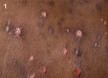
By March 2008, her condition had deteriorated, and she experienced lethargy, apathy, fevers, night sweats, weight loss, reduced rousability, abulia (impairment or loss of willpower) and seizures. The pruritus was pervasive and the scratching automatic. A past family history of human African trypanosomiasis (HAT) in her mother, which was diagnosed and treated in Uganda, was elicited. On examination, she had cachexia, and was drowsy, but rousable, and oriented to person, but not place or time. She had generalised itch with hyperpigmented, lichenified papules and excoriated nodules (Figure 1). Neurological examination showed symmetrical brisk reflexes, bilateral upper-limb cogwheeling, and myoclonic jerks involving her limbs, mouth and periocular muscles. She had palpable posterior cervical lymph nodes of less than 1 cm in diameter. Late-stage HAT was suspected, and she was admitted to hospital for further investigation. Differential diagnoses included other infective encephalitides (tuberculous and viral), vascular events and malignancies (lymphoma).
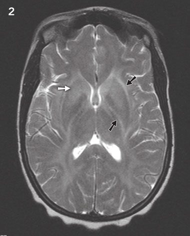
Investigations showed that the patient had microcytic anaemia (haemoglobin concentration, 96 g/L; reference range [RR], 115–145 g/L), which was attributed to a known α-thalassemia trait: her renal function and hepatic function were normal. Her erythrocyte sedimentation rate was 42 mm/h (RR, < 20 mm/h). Elevated concentrations of total serum protein (104 g/L; RR, 60–80 g/L), gammaglobulin (32 g/L; RR, 8–16 g/L), IgG (24.3 g/L; RR, 5.8–13.7 g/L) and IgM (7.8 g/L; RR, 0.3–1.7 g/L) were detected. A skin biopsy suggested lichen simplex chronicus. Initial peripheral blood smears, lymph node and bone marrow aspirates all tested negative for parasites. Examination of the cerebrospinal fluid (CSF) revealed a mononuclear pleocytosis of 100 × 106 cells/L (RR, < 5 × 10⁶ cells/L), an elevated protein level (0.9 g/L; RR, 0.15–0.45 g/L), markedly elevated level of IgM (0.36 g/L; RR, undetectable) and a low level of glucose (2.1 mmol/L; RR, 2.4–4.6 mmol/L); micro-organisms were not seen on examination of the centrifuged deposit. Gadolinium-enhanced magnetic resonance imaging (MRI) showed bilateral, symmetrical, widespread high-signal white matter change on T2-weighted imaging (Figure 2). Simultaneous T2-weighted images demonstrated high signal changes in the splenium, the brainstem and the cerebellar white matter (images not shown), and post-gadolinium images showed minimal leptomeningeal enhancement (images not shown). An electroencephalogram (EEG) showed diffuse delta-wave slowing consistent with a metabolic encephalopathy, with no evidence of ictal activity.
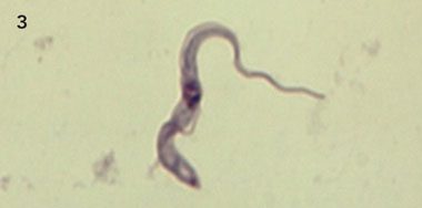
Despite initial negative results on CSF testing, HAT was suspected on clinical grounds. Thus, repeat large-volume CSF examination was performed. Direct examination of CSF was again negative, but double-centrifuged CSF microscopy showed trypanomastigotes (Figure 3), confirming the diagnosis of late-stage HAT.
Both subspecies of Trypanosoma brucei can be acquired in Uganda.1 We considered Trypanosoma brucei gambiense infection to be more likely in this patient as it is hyper-endemic in north-western Uganda and because of the subacute clinical presentation. However, as Trypanosoma brucei rhodesiense infection remained plausible, given the patient’s past travel to southern Uganda, subspecies testing was indicated. Lacking an Australian medical reference laboratory for this, we referred specimens to the Institute of Tropical Medicine (Antwerp, Belgium) for serological testing, and to a local reference laboratory for nucleic acid amplification testing.2 While awaiting results, the patient became uncommunicative, bed-bound and increasingly cachectic, making empiric trypanocidal therapy imperative. Treatment for late stage T. b. gambiense infection was commenced 19 days after admission, with intravenous eflornithine (obtained through the World Health Organization [WHO]) at a dose of 100 mg/kg every 6 hours for 2 weeks. This was tolerated without significant toxicity. At discharge on Day 39, the patient was orientated, communicative and walking independently despite persistence of slight limb hypertonia.
Subsequently, subspecies serology showed elevated titres to T. b. gambiense on a serum immunofluorescent antibody test (IFAT; titre, 1:1600; RR, negative) and a card agglutination antibody test (CAAT; titre, 1:32; RR, negative). The CSF IFAT titre to T. b. gambiense was 1: 8 (RR, negative). The local nucleic acid amplification test suggested T. b. rhodesiense (data not shown), but this was felt to be clinically discordant.
Three months after discharge, the patient was lucid, conversant in both English and her native tongue, neurologically intact and free from itch. Examination of her CSF showed improving pleocytosis with normal biochemical findings. MRI verified improvement; the post-contrast enhancement had resolved, but mild, diffuse cerebral atrophy was evident. She has resumed her studies and normal social activities and was relapse free at 16-month follow-up.
Discussion
Human African trypanosomiasis or sleeping sickness is caused by two subspecies of the haemoflagellate parasite Trypanosoma brucei — T. b. rhodesiense (east-African HAT) and T. b. gambiense (west-African HAT).1,3 The parasite is transmitted by the bite of tsetse flies (Glossina spp.).1 HAT is endemic only in sub-Saharan Africa, where the WHO estimates that between 300 000–500 000 people are currently infected, with 100 000 deaths directly attributed to this disease annually.3 Uganda is currently the only country where infection with both subspecies occurs. While the two foci of trypanosomiasis within Uganda appear separate, as a result of civil unrest, population displacement and the northward spread of wild and domestic animals from central and south-east Uganda, it is likely that these foci will soon converge.4-6 HAT is rarely diagnosed outside Africa. When T. b. gambiense infection is diagnosed, it is usually in the late (secondary) stage.1,7 To our knowledge, this is the first case of T. b. gambiense infection diagnosed in Australia.
HAT is a biphasic disease. Early-stage disease (Stage I) denotes the post-inoculation period followed by haemo-lymphatic spread.1 An inflammatory nodule or ulcer (chancriform) may be seen at the site of inoculation, more commonly with T. b. rhodesiense infection.1 Lymphatic spread results in lymphadenopathy, with posterior cervical lymphadenopathy (Winterbottom’s sign) typical in T. b. gambiense disease.1,3 Haematogenous spread produces fluctuating fevers, hepatosplenomegaly, serositis and myocarditis, which occur in both forms of the infection, but are more common in T. b. rhodesiense disease.1 In late-stage disease (Stage II), the parasite passes through the blood–brain barrier into the central nervous system, resulting in meningoencephalitis, which is invariably fatal if not treated.1,3 Headache, ataxia, itching, speech disturbance, behavioural change, extrapyramidal signs and mental state changes may manifest.1,3 Pineal and thalamic invasion leads to hypersomnolence and circadian rhythm disruption.3 The natural history of the diseases caused by the two subspecies varies significantly; T. b. gambiense infection is insidious (over months to years), whereas T. b. rhodesiense infection usually progresses over days to weeks.1
Dermatological manifestations of HAT are non-specific. Pruritus is a pervasive symptom, present in over 50% of cases, and is a feature of disease chronicity.8 Pruritus generally correlates with greater CSF pleocytosis.8 Parasternal and generalised pruritus are common, as is peripheral oedema. Transient urticarial and macular eruptions have also been described.1,9 In very chronic disease (of more than 24 months’ duration), pruritus may decrease, heralding a pre-terminal phase of illness.8
Definitive diagnosis of HAT requires direct visualisation of the parasites in blood, CSF or tissue. Detection of trypanosomes in the CSF confirms late-stage disease. False negative results are common because of the low number of parasites in T. b. gambiense infection.1 As in the case we present here, concentration techniques such as double-centrifugation of CSF increase the sensitivity of microscopy.10 Non-diagnostic CSF changes include: lymphocyte counts of > 5/μL, increased CSF protein and IgM concentrations, and morula (Mott) cells.9,11 Identifying the subspecies of trypanosomes is difficult because both subspecies are morphologically identical. The serological tests CAAT and IFAT are available for the detection of T. b. gambiense antibodies, and this is useful in screening high-prevalence populations.9 The use of nucleic acid tests for the subspeciation of T. b. rhodesiense and T. b. gambiense is an emerging technique, and has been used successfully in the research setting.5,6,9 However, these assays are not widely available.9 Obtaining a detailed travel history from patients and recording the timing of symptoms and signs over the course of the illness remain important for differentiating the two forms of the disease.
Treatment of HAT depends on the subspecies of infecting trypanosome and disease stage. Patients with early-stage disease should have CSF examination to exclude subclinical neurological involvement.9 In Australia, treatment is challenging because we have limited access to targeted therapies for HAT. The necessary drugs are accessible through the WHO. Treatments for early-stage HAT infections are parenteral pentamidine for T. b. gambiense disease and suramin for T. b. rhodesiense disease.1,6 For late-stage HAT, melarsoprol was formerly the treatment of choice.1 However, 3%–10% of patients treated with melarsoprol develop encephalopathy, which is fatal in 10%–70% of cases. Survivors of encephalopathy frequently have residual brain damage.12,13 Eflornithine is equally efficacious and less toxic than melarsoprol12,13 for late-stage T. b. gambiense infection, with lower treatment-related mortality (about 0.8%).13 Adverse effects of eflornithine include seizures, gastrointestinal upset and neutropenia, but treatment interruption is generally not required.1 Recently, one week of combination therapy with nifurtimox and eflornithine was shown to be equally effective, less toxic and easier to administer than eflornithine monotherapy for late-stage T. b. gambiense infection,14 and is now considered the treatment of choice for this infection.15 Recommended follow-up after treatment for late-stage HAT includes 3–6-monthly CSF examinations for 2 years.4
This case highlights the diagnostic and therapeutic difficulties in managing late-stage HAT in a non-endemic country. Clinicians need to be aware of infections that are non-endemic to Australia that can occur in recently arrived travellers and migrants. Appropriate treatment of HAT is life-saving and associated with good clinical outcomes.
Figure 1
 |
|
The patient’s rash on presentation in March 2008, showing hyperpigmented, lichenified papules and nodules with excoriation involving the trunk. |
Competing interests
None identified.
Acknowledgements
We thank Aesen Thambiran (Migrant Health Unit, Perth WA) for providing information about the patient prior to her arrival in Australia. We also thank Dr Pere P Simarro (Innovative and Intensified Disease Management Program, Control of Neglected Tropical Diseases Unit of the World Health Organization, Geneva, Switzerland), and Nyree Marr and Naomi Lillywhite (Pharmacy Department, Royal Perth Hospital, Perth, WA) for coordinating prompt access to eflornithine through the WHO. We also thank J Blum and C Hatz (FMH Innere Medizin und Tropen- und Reisemedizin, Schweizerisches Tropeninstitut, Basel, Switzerland); Marjan Van Esbroeck and P Büscher (Institute of Tropical Medicine, Antwerp, Belgium); Henk Schallig (Royal Tropical Institute, Amsterdam, the Netherlands) for assistance in the diagnosis and subspeciation of this parasite. We thank RC Thompson and Zablon Njiru from the WHO Collaborating Centre for the Molecular Epidemiology of Parasitic Infections, School of Veterinary and Biomedical Sciences, Murdoch University, Perth, WA for performing the rapid detection of the sub-genus Trypanozoon by loop-mediated isothermal amplification of parasite DNA from our patient.
References
- Brun R, Blum J, Chappuis F, Burri C. Human African trypanosomiasis. Lancet 2010; 375: 148-159. Epub ahead of print 14 Oct 2009. 0_i1092027
- Njiru ZK, Mikosza AS, Matovu E, et al. African trypanosomiasis: sensitive and rapid detection of the sub-genus Trypanozoon by loop-mediated isothermal amplification (LAMP) of parasite DNA. Int J Parasitol 2008; 38: 589-599. 0_i1092029
- Burri C, Brun R. Human African trypanosomiasis. In: Cook GC, Zumla AI, editors. Manson’s tropical diseases. 21st ed. Edinburgh: Elsevier, 2003: 1303-1323. 0_i1092031
- World Health Organization. Control and surveillance of African trypanosomiasis. Report of WHO expert committee. World Health Organ Tech Rep Ser 1998; 881: I-VI, 1-114. 0_i1092033
- Brun R, Balmer O. New developments in human African trypanosomiasis. Curr Opin Infect Dis 2006; 19: 415-420. 0_i1092035
- Picozzi K, Fevre E, Odiit M, et al. Sleeping sickness in Uganda: a thin line between two fatal diseases. BMJ 2005; 331: 1238-1242. 0_i1092037
- Lejon V, Boelaert M, Jannin J, et al. The challenge of Trypanosoma brucei gambiense sleeping sickness diagnosis outside Africa. Lancet Infect Dis 2003; 3: 804-808. 0_i1092039
- Blum J, Schmid C, Burri C. Clinical aspects of 2541 patients with second stage human African trypanosomiasis. Acta Trop 2006; 97: 55-64. 0_i1092041
- Chappuis F, Loutan L, Simarro P, et al. Options for field diagnosis of human African trypanosomiasis. Clin Microbiol Rev 2005; 18: 133-146. 0_i1092043
- Cattand P, Miezan BT, de Raadt P. Human African trypanosomiasis: use of double centrifugation of cerebrospinal fluid to detect trypanosomes. Bull World Health Organ 1988; 66: 83-86. 0_i1092045
- Lejon V, Büscher P. Cerebrospinal fluid in human African trypanosomiasis: a key to diagnosis, therapeutic decision and post treatment follow up. Trop Med Int Health 2005; 10: 395-403. 0_i1092047
- Chappuis F, Udayraj N, Stietenroth K, et al. Eflornithine is safer than melarsoprol for the treatment of second stage Trypanosoma brucei gambiense human African trypanosomiasis. Clin Infect Dis 2005; 41: 748-751. 0_i1092049
- Priotto G, Pinoges L, Fursa I, et al. Safety and effectiveness of first line eflornithine for Trypanosoma brucei gambiense sleeping sickness in Sudan: cohort study. BMJ 2008; 336: 705-708. 0_i1092051
- Priotto G, Kasparian S, Mutambo W, et al. Nifurtimox–eflornithine combination therapy for second-stage Trypanosoma brucei gambiense trypanosomiasis: a multicentre, randomised, phase III, non-inferiority trial. Lancet 2009; 374: 56-64. 0_i1092055
- World Health Organization. Report of the 17th expert committee on the selection and use of essential medicines. 23 to 27 March 2009. WHO Technical Report Series (12 February 2010). Geneva: WHO, 2010: 43-44. http://www.who.int/selection_medicines/committees/expert/17/WEB_TRS_DEC_2009.pdf (accessed Feb 2010).
Diagnosing Acute Kava Dermopathy: A Case Report of a Characteristic Cutaneous Eruption
Ali Abid, Nicholas Allen, Abeer Hagelamin, Christopher Henderson, Artiene Tatian
Localised Herpes Simplex Following Midline Laparotomy
Jessica S. Bulluss, Paul Chee, Matthew J. Verheyden
No silver lining with health misinformation: argyria caused by intentional silver consumption
Luke Collins, Logesh Palanikumar, Stephen Bacchi
Lingual Raynaud phenomenon
Michael Taggart, Mina John
Melasma in the male: a less well recognised entity
Tim Aung, Rowland Noakes
Age group‐specific changes in keratinocyte cancer treatment rates in Australia, 2012–2021: a retrospective cohort study based on MBS claims data
Catherine M Olsen, Nirmala Pandeya, Rachel E Neale, David C Whiteman