




Volume 192 - Issue 11
Hydroxychloroquine retinopathy: screening needed to prevent blindness
Authors: Elvis Ojaimi, Robyn H Guymer, Tien Y Wong and C Alex Harper
Med J Aust 2010; 192 (11): 668-669. || doi: 10.5694/j.1326-5377.2010.tb03676.x
Published online: 7 June 2010
Published online: 7 June 2010
To the Editor: Hydroxychloroquine is used infrequently (in less than 0.1% of Australians) for the long-term management of chronic conditions (eg, rheumatoid arthritis).1 Hydroxychloroquine retinopathy is a rare but sight-threatening side effect that is usually not reversible.2-5 Various eye screening recommendations for hydroxychloroquine toxicity have been proposed overseas,3-5 but there is no recommended consensus for eye screening in Australia. As a result, screening is currently not uniform or universal, and this sometimes leads to significant consequences.
A 36-year-old woman with a longer than 10-year history of hydroxychloroquine therapy (400 mg/day; body weight, 62–67 kg) for rheumatoid arthritis presented to her general practitioner after a year of central visual disturbance and photopsias (a sensation of flashes of light). She was referred to the Medical Retinal Clinic at the Royal Victorian Eye and Ear Hospital in August 2009 with suspected hydroxychloroquine toxicity. She had undergone screening by her optometrist for an initial period, but had not been screened within the past 4 years because she failed to attend; she appeared unaware of the potential serious side effect of the medication on her vision.
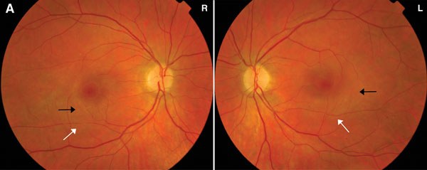
On examination, her visual acuity was 6/9 in the right eye and 6/12 + 2 letters in the left. Her colour vision (assessed by Ishihara plates for colour blindness) was normal. She had left vortex keratopathy (a whorl-like corneal epithelial deposit) and examination of her fundus revealed subtle macular pigment change and retinal arteriolar attenuation (Box, A). Fundus autofluorescence showed significant changes at the level of the retinal pigment epithelium (Box, B). Dense bilateral paracentral field defects were detected on visual field testing. The rheumatologist was notified of the hydroxychloroquine retinal toxicity, and therapy with the drug was ceased. At 6-month eye review, the patient’s vision was stable.
Recommendations on the timing of eye screening vary.3 The manufacturer’s product information recommends quarterly ophthalmological examinations, but this is impractical and not cost-effective.3,5 The Royal College of Ophthalmologists (RCO) in the United Kingdom found no evidence-based justification for a systematic screening program.4 They recommend ophthalmological referral only when there are visual symptoms (eg, distorted or patchy central vision, reading difficulties), or eye disease is detected at baseline and confirmed by an optometrist. In contrast, the American Academy of Ophthalmology (AAO) suggests a systematic approach, determined by risk status, that is based on factors such as hydroxychloroquine dose and duration of intake.5 Despite their differences, these protocols both aim to detect toxicity early and minimise the degree of visual loss, rather than to prevent visual loss. This is because there are currently no established criteria to identify toxicity at a reversible stage.4
We recommend adoption of the AAO or RCO protocols and alerting patients to the symptoms of toxicity. Careful counselling of patients at commencement of hydroxychloroquine treatment and on an ongoing basis is essential to promote early presentation and minimise toxicity. Hydroxychloroquine toxicity, although rare, can lead to severe loss of vision if therapy is not stopped. By stopping treatment, the condition may stabilise, and further irreversible vision loss can potentially be avoided.
Hydroxychloroquine retinopathy in a 36-year-old woman
 |
|
A: Subtle macular pigment change (black arrows) and some arteriolar narrowing (white arrows). 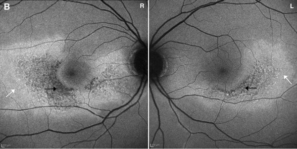 B: Autofluorescence imaging showing symmetrical changes at the level of the retinal pigment epithelium. Mottled loss of autofluorescence (black arrows) indicates loss of retinal pigment epithelium cells, and the adjacent increased autoflourescence (white arrows) indicates cell abnormality. |
Acknowledgements
We thank the Medical Photographic Imaging Centre at the Royal Victorian Eye and Ear Hospital for the images.
References
- Australian Government. Pharmaceutical Benefits Schedule Item Reports. Canberra: Medicare Australia. https://www.medicareaustralia.gov.au/cgi-bin/broker.exe?_PROGRAM=sas.pbs_item_standard_report.sas&_SERVICE=default&itemlst=%2701512N%27&ITEMCNT=1&_DEBUG=0&LIST=1512N&VAR=SERVICES&RPT_FMT=1&start_dt=200807&end_dt=200906 (accessed Apr 2010).
- Gass JDM. Stereoscopic atlas of macular diseases: diagnosis and treatment. 4th ed. St Louis: CV Mosby Company, 1997. 0_i1091894
- Yam JCS, Kwok AKH. Ocular toxicity of hydroxychloroquine. Hong Kong Med J 2006; 12: 294-304. 0_i1091896
- Royal College of Ophthalmologists. Hydroxychloroquine and ocular toxicity. Recommendations on screening. October 2009. London: RCO, 2009. http://www.rcophth.ac.uk/docs/publications/published-guidelines/Hydroxychloroquine_and_Ocular_Toxicity_final_Oct_2009.pdf (accessed Apr 2010).
- Marmor MF, Carr RE, Easterbrook M, et al. Recommendations on screening for chloroquine and hydroxychloroquine retinopathy: a report by the American Academy of Ophthalmology. Ophthalmology 2002; 109: 1377-1382. 0_i1091901
A Diagnostic Headache
Thomas Glynn, Michelle T. Leech, Stacy K. Goergen, Emily Lin, Nadeem Toodayan, Ralph Junckerstorff
Neisseria meningitidis: an uncommon cause of conjunctivitis
Chloe Story, Tina Marinelli, Simeon Crawford, Amrita Ronnachit
Dismantling barriers to research and clinical care for individuals with a vision impairment
Eden G Robertson, Kate Hetherington, Meredith Prain, Julia Hall, Leighton Boyd AM, Rosemary Boyd OAM, Emily Shepard, Hollie Feller, Sally Karandrews, Fleur O'Hare, Kanae Yamamoto, Matthew P Simunovic, Robyn V Jamieson, Alan Ma, Lauren Ayton AM, Anai Gonzalez‐Cordero
The New South Wales Pharmacy Trial for herpes zoster: on the nose?
Christian P Pappas, Timothy R Holmes, Minas T Coroneo
Skin fragility disorder misdiagnosed as child abuse: a cautionary tale
Heather G Mack, Fred K Chen, John R Grigg
Health economic aspects of inherited retinal diseases: looking for cost‐effective treatments
Benjamin Kamien, Rachael Heath Jeffery, Fred K Chen