




Volume 190 - Issue 4
Massive haemoptysis due to aortobronchial fistula caused by pulmonary hydatidosis
Authors: Stefan Buchholz, David Sowden, Troy Stapleton, Peter Pohlner and Craig Wright
Med J Aust 2009; 190 (4): 222. || doi: 10.5694/j.1326-5377.2009.tb02362.x
Published online: 16 February 2009
Published online: 16 February 2009
To the Editor: A 56-year-old woman was recently admitted with recurrent large-volume haemoptysis associated with left-sided tearing thoracic pain. Growing up on a sheep farm in rural New South Wales, she had been diagnosed at age 8 years with pulmonary hydatidosis, which remained dormant on periodic clinical assessments. However, 2 years before presentation she started to cough up gelatinous material containing scolices of Echinococcus granulosus. Surgery was declined at that time due to the anticipated complexity of the operation and associated high perioperative risk. Long-term anthelmintic therapy was commenced.
On admission, a computed tomography (CT) scan showed a contained aortic pseudoaneurysm (Box), consistent with rupture of the aorta into the hydatid cyst. Other images showed the cyst containing gas, indicating communication with the airway.
After stabilisation, the patient was transferred to a cardiothoracic centre. A left upper lobectomy with dissection and removal of the mediastinal cyst was undertaken through a median sternotomy, and the aortic fistula was successfully repaired using bovine pericardial strips. Intraoperatively, there was no evidence of pericardial involvement. Histopathological examination showed a disrupted and degenerate hydatid cyst without a germinal layer and no protoscolices. The patient received albendazole for 6 months after surgery and her recovery was uneventful.
Hydatid disease is caused by the intestinal parasitic tapeworm E. granulosus which, in Australia, is most prevalent in the eastern half of NSW at higher altitudes.1 Symptomatic intramural aortic-wall hydatidosis causing aortic-wall rupture and pseudoaneurysm formation has been described in fewer than a dozen cases.2
Hypotheses for arterial-wall invasion include dissemination during cardiac surgery; entry through vasa vasorum or pre-existing small intimal tears or aneurysms; or partial incorporation of the aortic wall into the hydatid pericyst.2,3
We identified four case reports in adults describing fistula formation between a pulmonary hydatid cyst and the aorta, including three European cases3-5 and one South African case (published twice).6,7 All patients were middle-aged men: two presented with chest pain and large-volume haemoptysis, one with anaphylactic shock and bilateral ischaemic lower limbs from aortic-wall hydatid cyst emboli, and one with a cyst eroding the abdominal aorta (found incidentally during surgery for a coeliac trunk aneurysm). One patient died during removal and another patient after removal of the primary cyst.
Haemoptysis in pulmonary hydatid disease is a common presenting symptom. Mechanisms include pressure erosion of a bronchus, obstructive infection, cyst rupture or — very rarely, and emphasised in our case — erosion of a major vascular structure.
Aortobronchial fistula resulting from pulmonary hydatidosis
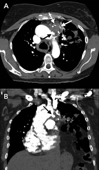
A: Contrast-enhanced thoracic computed tomography scan showing consolidation and cavitation within the left upper lobe. A multiloculated 4 cm diameter peripherally calcified hydatid cyst was present in the medial aspect of the left upper lobe (white arrows), penetrating under the aortopulmonary window. Contrast medium extended posteriorly into the base of the lesion, suggestive of an aortic leak (black arrow).
B: Coronal reconstruction of the aortic arch demonstrated a round collection of contrast medium with a 1.4 cm base (black arrow), consistent with a contained saccular aortic pseudoaneurysm.
References
- Jenkins DJ, Power K. Human hydatidosis in New South Wales and the Australian Capital Territory, 1987–1992. Med J Aust 1996; 164: 18-21. <eMJA full text>
- Aydin NB, Celik S, Suzer A, et al. Hydatid cyst in the wall of the ascending aorta. Asian Cardiovasc Thorac Ann 2006; 14: 153-154. 0_i1091867
- Oezer C, Akbas T, Yildiz A, et al. False aneurysm of the thoracic aorta due to hydatid cyst rupture. Eur J Radiol 2005; 54: 27-29. 0_i1091869
- Marti-Bonmati L, Touza R, Montes H. CT diagnosis of primary mediastinal hydatid cyst rupture into the aorta: a case report. Cardiovasc Intervent Radiol 1988; 11: 296-299. 0_pgfId-1691220
- Viver E, Bianchi L, Callejas JM, Martorell A. Hydatid cyst fistula into the aorta. J Cardiovasc Surg (Torino) 1989; 30: 624-626. 0_i1091872
- Nel JD, Kriegler SG, Van Vuuren WM, et al. An unusual cause of nearly fatal hemoptysis. Respiration 2001; 68: 635-636. 0_i1091874
- Harris DG, Van Vuuren WM, Augustyn J, Rossouw GJ. Hydatid cyst fistula into the aorta presenting with massive hemoptysis. J Cardiovasc Surg (Torino) 2001; 42: 565-567. 0_i1091878
Fatty Liver Disease in Australia: A Narrative Review on the Epidemiology, Natural History, Prognostication and Management in People With Metabolic Dysfunction
Karl Vaz, Daniel Clayton-Chubb, William W. Kemp, Stuart K. Roberts, Ammar Majeed
Sex Differences in Management and Outcomes of People With ST-Elevation Myocardial Infarction, New South Wales, 2011–2020: A Retrospective Cohort Study
Adam C. Livori, Jedidiah I. Morton, Dianna J. Magliano, Jonathan E. Shaw
Rethinking Rheumatic Fever Diagnosis: Progress, Pitfalls and the Path Towards a Definitive Test
Anna P. Ralph, Rachel Webb, Tom Parks, Nicole J. Moreland, Asha C. Bowen, Benjamin Jones, Emma Ndagire, Andrea Z. Beaton
Sex Differences in Management and Outcomes of People With ST-Elevation Myocardial Infarction, New South Wales, 2011–2020: A Retrospective Cohort Study
Samia Kazi, Simone Marschner, Clara K. Chow
Distribution of Cardiovascular Disease Risk Based on the Updated 2023 Guideline-Recommended Australian Cardiovascular Disease Risk Algorithm and Comparison With the 2012 Algorithm: An Observational Study
Nina Lazarevic, Meghana Bhat, Grace Joshy, Danielle C. Butler, Mark Woodward, Anushka Patel, Rod T. Jackson, Garry Jennings, Rosemary Wyber, Ellie Paige, Emily Banks
They Brought the Hospital to Me
Hugh N. Burrill