




Volume 188 - Issue 9
A case of primary cerebral vasculitis
Authors: Sanjaya S Herath, Dayna B Law, Peter J O Stride, Vernon J Heazlewood and Luke S Gaffney
Med J Aust 2008; 188 (9): 547-549. || doi: 10.5694/j.1326-5377.2008.tb01779.x
Published online: 5 May 2008
Published online: 5 May 2008
To the Editor: Primary cerebral vasculitis (PCV) is a potentially fatal disease. Early diagnosis and therapy are vital. We describe a case where confounding factors delayed diagnosis.
A 42-year-old woman presented with headache, nausea, vomiting, malaise and binocular blindness for 3 days. Two weeks previously, she had presented to the emergency department with headache and vomiting, but investigations, including computed tomography (CT) of the brain and lumbar puncture, gave normal results. She had a history of depression, was a smoker (20 pack-year history), and used cannabis regularly and alcohol occasionally, but denied other recreational drug use.
Her mood appeared depressed. Vital signs and findings from a general examination were normal. Eye movements were full, direct and indirect pupillary reflexes were intact, and optic fundi were normal. Results of a CT angiogram were reported as normal by a consultant radiologist. Results of blood tests, including inflammatory markers, and a repeat lumbar puncture, were unremarkable. A toxicology screen was not performed. Depression with conversion disorder was diagnosed, and admission with analgesia was advised. A neurologist’s review on Day 2 did not detect organic disease. The mental health team diagnosed severe depression and prescribed antidepressants.
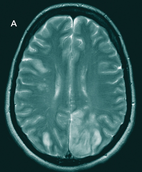
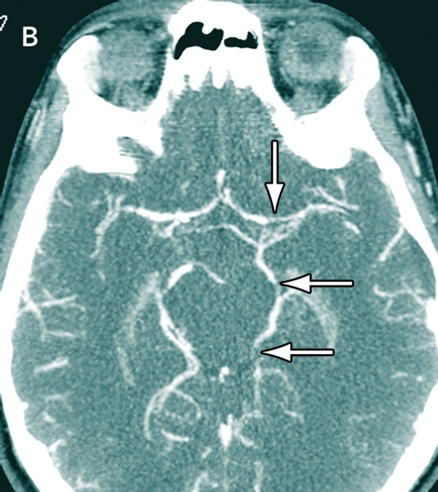
On Day 4, the patient’s condition deteriorated and she become non-communicative with signs of right hemiplegia. An electroencephalogram showed polyrhythmic generalised slow waves consistent with encephalopathy. She was transferred to a tertiary centre where magnetic resonance imaging (MRI) and CT angiography of the brain showed multiple bilateral infarcts (Figure, A) with beaded arteries, the classic appearance of vasculitis. She was given high-dose prednisolone and cyclophosphamide. Investigations were negative for causes of secondary vasculitis. Her condition continued to deteriorate and she died 8 days after admission. Autopsy was refused. Subsequent review of the second CT scan detected irregular cerebral vessels (Figure, B).
PCV is an uncommon disorder of the central nervous system, with unknown aetiology and no specific characteristic features, affecting small cerebral arteries but not extracranial vessels. Symptoms and signs vary but include headache, encephalopathy, seizures, personality change, weakness, and altered level of consciousness, as well as superimposed focal cranial neuropathy or hemiplegia. Recognition is difficult, but differentiation from reversible cerebral vasoconstriction syndrome is important.1,2
Brain biopsy is seen as the “gold standard” for diagnosing PCV. CT angiography may show diffuse or localised changes, with vessel beading, aneurysms, and luminal narrowing. MRI may show areas of white and grey matter infarction, or haemorrhage. MRI is more sensitive than CT, but less sensitive than CT angiography. Up to 100% of biopsy-positive cases appear abnormal on MRI. Suspected cases require careful clinical appraisal and either CT angiography or MRI, probably followed by an image-guided brain biopsy.3
Initial reported cases of PCV had a poor prognosis; most patients died within a few weeks.2 Immunosuppressive therapy with glucocorticoids and cyclophosphamide (as used in secondary severe vasculitis) may be beneficial, although there are no clinical trials.4 A future therapeutic alternative may be infliximab, which has been used successfully for one patient with cerebral vasculitis secondary to Behçet’s disease who had known elevated levels of tumour necrosis factor α.5
Despite increasing awareness and advances in angiography, PCV remains an uncommon diagnostic and therapeutic problem which should be considered in cases of severe, non-febrile neurological illness with stroke-like features.
Acknowledgements
We thank Dr Chris Staples, Neurologist, Redcliffe Hospital, for reviewing both the case and this manuscript.
References
- Calabrese LH, Mallek JA. Primary angiitis of the central nervous system. Report of 8 new cases, review of the literature, and proposal for diagnostic criteria. Medicine (Baltimore) 1988; 67: 20-39.
- Lie JT. Primary (granulomatous) angiitis of the central nervous system: a clinicopathologic analysis of 15 new cases and a review of the literature. Hum Pathol 1992; 23: 164-171. 0_i1091855
- Greenan TJ, Grossman RI, Goldberg HI. Cerebral vasculitis: MR imaging and angiographic correlation. Radiology 1992; 182: 65-72. 0_i1091857
- Fountain NB, Lopes MB. Control of primary angiitis of the CNS associated with cerebral amyloid angiopathy by cyclophosphamide alone. Neurology 1999; 52: 660-662. 0_i1091859
- Licata G. Antitumour necrosis factor alpha monoclonal antibody therapy for recalcitrant cerebral vasculitis in a patient with Behçet’s syndrome Ann Rheum Dis 2003; 62: 280-281. 0_i1091863
Motor Neuron Disease Mortality Trends in Australia From 1986 to 2023: A Population-Based Study
Carol M. Y. Lee, Rupendra N. Shrestha, Julian Gold, Mark Stevenson, Kelly L. Williams, Lyndal Henden, Sandrine K. K. Chan Moi Fat, Zoe Zussa, Amanda Wright, Martin Jude, Mark A. S. Laidlaw, Willem J. R. Fokkink, Dominic B. Rowe
Mild traumatic brain injury and concussion and persisting post‐concussion symptoms: new guidelines to support evidence‐based assessment and management in Australia and Aotearoa New Zealand
Karen M Barlow, Jennie L Ponsford, Alice Theodom, Gill Cowen, Gavin A Davis, Vicki Anderson, Franz E Babl, David Cole, Jennifer Cullen, Stuart R Dalziel, Melinda Fitzgerald, Howard Flavell, Caroline Yates, Rebecca Kimble, John H Olver, Rhonda Orr, Mark Ralfe, Michael Rose, Nick Rushworth, Julia Treleaven, Gary Browne, Nathan Delang, Sarah Harris, Gary Mitchell, Sean Tweedy
The prevalence of and variation in indicators of the quality and safety of long term aged care in Australia, 2019: a cross‐sectional population‐based study
Tesfahun C Eshetie, Gillian E Caughey, Catherine Lang, Olivia Ryan, Renuka Visvanathan, Craig Whitehead, Keith Evans, Janet K Sluggett, Jyoti Khadka, Carolyn Dawkins, Helena Williams, Miranda Starke, Sara Blunt, Anne Liddell, Megan Corlis, Anna Sheppeard, Penelope Lello, Marilyn Thien, Steven L Wesselingh, Maria C Inacio
Health and economic benefits of improving pre‐hospital identification of stroke in Australian women: a modelling study
Thomas Gadsden, Lei Si, Emily R Atkins, Cheryl Carcel, Xia Wang, Stephen Jan, Mark Woodward, Laura E Downey
Symptom burden, quality of life, and diagnostic journey of people with postural orthostatic tachycardia syndrome, Australia, 2021–24: a descriptive patient registry data study
Marie‐Claire Seeley, Gemma Wilson, Eric Ong, Amy Langdon, Jonathan Chieng, Danielle Bailey, Kristina Comacchio, Amanda Page, Dennis H Lau, Celine Gallagher
The intersection of rurality and dementia prevalence in Australia for Aboriginal and Torres Strait Islander and non‐Indigenous peoples
Antonia J Clarke, Marwan Ahmed, Judith M Katzenellenbogen, John Towney (Wiradjuri), Anna H Balabanski, Adrienne Withall (Dharawal Yuin), Kylie Radford, Amy Brodtmann