




Volume 188 - Issue 4
Perinephric haematopoiesis
Author: Philip Y-I Choi
Med J Aust 2008; 188 (4): 250. || doi: 10.5694/j.1326-5377.2008.tb01598.x
Published online: 18 February 2008
Published online: 18 February 2008
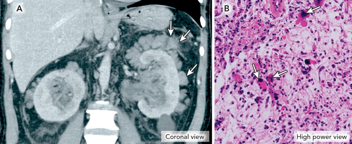
A 59-year-old man presented with persistent abdominal discomfort 12 months after splenectomy for thrombocytopenia to ameliorate progressive myelofibrosis. Physical examination revealed bilateral loin masses. Computed tomography of the abdomen identified perinephric cuffing by soft tissue nodular masses in the perinephric fat (Figure A). Core needle biopsy of the right perinephric fat revealed haematopoietic cells (Figure B).
Extramedullary haematopoiesis is not uncommon when intramedullary haematopoiesis is impaired.1-3 Common sites include the liver and spleen. It is unusual for extramedullary haematopoiesis to involve the perinephric fat. The role of splenectomy in accelerating the development of extramedullary haematopoiesis in uncommon sites has become increasingly apparent.1
Acknowledgements
I thank Professor Douglas Joshua, Institute of Haematology, Royal Prince Alfred Hospital, for guidance, and Dr Jim Raleigh, Department of Radiology, Royal Prince Alfred Hospital and Dr Geoffrey Watson, Department of Anatomical Pathology, Royal Prince Alfred Hospital for assistance with preparation of images.
References
- Gibbins J, Pankhurst T, Murray J, et al. Extramedullary haematopoiesis in the kidney: a case report and review of literature. Clin Lab Haematol 2005; 27: 391-394.
- Gryspeerdt S, Oyen R, Van Hoe L, et al. Extramedullary haematopoiesis encasing the pelvicalyceal system. CT findings. Ann Hematol 1995; 71: 53-56. 0_pgfId-1091831
- Woodward N, Ancliffe P, Griffiths MH, Cohen S. Renal myelofibrosis: an unusual case of renal impairment. Nephrol Dial Transplant 2000; 15: 257-258. 0_i1091833
Splenic injury in severe cases of the zoonoses Q fever and rickettsial infection: diagnostic challenges
Ashleigh Drury, Philippa Harrison, Aiveen Bannan
Health and economic benefits of improving pre‐hospital identification of stroke in Australian women: a modelling study
Thomas Gadsden, Lei Si, Emily R Atkins, Cheryl Carcel, Xia Wang, Stephen Jan, Mark Woodward, Laura E Downey
Updating the diagnosis and management of elevated serum ferritin levels in the era of routine ferritin testing of blood donors by Australian Red Cross Lifeblood
Gary D Zhang, James Chen, Daniel M Johnstone, Martin B Delatycki, Katie Allen, John K Olynyk
Early cardiovascular collapse after envenoming by snakes in Australia, 2005–2020: an observational study (ASP‐31)
Geoffrey K Isbister, Katherine Z Isoardi, Angela L Chiew, Shane Jenkins, Nicholas A Buckley
Updated recommendations for warfarin reversal in the setting of four‐factor prothrombin complex concentrate
Danielle Robinson, James McFadyen, Eileen Merriman, Chee Wee Tan, Ross Baker, Huyen Tran
Updating the diagnosis and management of iron deficiency in the era of routine ferritin testing of blood donors by Australian Red Cross Lifeblood
Gary D Zhang, Daniel Johnstone, Michael F Leahy, John K Olynyk