




Volume 187 - Issue 10
Magnetic resonance imaging for paraneoplastic dermatomyositis
Authors: Daniele Torchia, Emiliano Antiga, Letizia Ricupero, Marzia Caproni and Paolo Fabbri
Med J Aust 2007; 187 (10): 589. || doi: 10.5694/j.1326-5377.2007.tb01424.x
Published online: 19 November 2007
Published online: 19 November 2007
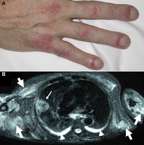
A 64-year-old man was referred for assessment of a symmetric violaceous rash of the dorsal aspect of the fingers (Figure, A), trunk, olecranon processes and malleoli. An oedematous, blue-purple discolouration of the eyelids was also apparent. The patient complained of severe weakness of the proximal muscles and dysphagia. T2-weighted magnetic resonance imaging showed muscular inflammation and a pulmonary mass (Figure, B) that was histologically identified as squamous cell carcinoma and staged as T3 N0 M0.
The dermatomyositis, confirmed by elevation of serum muscle enzymes and electromyography, improved clinically with oral prednisone plus hydroxychloroquine, and did not recur after surgical excision of the tumour.
Diagnosing Acute Kava Dermopathy: A Case Report of a Characteristic Cutaneous Eruption
Ali Abid, Nicholas Allen, Abeer Hagelamin, Christopher Henderson, Artiene Tatian
Localised Herpes Simplex Following Midline Laparotomy
Jessica S. Bulluss, Paul Chee, Matthew J. Verheyden
No silver lining with health misinformation: argyria caused by intentional silver consumption
Luke Collins, Logesh Palanikumar, Stephen Bacchi
Lingual Raynaud phenomenon
Michael Taggart, Mina John
Melasma in the male: a less well recognised entity
Tim Aung, Rowland Noakes
Age group‐specific changes in keratinocyte cancer treatment rates in Australia, 2012–2021: a retrospective cohort study based on MBS claims data
Catherine M Olsen, Nirmala Pandeya, Rachel E Neale, David C Whiteman