




Volume 178 - Issue 5
Human fasciolosis acquired in an Australian urban setting
Authors: Andrew J Hughes, Terry W Spithill, Rebecca E Smith, Craig S Boutlis and Paul DR Johnson
Med J Aust 2003; 178 (5): 244-245. || doi: 10.5694/j.1326-5377.2003.tb05174.x
Published online: 3 March 2003
Published online: 3 March 2003
To the Editor: Although liver flukes (genus, Fasciola) are parasites of livestock, human infection is a significant global health problem,1 albeit seldom seen in Australia.2 Infected livestock contaminate waterways with parasite eggs, leading to infection of snails that shed metacercariae on to vegetation, such as watercress.1,3,4 Adult parasites reside in and damage the bile ducts.1,3 Liver flukes could cause disease if introduced into the food chain.1
We report the first case in Australia of liver fluke infection (fasciolosis) in a patient with no history of farm or livestock contact. She probably acquired the disease from eating watercress purchased at a Melbourne market four to five months before symptom onset.
Computed tomography of the liver in a woman with fasciolosis
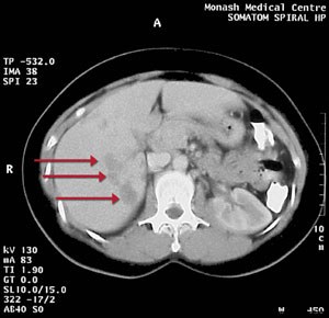
Low density lesions (arrowed) following intrahepatic ductal branches in the right lobe of the liver were consistent with Fasciola parasites causing biliary obstruction.
A 35-year-old woman presented in August 1998 with fever and right upper quadrant abdominal pain. Blood examination showed eosinophilia (2.5 x 10⁹/L; reference range [RR], < 0.6 x 10⁹/L]). Liver function tests gave normal results apart from elevated serum aspartate aminotransferase levels of 48 U/L (RR < 41 U/L). Abdominal computed tomography showed multiple low density lesions in the right lobe of the liver, with diameter up to 3 cm (Box). A fine needle aspirate showed no evidence of malignancy. Blood tests four weeks later revealed increasing eosinophilia (3.5 x 10⁹/L) and worsening liver function (serum levels: alanine aminotransferase, 163 U/L [RR, 7–56 U/L]; alkaline phosphatase, 126 U/L [RR, 30–120 U/L]; γ-glutamyl transferase, 98 U/L [RR, 5–45 U/L]).
A parasitic infection was suspected, but four faecal samples and serological tests for hydatids, Schistosoma, Strongyloides and Entamoeba spp. were negative. Coprological diagnosis of fasciolosis can be problematic, as eggs may be released intermittently and in small numbers, especially in low-intensity infection.1 Enzyme-linked immunosorbent assay (ELISA) using Fasciola hepatica antigen, performed at Westmead Hospital, Sydney, was borderline positive. However, ELISA for IgG4 antibodies against recombinant F. hepatica cathepsin L5 antigen, performed at Monash University, Melbourne, was strongly positive.
The patient was treated with two doses of triclabendazole (12 mg/kg body weight per dose) on successive days in October 1998. Abdominal pain subsided within two weeks, her appetite was restored, and eosinophil count and liver function normalised within four weeks. Computed tomography two months after treatment showed a reduction in size of the liver lesions. The patient remained well six months later.
This case demonstrates that fasciolosis may present to urban medical practitioners in Australia. Ingestion of watercress is an important clue to the aetiology.2 Serological diagnosis is possible before eggs appear in faeces using a new specific ELISA test that detects the IgG4 response to cathepsin L antigen.5
References
- Mas-Coma S, Bargues MD, Esteban JG. Human fasciolosis. In: Dalton JP, editor. Fasciolosis. Wallingford: CAB International, 1999: 411-434. CBBJFGDA
- Torresi J, Richards MJ, Taggart GJ, Smallwood RA. Fasciola hepatica liver infection in a Victorian dairy farmer. Med J Aust 1996; 164: 511. CBBDCDBD
- Boray JC. Experimental fascioliasis in Australia. Adv Parasitol 1969; 7: 95-210. CBBFJHJC
- McCausland I, Vandegraaff R, Nugent L. Fascioliasis in dairy cows on irrigated pasture. Aust Vet J 1980; 56: 324-326. CBBIGIBD
- O'Neill SM, Parkinson M, Strauss W, et al. Immunodiagnosis of Fasciola hepatica infection (fascioliasis) in a human population in the Bolivian Altiplano using purified cathepsin L cysteine proteinase. Am J Trop Med Hyg 1998; 58: 417-423. i1082305
Costs of Delivering the Hep B PAST Model of Care in Remote Australia: A Retrospective Pre–Post Study
Hoa Nguyen, Anh Le Tuan Nguyen, Paula Binks, Melita McKinnon, Emily Vintour-Cesar, Karen Wills, Nicola Stephens, Benjamin Cowie, Joshua S. Davis, Yuejen Zhao, Peter Nihill, Julie A. Campbell, Andrew J. Palmer, Jane Davies, Barbara de Graaff
Buruli Ulcer Acquired From Ringtail Possum Rescue
Daniel P. O'Brien, Naomi Clarke, Sara Bonnici, Mohammad A. Hussain
Q Fever Visceral Abscesses After Contact With Native Australian Marsupials
Samantha J. Laws, Enzo Binotto, James Stewart, Jennifer Ho, Trent Yarwood, Josh Hanson, Simon Smith
Hospital-Admitted Injection-Related Infections Among Incarcerated People Who Inject Drugs in Australia: A Retrospective Cohort Study
Andrew Palmer, Matthew Carter, Jeremy Yeo, Cecilia Shim, Jason Connor, Jeremy Hayllar, Gerald Holtmann, Naomi Moy, Elliott G. Playford, Naomi Runnegar, Paul J. Clark
The Live Attenuated Influenza Vaccine in Australia: An Additional Tool for Influenza Prevention
Cyra Patel, Alexis Pillsbury, Tran Nguyen, Xia Wang, Helen E. Quinn, Clayton K. Chiu, Allen C. Cheng, Katie L. Flanagan, Zhicheng Wang
Reflex Testing for Hepatitis D Infection: A Unique Opportunity to Reduce Hepatitis D-Related Chronic Liver Disease Deaths in Australia
Jessica Howell, Lauren Andersson, Miriam T. Levy, James O'Beirne, Leon Adams, Katharine Irvine, Avik Majumdar, Golo Ahlenstiel, Kathy Jackson, Krispin Hajkowicz, Joseph Doyle, Jane Davies, Sarah Cherian, Wayne Dimech, Alexander J. Thompson