




Medical education
Lessons from practice
Volume 219 - Issue 9
Good's syndrome associated with multiple basal cell carcinomas: a case report
Authors: Michelle Wu, Nicole Seebacher and Margit Polcz
Med J Aust 2023 || doi: 10.5694/mja2.52093
Published online: 9 October 2023
Published online: 9 October 2023
A 61-year-old man of European ancestry presented with five large fungating lesions
Clinical record
A 61‐year‐old man of European ancestry (Fitzpatrick skin type II) presented with five large fungating lesions (Box 1). These tumours had been progressively growing over a 12‐month period. The presentation of this patient was delayed as he had initially misattributed the lesions to ingrown hairs. He had no personal or family history of melanoma or non‐melanoma skin cancers. He had no personal medical history of autoimmune conditions, recurrent infections, or immunosuppression.
The five tumours ranged from 1cm to 10cm in diameter. He had no cervical, axillary, or inguinal lymphadenopathy. Punch biopsies of all five lesions confirmed basal cell carcinoma (BCC; Box 2). He subsequently had a chest computed tomography scan the following week to determine the extent of invasion of a large overlying BCC, which incidentally revealed an 8cm thymoma that was resected five months later.
He had several full skin checks over the course of the next ten months, which revealed a total of 13 additional BCCs and a squamous cell carcinoma (SCC) in situ (Box 2). He was started on nicotinamide 500mg twice daily. Genetic testing for Gorlin syndrome was not completed as he did not fulfill the major or minor criteria. Antinuclear antibodies were present at a titre of 1:80. Immunophenotyping revealed very low B cells and inversion of CD4+/CD8+ T cell compartments (Box 3). Functional evaluation of his humoral immunity with before and after antibody titres to the 23‐valent pneumococcal polysaccharide vaccine (Pneumovax; Merck Sharpe and Dohme) showed an inadequate response. Considering his medical history of a thymoma, he was diagnosed with Good's syndrome. He was subsequently commenced on monthly intravenous 35g of 10% immunoglobulin and full skin checks every six months.
Discussion
Good's syndrome (thymoma with immunodeficiency) is a rare condition characterised by thymoma associated with deficiency in humoral and cellular immunity, and was first reported by Robert Good in 1954.1 It most commonly affects adults aged in their 40s or 50s.2 The clinical manifestations are highly heterogenous, ranging from sinopulmonary infections, to chronic diarrhoea and haematological complications.2 Autoimmune conditions have been reported, most commonly with pure red blood cell aplasia, myasthenia gravis, and oral lichen planus. Management involves regular intravenous immunoglobin and treatment of associated infections and autoimmune disorders. Thymectomy can be considered to prevent local invasion and metastasis, but it does not improve immunodeficiency.3 Despite these therapeutic options, the prognosis of Good's syndrome remains poor, with a ten‐year overall survival rate of 33%.2
To our knowledge, this is the first case of Good's syndrome that has been associated with the development of multiple large BCCs. Although there has been mention of two previous cases of BCC in Good's syndrome, these were not noted to be aggressive or multiple as in our patient.3 Epidemiological data suggest a strong link between non‐melanoma skin cancers and the immune system.4 Immunocompromise is widely recognised as a risk factor for the development of non‐melanoma skin cancers, such as in acquired immunodeficiency syndrome, organ transplant recipients, and patients with chronic lymphocytic leukaemia.4 Moreover, non‐melanoma skin cancers are more likely to be of the more aggressive subtypes in the immunocompromised population.4 The SCC to BCC ratio (4:1) is often reversed in this population compared with the general population (1:4).5 Our case reports an SCC to BCC ratio of 1:18, which is an interesting finding that suggests a possible immunopathological interaction between Good's syndrome and BCCs.
Lessons from practice
- Multiple basal cell carcinomas may be an early marker for immunodeficiency.
- For patients presenting with multiple or large tumours, immunophenotyping should be considered to identify possible underlying immunodeficiency.
- Evaluation of antibody response may be a more useful marker of B cell function compared with absolute B cell counts.
Box 1 – Large fungating lesions in a 61‐year‐old man
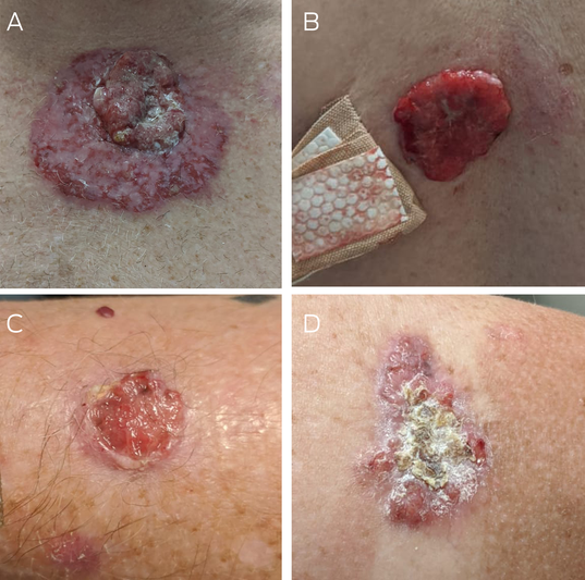
(A) Large exophytic nodule on central chest with overlying telangiectatic vessels and central ulceration measuring 100mm by 100mm. (B) Moist red nodule with rolled edges located on middle upper back measuring 55mm by 65mm. (C) Smaller erythematous nodule with rolled edges and central ulceration located on left medial forearm measuring 55mm by 25mm. (D) Large scaly plaque with irregular border located on right posterior shoulder measuring 45mm by 23mm.
Box 2 – Timeline of lesions and their respective histology and treatment
|
Date |
Location |
Histological type |
Treatment |
||||||||||||
|
|
|||||||||||||||
|
Week 0 (initial presentation) |
Central chest |
Infiltrating BCC |
WLE with SSG |
||||||||||||
|
Left medial forearm |
Nodular BCC |
WLE |
|||||||||||||
|
Right shoulder |
Nodular BCC |
WLE |
|||||||||||||
|
Left posterior shoulder |
Infiltrating BCC with perineural invasion |
WLE |
|||||||||||||
|
Central back |
Infiltrating BCC |
WLE with SSG |
|||||||||||||
|
Week 15 |
Right upper temple |
Superficial BCC |
SE |
||||||||||||
|
Right lower temple |
Nodular and partly infiltrating BCC |
SE |
|||||||||||||
|
Week 32 |
Left forearm |
Nodular BCC |
SE |
||||||||||||
|
Right shoulder |
Superficial BCC |
Topical imiquimod* |
|||||||||||||
|
Left wrist |
Superficial BCC |
SE |
|||||||||||||
|
Left upper chest upper region |
Superficial BCC |
Topical imiquimod* |
|||||||||||||
|
Left thigh |
SCC in situ |
SE |
|||||||||||||
|
Right medial calf |
Infiltrative BCC |
SE |
|||||||||||||
|
Left medial calf |
Superficial BCC |
SE |
|||||||||||||
|
Week 71 |
Right shoulder† |
Superficial BCC |
Topical imiquimod* |
||||||||||||
|
Left upper chest lower region |
Superficial BCC |
Topical imiquimod* |
|||||||||||||
|
Left thigh |
Superficial BCC |
Topical imiquimod* |
|||||||||||||
|
Left shin |
Superficial BCC |
Topical imiquimod* |
|||||||||||||
|
Left proximal arm |
Superficial BCC |
Topical imiquimod* |
|||||||||||||
|
Left upper chest upper region† |
Nodular BCC |
SE |
|||||||||||||
|
Week 82 |
Central forehead |
Nodular BCC |
SE |
||||||||||||
|
|
|||||||||||||||
|
BCC = basal cell carcinoma; SCC = squamous cell carcinoma; SE = surgical excision; SSG = split skin graft; WLE = wide local excision. * Topical imiquimod treatment regime involves daily application five days per week for six weeks. † Lesion previously treated with topical imiquimod 5%. |
|||||||||||||||
Box 3 – Laboratory parameters before immunoglobulin replacement
|
Results |
Values |
Reference range |
|||||||||||||
|
|
|||||||||||||||
|
B cell count (%) |
0.06×10⁹/L (2%) |
0.11–0.70×10⁹/L (7–27%) |
|||||||||||||
|
CD3+ T cell count (%) |
2.6×10⁹/L (84%) |
1.2–2.7×10⁹/L (49–84%) |
|||||||||||||
|
CD4+ T cell count (%) |
1.12×10⁹/L (36%) |
0.44–2.16×10⁹/L (28–63%) |
|||||||||||||
|
CD8+ T cell count (%) |
1.55×10⁹/L (50%) |
0.13–1.31×10⁹/L (10–40%) |
|||||||||||||
|
CD4+/CD8+ T cell ratio |
0.72 |
1.5–2.5 |
|||||||||||||
|
IgA level |
1.80g/L |
0.70–4.00g/L |
|||||||||||||
|
IgG level |
8.36g/L |
7.00–16.00g/L |
|||||||||||||
|
IgM level |
0.99g/L |
0.40–2.30g/L |
|||||||||||||
|
|
|||||||||||||||
|
|
|||||||||||||||
Competing interests
No relevant disclosures.
References
- Good RA, Varco RL. A clinical and experimental study of agammaglobulinemia. J Lancet 1955; 75: 245‐271.
- Kelesidis T, Yang O. Good's syndrome remains a mystery after 55 years: a systematic review of the scientific evidence. Clin Immunol 2010; 135: 347‐363.
- Zaman M, Huissoon A, Buckland M, et al. Clinical and laboratory features of seventy‐eight UK patients with Good's syndrome (thymoma and hypogammaglobulinaemia). Clin Exp Immunol 2019; 195: 132‐138.
- Griffiths CEM, Barfer J, Bleiker TA, et al; editors. Rook's textbook of dermatology, 4 volume set, 9th ed. Chichester (UK): Wiley Blackwell, 2016.
- O'Reilly Zwald F, Brown M. Skin cancer in solid organ transplant recipients: advances in therapy and management: part II. Management of skin cancer in solid organ transplant recipients. J Am Acad Dermatol 2011; 65: 263‐279.
Provenance: Not commissioned; externally peer reviewed.
Diagnosing Acute Kava Dermopathy: A Case Report of a Characteristic Cutaneous Eruption
Ali Abid, Nicholas Allen, Abeer Hagelamin, Christopher Henderson, Artiene Tatian
Localised Herpes Simplex Following Midline Laparotomy
Jessica S. Bulluss, Paul Chee, Matthew J. Verheyden
No silver lining with health misinformation: argyria caused by intentional silver consumption
Luke Collins, Logesh Palanikumar, Stephen Bacchi
Lingual Raynaud phenomenon
Michael Taggart, Mina John
Melasma in the male: a less well recognised entity
Tim Aung, Rowland Noakes
Age group‐specific changes in keratinocyte cancer treatment rates in Australia, 2012–2021: a retrospective cohort study based on MBS claims data
Catherine M Olsen, Nirmala Pandeya, Rachel E Neale, David C Whiteman