




Medical education
Lessons from practice
Volume 218 - Issue 7
Axial crystal arthropathy mimicking facet joint septic arthritis with epidural abscess
Authors: Hannah Poole, Amy Crowe and John Daffy
Med J Aust 2023; 218 (7): 301-303. || doi: 10.5694/mja2.51891
Published online: 17 April 2023
Published online: 17 April 2023
A 78-year-old man with hypertension, dyslipidaemia, hypothyroidism and gout presented with two weeks of migratory polyarthritis and fever
Clinical record
A 78‐year‐old man with hypertension, dyslipidaemia, hypothyroidism and gout presented with two weeks of migratory polyarthritis and fever. His previous attacks of gout occurred biannually, affected his fifth toe only, and were managed with short courses of colchicine. He was not receiving urate‐lowering therapy and did not have renal impairment.
He presented with worsening back pain, lower limb weakness, and new urinary incontinence. He was febrile (38°C) and his left hand and wrist were swollen and tender. He had no joint tenderness elsewhere and no tophi. Left wrist aspirate two days prior demonstrated monosodium urate (MSU) crystals, and colchicine 500μg twice daily was started. Serum urate was 0.37mmol/L (reference interval [RI], 0.21–0.42mmol/L).
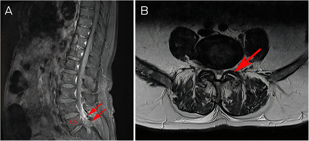
In the setting of fever, back pain and neurological compromise, an infectious epidural abscess was suspected. An urgent magnetic resonance imaging (MRI) scan of the spine was done, with findings suspicious for L4/L5 facet joint septic arthritis with 9 × 7mm epidural abscess at L5/S1 (Box 1). He underwent emergency L4/S1 laminectomy and evacuation of presumed abscess. Empiric antibiotics for spinal infection were commenced.
Fevers persisted and C‐reactive protein remained elevated (>200mg/L; RI, <5mg/L) despite abscess evacuation and ongoing broad‐spectrum antimicrobials. Crystal examination of the epidural fluid sample was requested, which revealed MSU and calcium pyrophosphate dihydrate (CPPD) crystals (Box 2). A diagnosis of concomitant axial gout and pseudogout with epidural tophaceous mass was made. On day 3 after the operation, prednisolone 30mg daily was commenced. Colchicine was continued throughout. Five days after the operation, bacterial cultures remained negative and antibiotics were ceased. Allopurinol 100mg was commenced with continuation of prednisolone.
Outpatient dual‐energy computed tomography (DECT) of the lumbar spine demonstrated bilateral facet joint degenerative changes without definitive evidence of uric acid deposition. Prednisolone was weaned to cessation without relapse of axial symptoms.
Discussion
In Australia, almost 5% of general practice attendees aged over 65 years have a history of gout.1 Prevalence is rising in high income countries, with an increase of 63.9% from 1997 to 2012 reported in the United Kingdom.2 Gout and pseudogout are inflammatory arthropathies caused by the precipitation and deposition of MSU and CPPD crystals respectively. Co‐deposition of MSU and CPPD crystals in a single joint is uncommon and, to our knowledge, has not previously been reported in the axial skeleton.3
Axial crystal arthropathy is difficult to differentiate from infective spondylodiscitis. Both conditions most commonly affect the lumbar spine, present with localised or radicular pain and can cause spinal cord or nerve root compression.4,5,6 In a review of 68 published cases of spinal gout, 55.9% presented with spinal cord compression, 25% with spinal nerve root compression or radiculopathy, and 66.2% required surgical management.5 The duration of symptoms varied from 24 hours to six years.5 Fever is more common in spinal infection, occurring in just over 50% of cases compared with 19.1% of axial gout cases.5,6
MRI is non‐specific for crystal deposition and its diagnostic accuracy for axial disease remains unclear.7 Reported findings include narrowing of the intervertebral space, punched‐out erosions, or extensive vertebral destruction as well as epidural tophaceous mass.8 MRI is not included in current diagnostic guidelines for gout, which rely on conventional radiography, ultrasound and DECT.9 The sensitivity of DECT increases with chronicity of gout with a range of 71–97% and specificity of 84–95%.9 Case reports have found that DECT can be useful in the diagnosis of spinal gout, but, as with peripheral disease, a negative scan does not exclude gout.7,9,10 The utility of DECT in CPPD is still under investigation, with early reports suggesting it is less reliable than in MSU detection.7 In this case, DECT was performed after the operation following the excision of tophaceous material, six weeks of oral prednisolone and complete resolution of axial symptoms, potentially limiting the yield.
Demonstration of MSU or CPPD crystals via polarised light microscopy is required for a definitive diagnosis of gout or pseudogout respectively.9 Crystal examination is not performed routinely for spinal samples. Hyperuricaemia is associated with incident gout but has low specificity for the diagnosis of gout and serum uric acid levels can be normal during acute episodes.9 A recent case series found that axial gout involvement was not associated with serum urate levels, duration of known gout, or peripheral joint involvement.4
Treatment of axial crystal disease is the same as peripheral disease. Options for acute flares include non‐steroidal anti‐inflammatories, colchicine or steroids.5 This should be followed by long term urate‐lowering medications in gout.3,5 Surgical decompression and excision of tophi are necessary in neurological compromise.5,8
The clinical presentation, imaging and operative findings in this case appeared consistent with facet joint septic arthritis with epidural abscess. The diagnosis and decision to treat as axial crystal arthropathy rather than infection required careful consideration and a combined specialist approach. To date, diagnosis of axial crystal disease has primarily occurred only in those requiring surgical decompression.8 In some cases diagnosis has only been made retrospectively following relapse of symptoms.10 It is likely that the prevalence of axial crystal disease is much higher than currently reported, with most cases unrecognised.
Lessons from practice
- Crystal arthropathy is common and prevalence continues to rise.
- Axial involvement is under‐recognised, difficult to diagnose, and can result in significant morbidity.
- Dual‐energy computed tomography (DECT) can aid in diagnosis, but definitive diagnosis relies on identification of crystals via polarised light microscopy. A negative DECT does not exclude gout. Crystal examination needs to be specifically requested for spinal samples.
- Treatment options include non‐steroidal anti‐inflammatory drugs, colchicine, or steroids. Surgical decompression is required in cases of neurological compromise.
Box 1 – Magnetic resonance imaging T1 weighted sagittal (A) and T2 weighted axial (B) images of the lumbar spine inflammatory change with contrast enhancement at the left L4/L5 facet joint (arrow) and small area of fluid signal in the posterior epidural space at L5/S1
Box 2 – Polarised light microscopy: (A) calcium pyrophosphate dehydrate (CPPD) crystals; rhomboid shape, weak positive birefringence (blue) when parallel to optical axis and monosodium urate crystals; needle shaped, strong negative birefringence (orange) when in parallel to optical axis × 100; (B) and (C) CPPD crystal (white arrow) × 400*
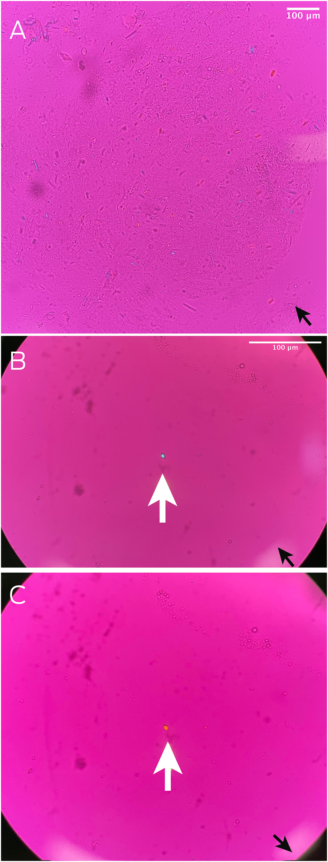
* Optical axis demonstrated by back arrows in lower right corner.
Competing interests
No relevant disclosures.
References
- Robinson PC, Taylor WJ, Dalbeth N. An observational study of gout prevalence and quality of care in a national Australian general practice population. J Rheumatol 2015; 42: 1702‐1707.
- Kuo CF, Grainge MJ, Mallen C, et al. Rising burden of gout in the UK but continuing suboptimal management: a nationwide population study. Ann Rheum Dis 2015; 74: 661‐667.
- Ankli B, Kyburz D, Hirschmann A, et al. Calcium pyrophosphate deposition disease: a frequent finding in patients with long‐standing erosive gout. Scand J Rheumatol 2018; 47: 127‐130.
- Jin HJ, Son ES, Kim DH. The frequency of axial deposition in Korean patients with gout at a tertiary spine center. Front Med (Lausanne) 2020; 7: 339.
- Elgafy H, Liu X, Herron J. Spinal gout: a review with case illustration. World J Orthop 2016; 7: 766‐775.
- Skaf GS, Domloj NT, Fehlings MG, et al. Pyogenic spondylodiscitis: an overview. J infect Public Health 2010; 3: 5‐16.
- Filippucci E, Reginato AM, Thiele RG. Imaging of crystalline arthropathy in 2020. Best Pract Res Clin Rheumatol 2020; 34: 101595.
- Zhou S, Xiao Y, Liu X, et al. Gout involved the cervical disc and adjacent vertebral endplates misdiagnosed infectious spondylodiscitis on imaging: case report and literature review. BMC Musculoskelet Disord 2019; 20: 425.
- Richette P, Doherty M, Pascual E et al. 2018 Updated European League Against Rheumatism evidence‐based recommendations for the diagnosis of gout. Ann Rheum Dis 2020; 79: 31‐38.
- Glick L, Fitzpatrick LA, Detsky AS, Rush RP. Looking back. New Engl J Med 2022; 386: 1742‐1749.
Provenance: Not commissioned; externally peer reviewed.
Patterns of Osteoporosis Treatment Initiation Following Low-Trauma Fracture: A Population-Based Retrospective Cohort Study in New South Wales, Australia, Using Linked Administrative Health Data
Mike Lin, Huy Nguyen, Thach Tran, Robert D. Blank, Dana Bliuc, Jacqueline R. Center
Rapid Growth in Diagnostic CT Knee Imaging in Australia: Implications for Imaging Stewardship
Hanwen Chen, Rachelle Buchbinder, Sean Docking, Ian A Harris
Spinal cord stimulation patterns of care, re‐interventions, and costs for private health insurers, Australia, 2011–22: a retrospective observational study
Caitlin MP Jones, Christopher G Maher, Rachelle Buchbinder, Ian A Harris, Chung‐Wei Christine Lin, Christopher Hayes, Alexandra Gorelik
The incidence of and risk factors for hospitalisations and amputations for people with diabetes‐related foot ulcers in Queensland, 2011–19: an observational cohort study
Yuqi Zhang, Susanna M Cramb, Steven M McPhail, Rosana Pacella, Jaap J Netten, Ewan M Kinnear, Peter A Lazzarini
Bone health perspectives among Indigenous people: a qualitative study
Troy Walker (Yorta Yorta), Karan P Singh, Vanessa Gan, Brooke Conley (Ngiyampaa), Jessica Bravo, Nigel Smith (Weilwan), April Clarke (Eastern Maar, Kirrae Whurrung, Djap Wurrung), Jackson Baker, Louise J Maple‐Brown, Robin M Daly, Jennifer Browne, Jesse Zanker, Cat Shore‐Lorenti, David Scott, Peter R Ebeling, Ayse Zengin
I am a Civil War
Jennifer G Mowbray