




Medical education
Lessons from practice
Volume 217 - Issue 11
Peeling away from hypervitaminosis A: the importance of close monitoring in patients taking high dose vitamin A supplementation
Authors: Jessica A Eldredge, Noel E Cranswick and Kathleen H McGrath
Med J Aust 2022; 217 (11): 582-583. || doi: 10.5694/mja2.51776
Published online: 12 December 2022
Published online: 12 December 2022
A 14-month-old girl was noted to have exfoliation of her peripheries on day 19 after orthotopic liver transplant for end-stage liver disease from extrahepatic biliary atresia
Clinical record
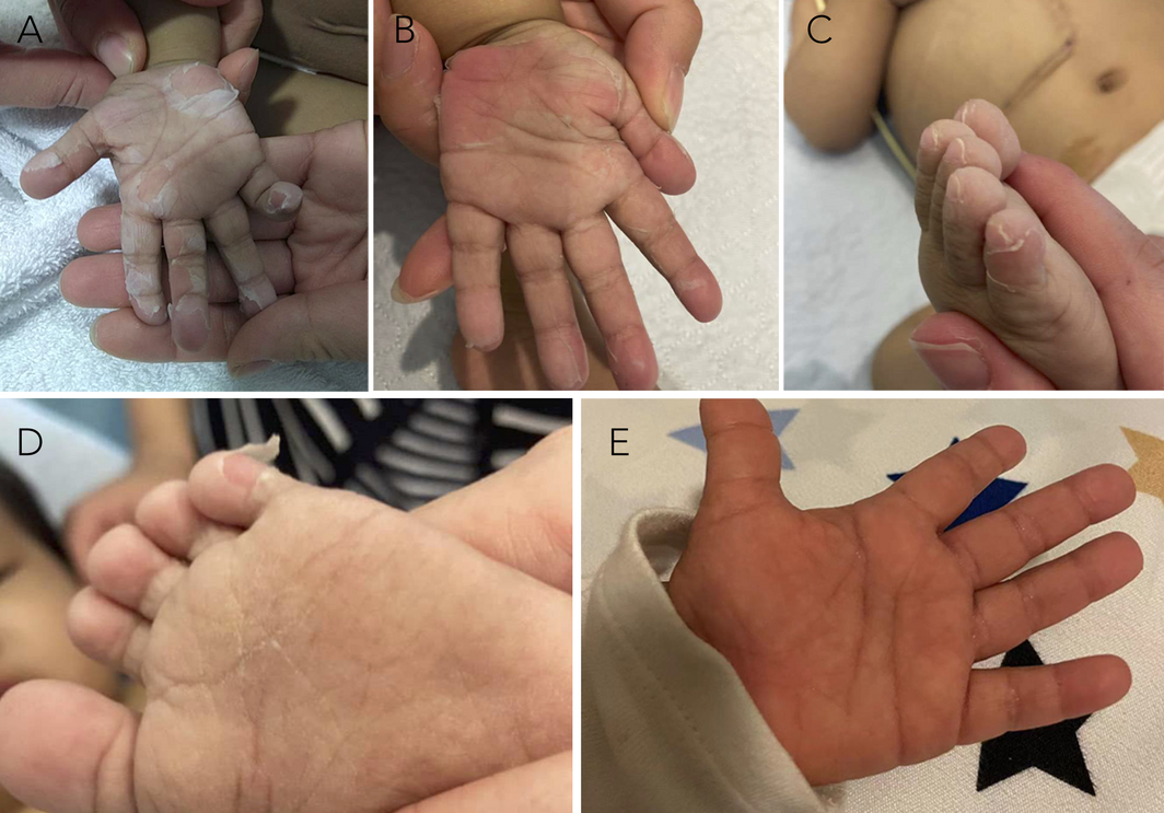
A 14‐month‐old girl was noted to have exfoliation of her peripheries on day 19 after orthotopic liver transplant for end‐stage liver disease from extrahepatic biliary atresia. Initially peeling of the skin over her fingertips was seen. This progressed over one week to involve the interdigital spaces on her hands, palmar surfaces (Box, A and B) and feet (Box, C and D). There was no mucosal or genital involvement. Subtle irritability was reported by her parents, which was felt to be multifactorial given her prolonged hospital admission. There was no previous history of rash, vomiting or diarrhoea, and her post‐transplant recovery was otherwise uncomplicated. Her immunosuppression regimen included tacrolimus, azathioprine and prednisolone. Other regular medications included aspirin, amlodipine, omeprazole, trimethoprim with sulfamethoxazole, acyclovir, nystatin, zinc, and fat‐soluble vitamin replacement, as per local institutional transplant protocol. Blood tests revealed a vitamin A concentration of 3.5μmol/L based on high performance liquid chromatography assay (range, 0.7–1.5μmol/L). This was markedly increased from her pre‐transplant measurement of 0.5μmol/L a month prior, when vitamin A supplementation had been increased.
Following a putative diagnosis of hypervitaminosis A, her vitamin A supplementation was ceased. There was subsequent improvement of her peripheral changes within days, with total resolution of her skin findings by day 8 (Box, E). Her repeat vitamin A concentration 5days after cessation was 2.2μmol/L. Naranjo score was 8, implying probable adverse drug reaction. This scale, with ranges from 0 to ≥9, is a validated method to assess drug causality.1
Discussion
Vitamin A refers to a group of fat‐soluble retinoids key to cellular differentiation, immune function, vision, and reproductive and bone health.2 Preformed vitamin A (retinol and retinyl esters) are found in animal products, and provitamin A carotenoids are derived from plant‐based dietary sources.2,3 After intestinal hydrolysis and esterification in the enterocyte, retinol esters are packaged into chylomicrons, which travel via lymphatic and venous circulation to the liver.2 Most total body vitamin A is stored in hepatic stellate cells, and are mobilised back into the serum bound via retinol‐binding proteins.2,3
Vitamin A deficiency is the leading cause of preventable blindness in the world and remains a public health priority in children in low income countries. Children with chronic cholestatic liver disease are at high risk of vitamin A deficiency due to reduced secretion of bile acids, which are necessary for enteral micelle formation. Other groups vulnerable to deficiency include very low birth weight and preterm infants, and children prone to fat‐soluble vitamin malabsorption (eg, children with cystic fibrosis). Serum vitamin A levels may be unreliable, unless deficiency is severe, with depleted hepatic stores. However gold standard retinol dose response testing is often clinically impractical.3 Routine supplementation dosing varies based on age and clinical indication. In children with cholestasis, a dose of 5000–2500 units or 1500–7500μg retinol activity equivalents (RAE) per day is suggested.3,4
Hypervitaminosis A is an uncommon but serious manifestation of excessive or prolonged supplementation of retinol derivatives via complementary, over‐the‐counter or prescribed means.5 When retinol‐binding proteins are oversaturated, excess retinol esters bind to plasma lipoproteins causing cell membrane toxicity and multisystem sequelae.2,5
Acute toxicity may present with nausea, vomiting, diarrhoea, and manifestations of raised intracranial pressure. A pruritic erythroderma with exfoliative dermatitis can be observed.2 Signs of subacute or chronic hypervitaminosis A may be more subtle and thus under‐recognised, particularly in children.5 Common symptoms include irritability and headache. Desquamation of palms and soles may be identified early, and alopecia can be seen.2 Hypercalcaemia and bone pain may evolve with skeletal changes, including hyperostosis and microfractures.2,5 Transaminitis and hepatosplenomegaly are associated, with rare progression to hepatic fibrosis, cirrhosis and liver failure.3 Raised intracranial pressure, stupor and death can be the ultimate outcome if hypervitaminosis A continues unrecognised.3
Biochemical interpretation of serum vitamin A concentration can be complex. If there is clinical suspicion of vitamin A excess, local assay reference ranges should be used as a guideline only.5 No antidote for hypervitaminosis A exists. Prevention and early recognition of associated clinical manifestations is key. The half‐life of vitamin A is 10–12days, and symptoms resolve rapidly with withdrawal of medication.2 This case highlights the importance of serial serum monitoring and heightened clinical awareness of hypervitaminosis A in patients taking high dose supplementation. This is particularly relevant when underlying risk factors for vitamin A deficiency have been addressed, such as in the case of liver transplantation.
Lessons from practice
- Hypervitaminosis A is an uncommon consequence of high or prolonged oral vitamin A retinol derivative dosing, which can result in hepatotoxicity and death.
- Subacute and chronic hypervitaminosis A can be associated with neurological, dermatological, skeletal and gastrointestinal manifestations, which may be subtle in children.
- No antidote for vitamin A toxicity is available, thus prevention and heightened awareness are key.
- Patients taking high dose supplementation should undergo serial monitoring of serum vitamin A levels, especially if the underlying risk factors for vitamin A deficiency have resolved. Serum concentrations should always be interpreted with the clinical picture in mind.
Box – Dermatological manifestations of hypervitaminosis A: (A) early palmar exfoliation and peeling of the fingertips; (B) palmar changes several days later; (C and D) peeling of skin in interdigital spaces and surfaces of toes; (E) resolution of symptoms after withdrawal of vitamin A supplementation
Competing interests
No relevant disclosures.
References
- Naranjo CA, Busto U, Sellers EM et al. A method for estimating the probability of adverse drug reactions. Clin Pharmacol Ther 1981; 30: 239‐245.
- Smith FR, Goodman DS. Vitamin A transport in human vitamin A toxicity. N Engl J Med 1976; 294: 805‐808.
- Nightingale S, Ng VL. Optimizing nutritional management in children with chronic liver disease. Pediatr Clin North Am 2009 Oct; 56: 1161‐1183.
- Pediatric and neonatal Lexi‐drugs: vitamin A [website]. Hudson (OH): Wolters Kluwer UpToDate, 2022. https://online.lexi.com/ (viewed Apr 2022).
- Coghlan D, Cranswick NE. Complementary medicine and vitamin A toxicity in children. Med J Aust 2001; 175: 223‐224.
Provenance: Not commissioned; externally peer reviewed.
Gabapentinoid Poisoning Presentations to Victorian Emergency Departments: A 15-Year Trend Analysis
Bishaal Tej Gurung, Amy McNeilage, Angus Skeen, Tina Lam, Joanna F. Dipnall, Jane Hayman, Suzanne Nielsen, Ting Xia
Self-Poisoning With Prazosin and Its Off-Label Use in Australia, 2014–2024: Analysis of NSW Poisons Information Centre Data
Olivia B. E. Lal, Nicholas A. Buckley, Rose Cairns
The risk of death after hospitalisation following intentional self‐poisoning: a retrospective observational study (PAVLOVA‐2)
Firouzeh Noghrehchi, Nicholas A Buckley, Rose Cairns
The public health impacts of mining in Australia
Javier Cortes‐Ramirez, Ruby N Michael, Leisa‐Maree Toms, Melissa Haswell
No silver lining with health misinformation: argyria caused by intentional silver consumption
Luke Collins, Logesh Palanikumar, Stephen Bacchi
Hyperkalaemic cardiac arrest due to cream of tartar ingestion
Daniel Yee Lee Ng, Laksmi Govindasamy, Andrew Hughes, Hwee Min Lee