




Medical education
Snapshot
Volume 217 - Issue 11
Cutaneous horn due to cutaneous squamous cell carcinoma
Authors: Li‐wen Zhang and Tao Chen
Med J Aust 2022; 217 (11): 586-586. || doi: 10.5694/mja2.51781
Published online: 12 December 2022
Published online: 12 December 2022
A 91-year-old man presented with a more than 10-year history of a slow growing horn- like lesion on the right side of the face
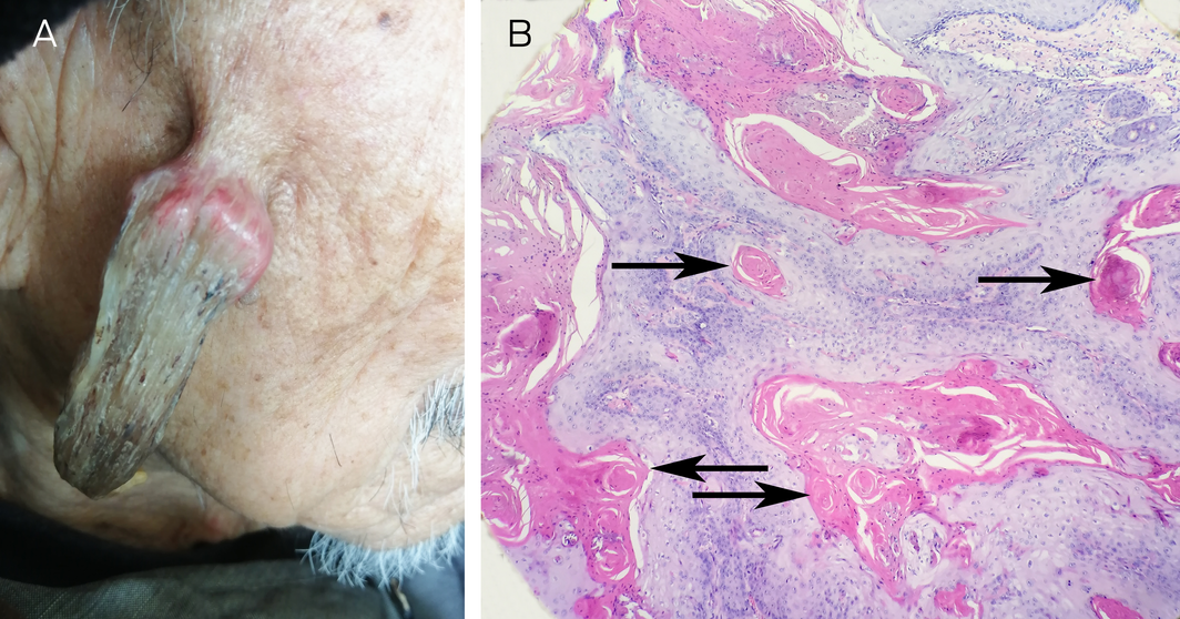
A 91‐year‐old man presented with a more than 10‐year history of a slow growing horn‐like lesion on the right side of the face. The patient denied pruritus, pain, or any other symptoms. Physical examination revealed a grey‐brown, firm, keratotic cutaneous horn 8cm long (Figure, A). The lesion was entirely excised surgically. Histopathology showed atypical keratinocytes and horn pearls within the dermis, with hyperchromatic and pleomorphic nuclei (Figure, B, arrows). A diagnosis of cutaneous horn due to well differentiated squamous cell carcinoma was made. Cutaneous horn is a reactive cutaneous growth caused by a variety of benign or malignant primary diseases, including squamous cell carcinoma, actinic keratosis, keratoacanthoma, Bowen disease, basal cell carcinoma, Kaposi sarcoma, viral warts, and seborrhoeic keratosis.1,2 Therefore, pathological examination is necessary for identifying primary disease.1,2 The most common treatment option is surgery. Other options are available if the lesion is small with a low suspicion of malignancy, such as electrocautery, cryotherapy, carbon dioxide laser, and Nd YAG laser.3
Competing interests
No relevant disclosures.
References
- Mantese SA, Diogo PM, Rocha A, et al. Cutaneous horn: a retrospective histopathological study of 222 cases. An Bras Dermatol 2010; 85: 157‐163.
- Yu RC, Pryce DW, Macfarlane AW, Stewart TW. A histopathological study of 643 cutaneous horns. Br J Dermatol 1991; 124: 449‐452.
- Shahi S, Bhandari TR, Pantha T. Verrucous carcinoma in a giant cutaneous horn: a case report and literature review. Case Rep Otolaryngol 2020; 2020: 7134789.
Diagnosing Acute Kava Dermopathy: A Case Report of a Characteristic Cutaneous Eruption
Ali Abid, Nicholas Allen, Abeer Hagelamin, Christopher Henderson, Artiene Tatian
Localised Herpes Simplex Following Midline Laparotomy
Jessica S. Bulluss, Paul Chee, Matthew J. Verheyden
No silver lining with health misinformation: argyria caused by intentional silver consumption
Luke Collins, Logesh Palanikumar, Stephen Bacchi
Lingual Raynaud phenomenon
Michael Taggart, Mina John
Melasma in the male: a less well recognised entity
Tim Aung, Rowland Noakes
Age group‐specific changes in keratinocyte cancer treatment rates in Australia, 2012–2021: a retrospective cohort study based on MBS claims data
Catherine M Olsen, Nirmala Pandeya, Rachel E Neale, David C Whiteman