




Medical education
Lessons from practice
Volume 217 - Issue 1
Necrotising autoimmune myopathy: a differential diagnosis for deranged liver function tests
Authors: William Luu, Joanne Sy and Queenie Luu
Med J Aust 2022; 217 (1): 20-22. || doi: 10.5694/mja2.51609
Published online: 4 July 2022
Published online: 4 July 2022
A 42-year-old man presented with elevated alanine aminotransferase and aspartate aminotransferase on a blood test screening for viral hepatitis
Clinical record
A 42‐year‐old man presented with elevated alanine aminotransferase (ALT; 81U/L; reference interval [RI], <55U/L) and aspartate aminotransferase (AST; 97U/L; RI, <45U/L) on a blood test screening for viral hepatitis. He was otherwise well. He was referred to a hepatologist, who found an elevated creatine kinase (CK) level of 3957U/L (RI, <250U/L), which increased to 5318U/L after 2 months, prompting a rheumatology referral. Repeat blood tests showed the CK level doubling within 2 months to 9690U/L, and ALT and AST increased to 230U/L and 269U/L respectively.
History revealed mild fatigue at the end of the day. His partner reported that he was unable to keep up when running together. He denied symptoms of weakness, dysphagia, breathlessness, fevers, weight loss, or night sweats. There was no history of statin use.
He was self‐employed and an ex‐smoker, had one standard drink on most days and walked 10000–15000 steps daily. There was no relevant family history.
On examination, his quadriceps muscles were disproportionately small compared with the rest of his musculature. He had normal axial, upper limb and lower limb power. His chest was clear on auscultation and there was no rash.
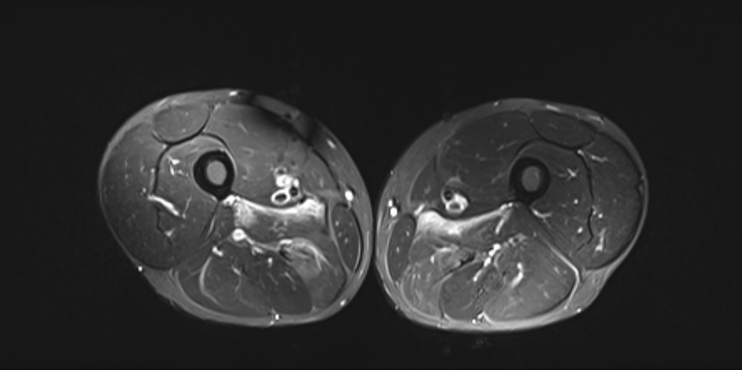
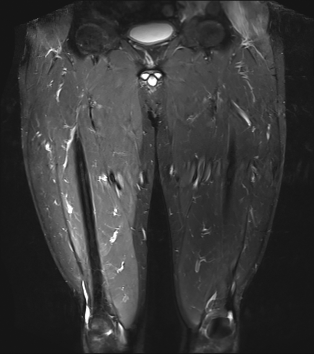
Further blood tests revealed normal troponin, C‐reactive protein and erythrocyte sedimentation rate. Mild subclinical hyperthyroidism was detected, with thyroid‐stimulating hormone 0.36mIU/L (RI, 0.4–3.5mIU/L), free triiodothyronine (T3) 5.1pmol/L (RI, 2.6–6.0pmol/L) and free thyroxine (T4) 15.4pmol/L (RI, 9.0–19.0pmol/L). Myositis panel revealed a positive anti‐signal recognition particle (SRP) antibody. Electromyography of upper and lower limbs was normal (Box 1). Magnetic resonance imaging (MRI) scan of the thighs confirmed diffuse myoedema (Box 2 and Box 3). Necrotising autoimmune myopathy (NAM) was confirmed on muscle biopsy of the right vastus lateralis (Box 4).
The diagnosis was made in February 2020, during the coronavirus disease 2019 (COVID‐19) pandemic. The patient had significant concerns about immunosuppression but agreed to prednisone 50mg daily and methotrexate 20mg weekly orally. The route of administration of methotrexate was later changed to subcutaneous after a few months to increase drug bioavailability.
After 10 months of treatment, AST and ALT normalised, and CK decreased from 9690U/L to 299U/L. He reported reduced fatigue and improved sleep. He remains on subcutaneous methotrexate 20mg weekly and prednisone 2.5mg daily.
Discussion
Idiopathic inflammatory myopathies describe a diverse family of systemic autoimmune diseases predominantly affecting skeletal muscle. This includes polymyositis, dermatomyositis, inclusion body myositis, and NAM (also known as “immune‐mediated necrotising myositis”).1
NAM was previously classified under polymyositis. However, it is given individual recognition because of the distinct muscle biopsy findings of muscle cell necrosis and minimal inflammation.2 Even though NAM is associated with statin use, connective tissue diseases and malignancy, most cases are idiopathic.3 NAM has three subtypes: anti‐SRP myopathy, anti‐hydroxy‐3‐methylglutaryl‐coenzyme A reductase (anti‐HMGCR) myopathy, and autoantibody‐negative NAM.2
While the classical presentation is subacute symmetrical proximal limb weakness, patients can also experience myalgia, neck weakness, dysphagia, dyspnoea, fatigue and weight loss.4 Any proximal muscle weakness should prompt a check of CK levels.2 Serum transaminases are elevated in idiopathic immune myositis due to their existence in extrahepatic tissues such as muscle.4 Early referral and diagnosis is important to improve outcomes and prognosis.
Electromyography can help distinguish myopathy from neuropathic causes of proximal weakness such as myasthenia gravis or neuropathy,2 although it was normal in our patient.
MRI cannot differentiate NAM from other types of idiopathic inflammatory myopathies, but it can identify the affected site for muscle biopsy and show disease activity and chronic damage. Myoedema is indicated by hyperintensity on short tau inversion recovery (STIR) sequences, while T1‐weighted sequences can help evaluate the degree of fatty replacement, which is a marker of irreversible chronic destruction.2
Muscle biopsy is important to confirm the diagnosis. NAM typically has myofibre necrosis and regeneration, with absent or minimal lymphocytic infiltrate. Around 50% of cases have multifocal upregulation of class I major histocompatibility complex, as seen in our patient.2
Induction treatment often involves prednisone or pulsed methylprednisone. Commonly used steroid‐sparing agents include azathioprine and methotrexate. Other agents include rituximab and intravenous immunoglobulin. Maintenance therapy is tailored to the patient’s treatment response. Our patient has responded well to prednisone and methotrexate.
Anti‐SRP myopathy has one of the worst muscle prognoses compared with other idiopathic inflammatory myopathies.2 The debilitating muscle weakness is often refractory to treatment,5 with around 50% of patients with anti‐SRP remaining afflicted after 2years of treatment. Extramuscular manifestations, in particular pulmonary involvement, are more likely to occur in anti‐SRP myopathy compared with anti‐HMGCR myopathy. Furthermore, younger age is a worse prognostic factor.2 Our patient had not developed significant muscular symptoms or extramuscular involvement at the time of diagnosis.
Lessons from practice
- Check the creatine kinase level in patients presenting with musculoskeletal symptoms and deranged liver function tests, and refer for further assessments as appropriate.
- Magnetic resonance imaging can confirm evidence of myoedema and identify the site for muscle biopsy.
- Muscle biopsy is recommended to confirm diagnosis of idiopathic inflammatory myopathies.
- Anti‐signal recognition particle (SRP) myopathy has a very poor prognosis of long term disability, in particular for younger patients, and hence requires early diagnosis and treatment.
Box 1 – Electromyography (EMG) investigation
Muscles investigated on EMG included:
• Right deltoid
• Right triceps brachii
• Right extensor digitorum communis
• Left trapezius
• Left biceps brachii
• Left first dorsal interosseous
• Right thoracic paraspinals (mid)
• Left iliopsoas
• Right iliopsoas
• Right vastus medialis
• Left vastus medialis
• Right tibialis anterior
• Left tibialis anterior
• Right tibialis posterior
• Left gastrocnemius (medial head)
Box 2 – Axial short tau inversion recovery (STIR) magnetic resonance imaging of proximal lower limbs showing diffuse hyperintensity in the medial thighs
Box 3 – Coronal short tau inversion recovery (STIR) magnetic resonance imaging of proximal lower limbs showing diffuse hyperintensity in the right thigh
Box 4 – Muscle biopsy showing rare necrotic and regenerating myofibres, with minimal focal inflammation and patchy upregulation of human leucocyte antigen class 1 (HLA‐1)
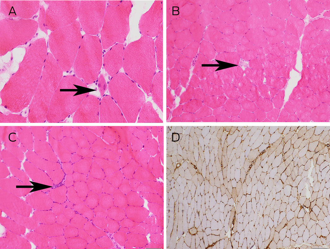
(A) Haematoxylin and eosin stain (×400 magnification) showing a regenerating myofibre (arrow). (B) Haematoxylin and eosin stain (×200 magnification) showing a necrotic myofibre (arrow). (C) Haematoxylin and eosin stain (×200 magnification) showing an isolated focus of chronic inflammation (arrow). (D) Immunohistochemical staining (×100 magnification) showing patchy upregulation of HLA‐1.
Competing interests
No relevant disclosures.
References
- Luu Q, Day J, Hall A, et al. External validation and evaluation of adding MRI or extended myositis antibody panel to the 2017 EULAR/ACR myositis classification criteria. ACR Open Rheumatol 2019; 1: 462‐468.
- Pinal‐Fernandez I, Casal‐Dominguez M, Mammen AL. Immune‐mediated necrotizing myopathy. Current Rheumatol Rep 2018; 20: 21.
- Suzuki S, Nishikawa A, Kuwana M, et al. Inflammatory myopathy with anti‐signal recognition particle antibodies: case series of 100 patients. Orphanet J Rare Dis 2015; 10: 61.
- Liang C, Needham M. Necrotizing autoimmune myopathy. Curr Opin Rheumatol 2011; 23: 612‐619.
- Aggarwal R, Oddis CV, Goudeau D, et al. Anti‐signal recognition particle autoantibody ELISA validation and clinical associations. Rheumatology 2015; 54: 1194‐1199.
Provenance: Not commissioned; externally peer reviewed.
Shifting focus to adolescent wellbeing and inclusive participation in the digital age
Allyson R Todd, Elena Wang, Stephanie R Partridge
The clinical presentation, investigation, and management of women diagnosed with endometriosis in Australian general practices, 2011–2021: an open cohort study
Danielle Mazza, Kailash Thapaliya, Sharinne B Crawford, Alissia Hui, Maryam Moradi, Luke E Grzeskowiak
The impact of the Breast Screen NSW transition from film to digital mammography, 2002–2016: a linked population health data analysis
Rachel Farber, Nehmat Houssami, Katy J L Bell
Non‐technical errors associated with deaths in surgical care, Australia, 2012–2019, by surgical specialty (Australian and New Zealand Audit of Surgical Mortality): a retrospective cohort study
Jesse Ey, Victoria Kollias, Octavia Lee, Kelly Hou, Matheesha Herath, John B North, Ellie Treloar, Suzanne Edwards, Martin Bruening, Adam J Wells, Guy J Maddern
The CURE Asthma roadmap
Gary P Anderson, Anthony Flynn, Phil G Bardin, John D Blakey, Shyamali C Dharmage, Paul Foster, Peter G Gibson, Adam Jaffe, Alan James, Christine R Jenkins, Sundram Sivamalai, Peter D Sly, Guy B Marks, Vanessa M McDonald, Judy Wetttenhall
Evidence from Australian cohort studies about asthma trajectories and transitions across the life course: a narrative review
Caroline Lodge, Xin Dai, Ingrid A. Laing, Michael P Menden, Anthony Flynn, Gary P Anderson, Sarath Ranganathan, Shyamali C Dharmage