




Volume 216 - Issue 8
From wipeout to drill out: a history of exostosis management and Australian surfing
Authors: Alon Taylor, Hannah North, Narinder P Singh and Paul A Fagan
Med J Aust 2022; 216 (8): 401-404. || doi: 10.5694/mja2.51496
Published online: 2 May 2022
Published online: 2 May 2022
The evolution of treatment for a distinctly Australian affliction
The evolution of treatment for a distinctly Australian affliction
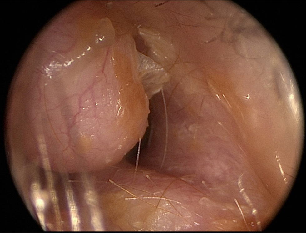
External auditory canal exostoses are benign, localised bony growths that form in the ear canal. They are classically described as reactive lesions caused by repeated exposure to cold water and wind. This results in chronic inflammation of the periosteum of the external canal leading to the formation of multiple, nodular bony outgrowths of the medial canal that are covered by thin skin. Exostoses tend to remain asymptomatic until they grow to significantly narrow the external auditory canal (Box 1). At this stage, they cause water trapping and hydration of the external canal skin, resulting in recurrent otitis externa. Acute otitis externa (often referred to as swimmer’s ear) is a common complication of exostoses, with management consisting of ear toileting, local antibiotic therapy (drops and ointment), and maintaining a dry ear. In its latter stages, exostoses may completely occlude the external canal, and patients typically present following repeated infection or hearing loss. Anatomically, exostoses typically occur in groups of three: anteriorly, posteriorly and superiorly based. These correspond to the three ossification centres of the tympanic ring, which is the bony structure which supports the ear drum, and the site from which exostoses arise.1
Exostoses are associated with people repeatedly exposed to cold water and wind, such as ocean swimmers and, in particular, surfers. The association with surfing led to the commonly used term “surfer’s ear”, credited to Californian otolaryngologist Daniel Seftel in 1977.2 Surfing was brought to Australia by Hawaiian Olympic swimmer and surfer Duke Kahanamoku, who demonstrated the ancient Hawaiian board riding technique to Sydneysiders at Freshwater Beach in 1915.3 Since then, surfing has evolved into a national obsession with about 2.7 million Australians participating in the sport.4 Australia is home to numerous accessible surfing locations, which has resulted in a high prevalence of external canal exostoses in both Australian recreational and professional surfers, with studies suggesting over 70% of Australian surfers demonstrate some evidence of the phenomenon.5
Historical management of exostoses
Despite the recent association with surfing, external canal exostoses have been identified in prehistoric Neanderthal skulls dating back 45 000 years, found in the vicinity of rivers and lakes.6 A current hypothesis is that exostoses offered an evolutionary advantage by protecting the middle ear and tympanic membrane during swimming and diving, allowing early Homo species to live and hunt in semi‐aquatic environments.6
The first printed clinical record of external canal exostoses is attributed to German physician Johan von Autenriech who reported them in the medical literature in 1809.1 However, there was little clinical awareness of the entity until Joseph Toynbee, a British otologist, described them in depth in 1849. He was the first to note their typical “shining white, and polished like ivory” appearance and suggested they were secondary to the “congestion of the mucous membranes of the ear as a result of rather free living”, noting most patients were “in the habit of drinking wine”.7
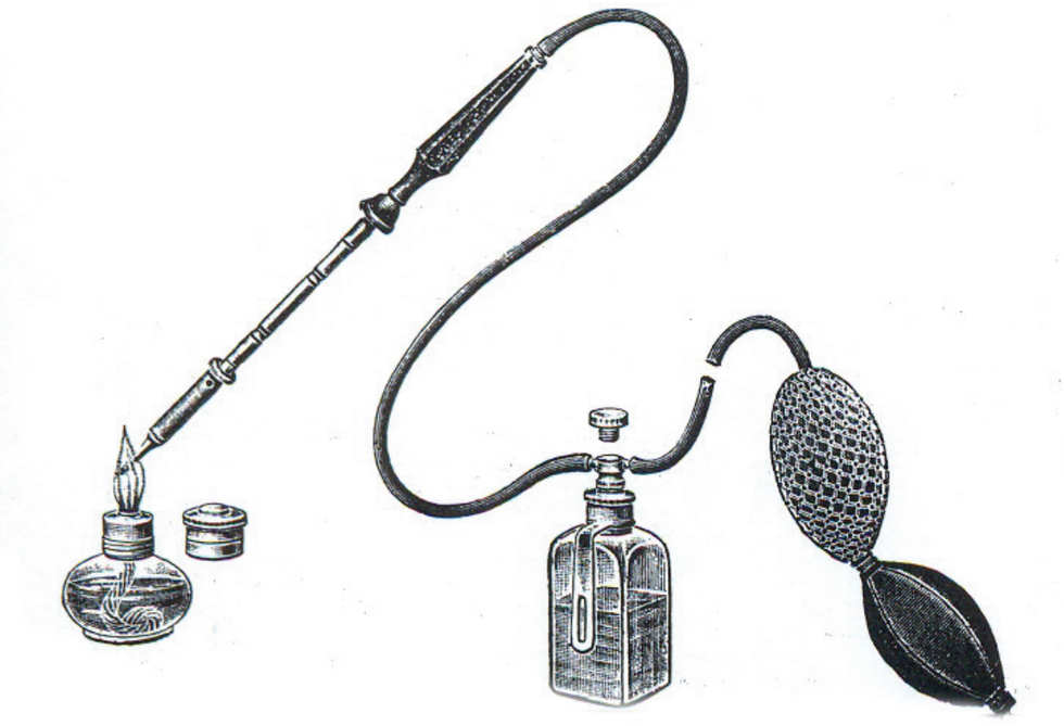
In the pre‐surgical era, physicians employed an array of concoctions to treat exostoses, including topical tincture of iodine, applying leeches to the external ear and dilatation with a bougie.8 Electrocautery was described as a method of treatment by British otologist Thomas Clark in 1873.9 Clark introduced three needles into the exostosis of a 15‐year‐old girl and applied a continuous current for three minutes. He remarked that it “produced a large coagulum at the point of entrance of the needles, and rendered the whole tumour white”, and he was able to excise it three weeks later with scissors.9 Cautery was the technique used in the first documented account of removal of exostoses in the Australian literature. In the Australian Medical Journal in 1894, a Melbourne physician reported the case of a 33‐year‐old man with occlusive right‐sided exostoses with concurrent infection of the outer and middle ear.10 He described using Paquelin’s cautery to remove the exostosis — a technique whereby a platinum tipped cautery device was heated over a flame, with the temperature maintained through the flow of a petroleum ether to the cautery end via a bellow system (Box 2).11
Exostosis removal with a chisel or osteotome was the first surgical technique reported. The description from Carl von Lincke in 1845 was the first account of using a fine, narrow, hollow chisel.8 By the end of the 1800s, the chisel and gouge technique was widely utilised in ear surgery, both in managing exostoses and in mastoid surgery.
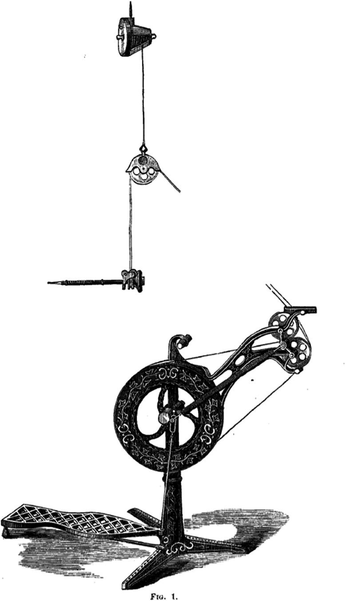
The proliferation of the dental drill in the late 1800s paved the way for a new surgical technique. In 1876, British otologist William Dalby and American otologist Arthur Mathewson were the first to use the dental drill to remove exostoses. Dalby described borrowing a drill from a dental colleague to make several passes through the exostoses of a man with a “thick fetid purulent discharge from the right ear” obstructed by exostoses.12 Mathewson described using a foot‐powered suspension dental drill (Box 3) with progressively larger drill passes through the exostoses and combining them to create a patent canal.13
The era of modern surgical exostectomy has been characterised by improvements in anaesthetic techniques, the adoption of binocular operative microscopy, the availability of micro‐instrumentation, and more evolved drilling systems and burrs.14 Modern drill canalplasty is associated with fewer tympanic membrane perforations, temporomandibular joint prolapse and fracture of the anterior canal wall, compared with osteotome techniques. The adoption of the piezoelectric bone drill over the past 15 years has allowed otolaryngologists to selectively aspirate bone while preserving the soft tissues of the ear canal and the tympanic membrane. This is particularly relevant in exostectomy, where damage to the delicate external canal skin by traditional rotatory drills may result in postoperative canal stenosis with associated morbidity.15
Surfer’s ear in Australia
Otological sequelae from surfing are common. In a cross‐sectional observational study of 113 Australian surfers, more than half reported seeking medical attention for ear symptoms, including frequent otitis externa, hearing deterioration and water trapping.16 Exostoses are particularly prevalent among cold water surfers, with the phenomenon demonstrating a dose–response relationship.17 Victoria’s Surf Coast Shire, seat of what is arguably the spiritual home of surfing in Australia, Bells Beach, is teeming with exostosis‐afflicted surfers owing to cold water temperatures (with an average of 12.8°C during favourable winter swells), high quality surf spots and an enthusiastic local surfing community. Hurst and colleagues reported the prevalence of exostoses in Bells Beach local surfers in 2003.5 After examining 300 surfers and spectators attending the annual Bells Beach surf contest, some degree of obstruction was found in 228 (76%) participants, and significant obstruction (defined as two‐thirds or more occlusion of the ear canal) was noted in 100 (33%) participants. The researchers found that the extent of exostoses in the right ear was significantly greater than that in the left. Surfers spend most of their time facing out to sea while waiting for a wave (which is towards the south at Bells Beach), while the predominant wind on the Surf Coast is from the west to northwest. As a result, the right ears of Bells Beach surfers are exposed to a greater degree of wind and cold water. This finding supports the thesis that exostoses develop secondary to the evaporate cooling effect and has been replicated in other cold water locations such as in California.18
The Gold Coast, and Queensland more broadly, is home to a large surf community who enjoy warmer water temperatures compared with Victoria (mean water temperature is 20–28°C). An observational study of Gold Coast surfers found a high prevalence of external canal exostoses, similar to that of Victorian cold water surfers, despite the warmer conditions.19 However, in Gold Coast surfers, the prevalence of severe exostoses (defined as obstructing more than two‐thirds of the canal) was half that of Victorian surfers, with no statistical difference in the severity of exostoses between the right and left ears.19
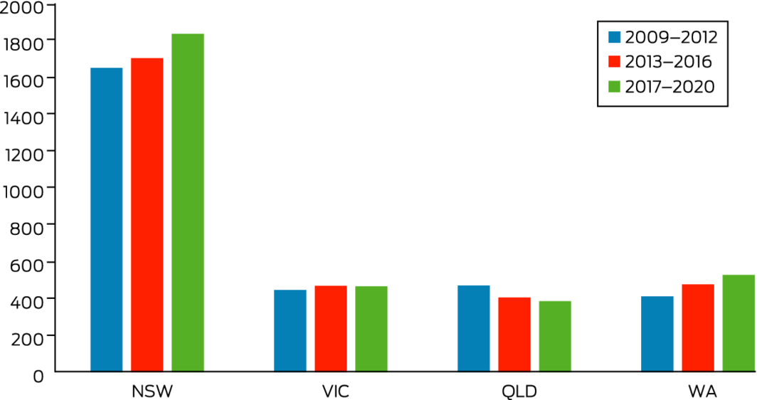
Australia likely performs the highest number of surgical exostectomies per capita worldwide (Box 4). Based on Australian Institute of Health and Welfare data, New South Wales surgeons perform the largest number of operations, having undertaken an average of 472 procedures per year (equating to 5.6 per 100 000 citizens) over the 2009–2020 period.20 This is likely due to the state’s large surfing population with rideable waves found along the entire coastline from the Victorian to Queensland border. Interestingly, Queensland surgeons, despite the state’s large surfing population, perform a relatively small number of surgeries per capita (2.4 per 100 000 citizens). This may be because warmer climates result in a lower prevalence of severe disease requiring surgery. Meanwhile, despite the frigid conditions in Victoria, the total number of exostectomies is also relatively low. This may reflect the likely smaller size of the Victorian surfing community, as there are less surfable waves and only the most committed surfers brave the cold temperatures, although data supporting this are lacking.
The literature is conflicted as to whether ear protection prevents exostoses. Evidence suggests that protective earplugs decrease the rate of development of exostoses and prolong the recurrence‐free period after surgery.21 However, some studies have reported no difference in the severity of exostosis in cold water surfers who wear protective earplugs compared with those who do not.22 External canal exostoses are a common finding in Australian surfers, with the ailment being a common cause of ear symptoms and presentation to medical care. Referral to an otolaryngologist is warranted in symptomatic exostoses (such as water trapping or recurrent infections) in the presence of significant hearing loss or if suspicion of a neoplastic process exists. Today’s otolaryngologists owe a debt of gratitude to their surgical predecessors and to the development of anaesthetic, microsurgical and drill technology, with modern day surgical treatment for exostoses being safe, effective and well tolerated.
Box 1 – Otoscopic image showing severe left external canal exostoses with near complete occlusion of the external auditory canal
Clinical photograph courtesy of Paul Fagan.
Box 4 – Number of exostectomies performed, by selected state, 2009–2020
Data source: Australian Institute of Health and Welfare.20
Competing interests
No relevant disclosures.
References
- DiBartolomeo JR. Exostoses of the external auditory canal. Ann Otol Rhinol Laryngol Suppl 1979; 88(6 Pt 2 Suppl 61): 2–20.
- Seftel DM. Ear canal hyperostosis — surfer’s ear. An improved surgical technique. Arch Otolaryngol 1977; 103: 58–60.
- Clarke P, Appel G. Bombora — the story of Australian surfing [film]. Australian Broadcasting Corporation, 2009.
- Surfing Australia. Annual report 2013. https://assets.surfinginaustralia.com/wp‐content/uploads/sites/6/2021/12/08103554/2013‐Annual‐Report‐no‐FR.pdf (viewed Nov 2021).
- Hurst W, Bailey M, Hurst B. Prevalence of external auditory canal exostoses in Australian surfboard riders. J Laryngol Otol 2004; 118: 348–351.
- Rhys‐Evans PH, Cameron M. Surfer’s ear (aural exostoses) provides hard evidence of man’s aquatic past. Hum Evol 2014; 29: 75–90.
- Toynbee J. On osseous tumours growing from the walls of the meatus externus of the ear. Prov Med Surg J 1849; 13: 533–537.
- Mudry A, Hetzler D. Birth and evolution of chiselling and drilling techniques for removing ear canal exostoses. Otol Neurotol 2016; 37: 109–114.
- Clark TE. Electrolysis of exostosis of the ear. Br Med J 1873; 2: 656–657.
- A case of exostosis and mastoid necrosis. Aust Med J 1894; 16.
- Helling TS, Azoulay D. Historical foundations of liver surgery. Cham: Springer, 2020.
- A discussion on the removal of bony growths from the external auditory canal. Br Med J 1889; 2: 704–709.
- Mathewson A. A case of exostosis of the external auditory meatus drilled out by the “dental engine”. Edinb Med J 1876; 23: 467–468.
- Fagan PA. The surgical removal of aural exostoses. J Otolaryngol Soc Aust 1990; 6: 220–221.
- Meller C, Havas TE. Piezoelectric technology in otolaryngology, and head and neck surgery: a review. J Laryngol Otol 2017; 131 Suppl S2: S12–S18.
- Simas V, Hing W, Rathbone E, et al. Auditory exostosis in Australian warm water surfers: a cross‐sectional study. BMC Sports Sci Med Rehabil 2021; 13: 52.
- Wong BJF, Cervantes W, Doyle KJ, et al. Prevalence of external auditory canal exostoses in surfers. Arch Otolaryngol Head Neck Surg 1999; 125: 969–972.
- King JF, Kinney AC, Iacobellis SF, et al. Laterality of exostosis in surfers due to evaporative cooling effect. Otol Neurotol 2010; 31: 345–351.
- Simas V, Hing W, Furness J, et al. The prevalence and severity of external auditory exostosis in young to quadragenarian‐aged warm‐water surfers: a preliminary study. Sports 2020; 8: 1–9.
- Australian Institute of Health and Welfare. Procedures data cubes, 2009–2020. https://www.aihw.gov.au/reports/hospitals/procedures‐data‐cubes/contents/data‐cubes (viewed Nov 2021).
- Lambert C, Marin S, Esvan M, Godey B. Impact of ear protection on occurrence of exostosis in surfers: an observational prospective study of 242 ears. Eur Arch Otorhinolaryngol 2021; 278: 4775–4781.
- Chaplin JM, Stewart IA. The prevalence of exostoses in the external auditory meatus of surfers. Clin Otolaryngol Allied Sci 1998; 23: 326–330.
Provenance: Not commissioned; externally peer reviewed.
Watchful Waiting Compared With Immediate Antibiotics for Urban Aboriginal and Torres Strait Islander Children With Uncomplicated Acute Otitis Media (WATCH): A Non-Inferiority Randomised Controlled Trial
Jennifer S. Reath, Hasantha Gunasekera, Sanja Lujic, Amanda J. Leach, Letitia Campbell, Robyn Walsh, Tim Usherwood, Geoffrey K. Spurling, Claudette A. Tyson, Deborah A. Askew, Kelvin Kong, Chelsea J. Watego, Peter Morris, Wendy Hu, Penelope A. Abbott
Climate and environmental crisis: effects on ear and hearing health in Australia and for Aboriginal and Torres Strait Islander peoples
Georgia M Tongs (Wiradjuri), Isabella Ludbrook, Jennifer H Martin, Robert Eisenberg, Kelvin M Kong (Worimi)
The New South Wales Pharmacy Trial for herpes zoster: on the nose?
Christian P Pappas, Timothy R Holmes, Minas T Coroneo
Antibiotics or watchful waiting for acute otitis media in urban Aboriginal and Torres Strait Islander children?
Valerie M Swift, Christopher G Brennan‐Jones
The views of parents and carers on managing acute otitis media in urban Aboriginal and Torres Strait Islander children: a qualitative study
Jennifer S Reath, Sarah O'Brien, Letitia Campbell, Hasantha Gunasekera, Claudette A Tyson, Deborah A Askew, Wendy Hu, Tim Usherwood, Kelvin Kong, Peter Morris, Amanda J Leach, Robyn Walsh, Penelope A Abbott
Routine ear health and hearing checks for Aboriginal and Torres Strait Islander children aged under 6 years attending primary care: a national consensus statement
Samantha Harkus, Vivienne Marnane, Isabel O'Keeffe, Carmen Kung, Meagan Ward, Neil Orr, John Skinner, Kelvin Kong, Lose Fonua, Michelle Kennedy, Mary Belfrage