




Medical education
Lessons from practice
Volume 216 - Issue 8
Rhabdomyolysis due to jellyfish envenomation in Western Australian waters
Authors: Anthony C Rengel, Sue Davel, Sarah O’Connor and Nicholas Medcraft
Med J Aust 2022; 216 (8): 397-399. || doi: 10.5694/mja2.51478
Published online: 2 May 2022
Published online: 2 May 2022
A 25-year-old woman presented via ambulance following a jellyfish sting
Clinical record
A 25‐year‐old woman presented via ambulance following a jellyfish sting. She felt a painful burning sensation across the left torso while swimming during an autumn afternoon in Perth, Australia. Linear welts developed over the next few hours. At 4 am, she was woken by pain in the left thigh which progressed over the next 12 hours to whole body myalgias, weakness and contractures in her toes. An ambulance was called and she was found by paramedics in significant pain, tachycardic, hypertensive and requiring assistance to mobilise. Paracetamol and methoxyflurane were administered en route to the hospital.
No fevers, dyspnoea, dysphagia, vomiting, diarrhoea, dysuria, chest pain or headache was experienced. She had no significant past medical conditions or known drug allergies. Her regular medications included the combined oral contraceptive pill, iron tablets and a mushroom extract. She worked in education, was nulliparous and a non‐smoker, with occasional alcohol intake.
On examination, blood pressure was 115/83 mmHg, heart rate was regular at 98 beats per minute, respiratory rate was 16 breaths per minute, oxygen saturation on room air was 98%, and she was afebrile. Cardiorespiratory exam was normal. Abdomen was soft and non‐tender, with linear excoriations seen on the left hip and torso. Her neurological examination revealed mild global reduction in power across all muscle groups with otherwise intact tone, sensation and reflexes. Her urine was dark cola coloured.
Initial investigations are shown in Box 1, with creatine kinase (CK) and renal function trends presented in Box 2. CK was significantly elevated at 68 900 IU/L (< 200 IU/L), confirming rhabdomyolysis. Urinalysis tested positive for blood and protein, with no organisms grown on culture. Transaminitis was noted on liver function testing. Following toxicology advice, she was admitted for intravenous rehydration, sodium bicarbonate, analgesia and repeated blood tests. A total of 18 L of intravenous fluid were administered across the admission, with urine output >200 mL/h.
Besides a non‐specific speckled antinuclear antibody (ANA) pattern with 1:80 titre, the autoimmune screen and myositis panel was negative. After 4 days, the CK had fallen significantly and pain had improved. She was discharged home, advised to repeat the CK test with a general practitioner, to do no exercise for 2 weeks and, as a precaution, to stop using the mushroom extract. Following 5 weeks of convalescence, she reported improvement in her muscle strength, with normalisation of her CK and liver function.
Discussion
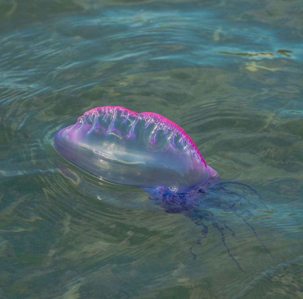
Jellyfish envenomation in southern Western Australia is usually attributed to the cnidarians Carybdea xaymacana and Chrysaora kynthia during summer months, while hydrozoans, including the common bluebottle Physalia utriculus and the Portuguese man o’ war Physalia physalis (Box 3), are responsible for cases during autumn and winter.1 Physalia envenomation presents with rapid development of linear erythema and scattered papules, which progress to a characteristic “string of beans” appearance of centrally blanched oval weals and surrounding erythema.1
While symptoms from P. utriculus stings commonly resolve within 24 hours, P. physalis stings are more persistent and severe.1 They are associated with systemic manifestations including abdominal pain, vomiting, myalgias and cramping, with neurological or cardiorespiratory manifestations reported in severe cases.1,2 Although the species was not visualised in our case and the dermatological presentation was non‐specific, the season and the toxidrome is suggestive of Physalia sp.
Our patient was taking a mushroom extract for several months before presentation and was asymptomatic during this time. Wild mushrooms are associated with multi‐organ failure and delayed onset myopathy following ingestion;3 however, only common edible and medicinal species were identified in this particular extract (lion’s mane, Cordyceps, reishi, turkey tail and shiitake). In view of her acute clinical course, mushroom toxicity was considered unlikely.
Rhabdomyolysis from Physalia envenomation has been previously reported in a 23‐year‐old Australian man in New Caledonia.4 He had a previous left nephrectomy with preserved renal function. The sting caused severe pain for 10 minutes before resolving, developing left‐sided chest and hypochondrial pain with dark urine the next day. The electrocardiogram was normal, but serum creatinine was 150 μmol/L (reference interval [RI], 45–90 μmol/L), potassium 5.4 mmol/L (RI, 3.5–5.2 mmol/L) and CK 4000 IU/L (RI, < 200 IU/L). Repeat investigations identified transaminitis and persistent rhabdomyolysis (CK 10 000 IU/L) — as seen in our case. Clinical improvement occurred over 48 hours with close monitoring and rehydration. While no myalgia or cramping was reported, hypnagogic myoclonus was observed.
The acute kidney injury in this patient is in keeping with a case report of 4‐year‐old child following P. physalis envenomation.5 We note that no kidney injury occurred in our case, due to the early identification of rhabdomyolysis and prompt treatment with intravenous fluid.
Our case represents another incidence of rhabdomyolysis occurring as a complication of potential Physalia envenomation in the southern hemisphere. As jellyfish envenomation is a common presentation in Australia, in any individual with symptoms exceeding 24 hours, investigation for rhabdomyolysis should be considered.
Lessons from practice
- Jellyfish envenomation is common in Australian waters throughout the year.
- Rhabdomyolysis is a rare complication of jellyfish envenomation.
- In cases where localised symptoms are accompanied by myalgia, urine discolouration or muscle weakness, rhabdomyolysis should be considered.
- Prompt treatment is required to prevent acute renal failure.
Box 1 – Initial blood tests at presentation
|
|
Result (reference interval) |
||||||||||||||
|
|
|||||||||||||||
|
Hb (g/L) |
162 (115–155) |
||||||||||||||
|
HCT |
0.463 |
||||||||||||||
|
Platelets (× 109/L) |
348 (150–400) |
||||||||||||||
|
WCC (× 109/L) |
18.5 (4.0–11.0) |
||||||||||||||
|
Neutrophils |
16.8 (1.8–7.5) |
||||||||||||||
|
Lymphocytes |
0.7 (1.3–4.0) |
||||||||||||||
|
Monocytes |
0.6 (0.1–1.2) |
||||||||||||||
|
Eosinophils |
0 (< 0.7) |
||||||||||||||
|
Basophils |
0 (< 0.4) |
||||||||||||||
|
Magnesium (mmol/L) |
0.95 (0.7–1.2) |
||||||||||||||
|
Phosphate (mmol/L) |
1.41 (0.75–1.50) |
||||||||||||||
|
Calcium (mmol/L) |
2.31 (2.10–2.60) |
||||||||||||||
|
Albumin (g/L) |
44 (38–50) |
||||||||||||||
|
INR |
1 (0.8–1.2) |
||||||||||||||
|
aPTT (seconds) |
26 (24–34) |
||||||||||||||
|
Troponin (ng/L) |
8 (< 11) |
||||||||||||||
|
Bilirubin (μmol/L) |
14 (< 16) |
||||||||||||||
|
ALP (IU/L) |
42 (30–110) |
||||||||||||||
|
GGT (IU/L) |
14 (< 31) |
||||||||||||||
|
ALT (IU/L) |
904 (< 31) |
||||||||||||||
|
|
|||||||||||||||
|
ALP = alkaline phosphatase; ALT = alanine aminotransferase; aPTT = activated partial thromboplastin time; GGT = γ‐glutamyl transferase; Hb = haemoglobin; HCT = haematocrit; INR = international normalised ratio; WCC = white cell count. |
|||||||||||||||
Box 2 – Trending creatine kinase (CK), electrolytes and renal function tests during admission
|
|
Day 1 pm |
Day 2 am |
Day 2 pm |
Day 3 am |
Day 3 pm |
Day 4 am |
Day 5 am |
Reference interval |
|||||||
|
|
|||||||||||||||
|
CK (IU/L) |
68 900 |
63 093 |
81 868 |
47 013 |
43 791 |
17 798 |
5484 |
< 200 |
|||||||
|
Na (mmol/L) |
137 |
140 |
141 |
143 |
142 |
144 |
143 |
135–145 |
|||||||
|
K (mmol/L) |
4.00 |
4.10 |
4.20 |
4.10 |
4.00 |
3.70 |
3.60 |
3.5–5.2 |
|||||||
|
CL (mmol/L) |
104 |
109 |
111 |
107 |
106 |
107 |
104 |
95–110 |
|||||||
|
HCO3 (mmol/L) |
21 |
20 |
23 |
29 |
29 |
30 |
33 |
22–32 |
|||||||
|
Urea (mmol/L) |
5.60 |
4.70 |
4.20 |
3.20 |
3.50 |
2.50 |
2.70 |
3.0–8.0 |
|||||||
|
Creatinine (μmol/L) |
52 |
52 |
59 |
48 |
48 |
52 |
55 |
45–90 |
|||||||
|
|
|||||||||||||||
|
CL = chloride; HCO3 = bicarbonate; K = potassium; Na = sodium. |
|||||||||||||||
Competing interests
No relevant disclosures.
References
- Tibballs J. Australian venomous jellyfish, envenomation syndromes, toxins and therapy. Toxicon 2006; 48: 830‐859.
- Marsh LM, Slack‐Smith SM. Field guide to sea stingers and other venomous and poisonous marine invertebrates of Western Australia. Perth: Western Australian Museum, 2010.
- Bedry R, Baudrimont I, Deffieux G, et al. Wild‐mushroom intoxication as a cause of rhabdomyolysis. N Engl J Med 2001; 345: 798‐802.
- Maldonado E, Maillaud C, Barguil Y, Labadie M. Rhabdomyolysis during envenomation by Physalia sp envenomation in New Caldonia. Med Sante Trop 2017; 27: 105‐108.
- Guess HA, Saviteer PL, Morris CR. Hemolysis and acute renal failure following a Portuguese man‐of‐war sting. Pediatrics 1982; 70: 979‐981.
Provenance: Not commissioned; externally peer reviewed.
‘Once Bitten’: A Case of Tetanus Following Snakebite in Australia
Adrian A. S. Doherty, Naomi N. E. Clarke, Sarah Fletcher, Sarah Huffam, Camille V. Awburn, Eugene Athan
Epidemiology and Quality of Care for Aboriginal and Torres Strait Islander Peoples Treated by Emergency Medical Services for Stroke in Victoria, Australia: Retrospective Analysis of Linked Data
Candice Menezes, Ziad Nehme, Luke J. Burchill, Tegwyn McManamny, Angela Dos Santos, Michelle Crilly, Luke P. Dawson, Benjamin Clissold, David Anderson, Emily Nehme
Intranasal Adrenaline Use for the Management of Insect Venom-Induced Anaphylaxis
Thomas J. Young, Wilhelmina R. F. Johnson, Braydon L. Pennington, Troy Wanandy, Wun Y. Lau, Thanh-Thao A. Le
Gabapentinoid Poisoning Presentations to Victorian Emergency Departments: A 15-Year Trend Analysis
Bishaal Tej Gurung, Amy McNeilage, Angus Skeen, Tina Lam, Joanna F. Dipnall, Jane Hayman, Suzanne Nielsen, Ting Xia
They Brought the Hospital to Me
Hugh N. Burrill
Predictors of Ambulance Transport to Hospital for Older People Living in Tasmanian Aged Care Facilities: A Retrospective Cohort Study
Sharon Andrews, Pieter F. Fouche, Belinda Flanagan, Michael McDermott, Melanie Greenwood