




Medical education
Lessons from practice
Volume 216 - Issue 4
Dust‐related diffuse fibrosis in a coal mine worker from New South Wales
Authors: Clare Wood, Elizabeth J Silverstone and Deborah H Yates
Med J Aust 2022; 216 (4): 183-185. || doi: 10.5694/mja2.51418
Published online: 7 March 2022
Published online: 7 March 2022
A 77-year-old never-smoker presented with an abnormal screening chest x- ray
Clinical record
A 77‐year‐old never‐smoker presented with an abnormal screening chest x‐ray. He was still working, having been employed in coal mining for more than 40 years. He had spent more than 30 years working on the coal face and had also worked on roof bolting, in mine development and on ventilation support. Recently, he had worked as a coal mines inspector, a job for which he had been chosen because of his broad experience. He had undergone periodic medical examinations during all his employment without a previously abnormal result. The patient’s last chest x‐ray had been 4 years before his diagnosis. Although results had been reported as normal at the time, review of this image had later shown early lower zone interstitial changes (International Labour Organization category 0/1), and the recent chest x‐ray showed category 1/1 changes (subtle abnormalities).
He reported mild exertional dyspnoea on climbing hills over the past 2 years and occasionally coughed up phlegm. He had no other respiratory history or medical history. Clinical examination showed an oxygen saturation of 97% on room air, and bilateral lower zone scanty late inspiratory crackles, but was otherwise normal.
The patient’s spirometry had also been measured 4 years previously for his periodic examination and had been believed to be at the lower limits of normal. He underwent full lung function testing, which is more sensitive for early respiratory disease, and typically shows a loss of diffusion capacity of the lungs for carbon monoxide and fall in total lung capacity before spirometric abnormality is detected. Full lung function showed a restrictive ventilatory defect (forced expiratory volume in 1 second/forced vital capacity, 1.81/2.31 L (55/54% predicted) and a total lung capacity of 3.48 L (55% predicted). Diffusion capacity of the lungs for carbon monoxide was reduced at 53% predicted.
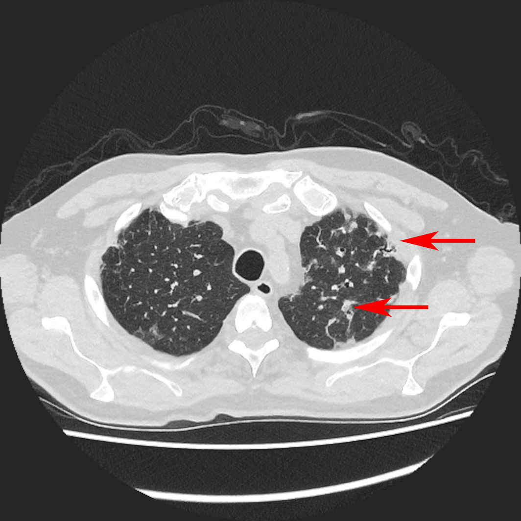
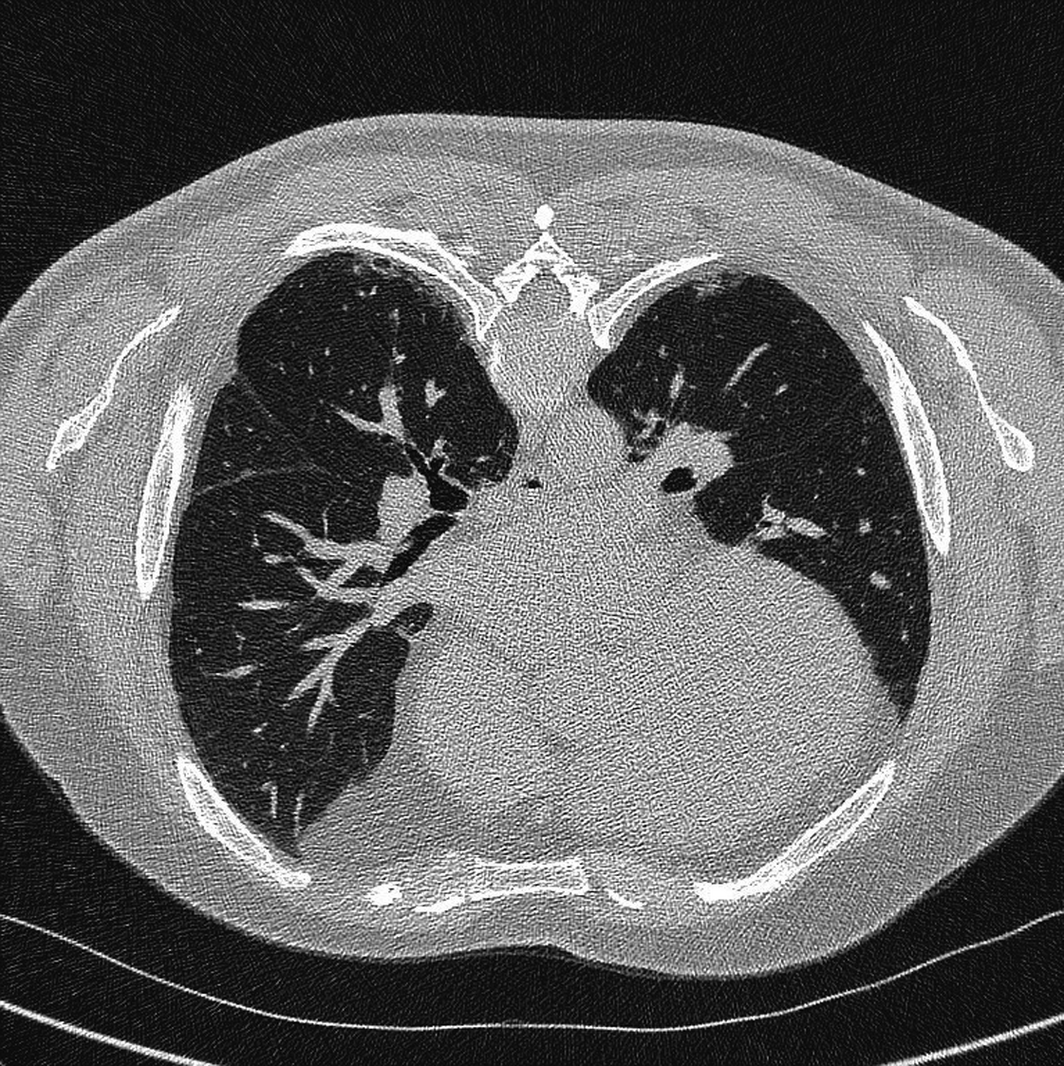
A computed tomography scan revealed interstitial pulmonary fibrosis in a usual interstitial pneumonia pattern (Box 1 and Box 2), with scanty small intraparenchymal nodules in both upper lobes. Auto‐antibody test results were all negative. He was diagnosed with dust‐related diffuse fibrosis.
Discussion
Dust‐related diffuse fibrosis (DDF) is an interstitial lung disease which occurs in dust‐exposed workers. It is probably under‐recognised in Australia, although good prevalence data are lacking.1 DDF represents a significant proportion of coal mine lung dust diseases and is classified as a type of pneumoconiosis, or pulmonary fibrosis secondary to dust inhalation (pneumon = lung; konis = dust [Greek]).2 However, the clinical picture differs from classical coal workers’ pneumoconiosis and clinicians therefore may not make the link with dust exposure, leading to a potential misdiagnosis of idiopathic pulmonary fibrosis (IPF).
The existence of lower zone interstitial pulmonary fibrosis is very well documented in coal mine workers,2,3,4 occurring in autopsy studies in up to 16% of miners, where it can occur alongside classical coal workers’ pneumoconiosis.3,4,5 In Australia, a recent review of coal mine lung disorders in Queensland confirmed DDF in 6%.1,6 A study of miners in the United States Coal Workers’ Health Surveillance Program found that 38% of coal miners had radiographic findings of interstitial lung disease, with irregular opacities of mainly lower zone predominance (upper, 21%; middle, 38%; lower, 41%).5
In contrast to classical coal workers’ pneumoconiosis, DDF has radiographic appearances of irregular (rather than rounded) opacities, predominantly in the lower zones. Clinical examination reveals lower zone late inspiratory crackles, unlike in classical coal workers’ pneumoconiosis, and clubbing is unusual. Radiologically, the classical nodular changes of coal workers’ pneumoconiosis do not always occur and are not required for making the diagnosis.2 Unless a thorough occupational history is obtained going back many years, DDF can easily be missed. Misdiagnosis can also occur because the average age of presentation is in workers aged in their 50s or 60s, similar to the age of presentation for IPF, and at a time when the patient may no longer be working. However, IPF implies the absence of a relevant occupational exposure and can only be diagnosed after exclusion of known causes of pulmonary fibrosis.7 Pulmonary function testing reveals a low diffusion capacity and a restrictive impairment identical to IPF, or a mixed pattern identical to combined pulmonary fibrosis and emphysema.
Pathological findings have been described for many years, and include interlobular septal thickening, often accompanied by emphysema, with numerous silica or silicate particles in the fibrotic septa and intra‐alveolar macrophages containing silica or silicate particles.2,3,4,8 Honeycombing may occur. Surprisingly, diffuse black pigmentation is not always present, with up to half of cases failing to show any pigmentation.2,3,5 Polarised light microscopy is needed to identify silica or silicate particles in the lungs, and then demonstrate alveolar macrophages laden with anthracotic pigment and birefringent silica or silicate particles. The histological precursor to the development of coal mine DDF is probably desquamative chronic interstitial pneumonia.8 The contribution of smoking compared with coal mine dusts is now believed to be negligible.2,5,8
DDF is more frequent in coal miners with many years of dust exposure and in older workers.2,3,4,5 Its prognosis appears to be better than that of IPF, but is yet poorly described.3 The efficacy of modern treatments including antifibrotic treatments (eg, nintedanib, perfenidone) has not yet been studied, but a new treatment trial using nintedanib (https://clinicaltrials.gov/ct2/show/NCT04161014) has recently commenced in New South Wales.
We present this case in order to highlight DDF as an under‐recognised disorder and to point out the ease with which it can be mistaken for other interstitial lung disorders. Enhanced respiratory surveillance using computed tomography and complex lung function testing, as recommended by the Thoracic Society of Australia and New Zealand, is likely to discover many more such cases.9 We encourage clinicians to take a careful occupational history such as that recommended by the US Centers for Disease Control and Prevention (https://www.atsdr.cdc.gov/csem/exphistory/docs/CSEMExposHist‐26‐29.pdf) and to remember that a history of coal mine or other dust exposure, even from many years before, may have considerable implications both for diagnosis and treatment in such patients.
Lessons from practice
- ▪ Coal mine dust‐related diffuse fibrosis (DDF) is an interstitial pulmonary fibrosis which is under‐recognised in Australia.
- ▪ DDF presents at an identical age to idiopathic pulmonary fibrosis and may have identical clinical and radiological findings.
- ▪ DDF is easily missed unless a thorough occupational history is taken to include dust exposures from first starting work to retirement.
- ▪ The long term prognosis for DDF is not well documented, but is likely to be better than for idiopathic pulmonary fibrosis and may well be improved by using modern antifibrotic treatments (eg, nintedanib, perfenidone).
Competing interests
Deborah Yates has assessed and treated patients with coal mine dust lung diseases who have been referred by Coal Services NSW. These referrals have all been for clinical purposes and billed as such.
References
- Sim M, Glass D, Hoy R, et al. Review of respiratory component of the Coal Mine Workers’ Health Scheme for the Queensland Department of Natural Resources and Mines: final report. Monash Centre for Occupational and Environmental Health and University of Illinois at Chicago, 2016. www.resources.qld.gov.au/__data/assets/pdf_file/0009/383940/monash‐qcwp‐final‐report‐2016.pdf (viewed Apr 2021).
- Cohen RA, Go LH, Green FH. Coal mine lung dust disease. In: Taylor AN, Cullinan P, Blanc P, Pickering A editors. Parkes’ occupational lung disorders. 4th ed. CRC Press, 2017: pp. 207–224.
- Cockroft AE. The importance of irregular opacities on the chest radiographs of coalworkers [MD thesis]. London University, 1984. https://spiral.imperial.ac.uk/bitstream/10044/1/38030/2/Cockcroft‐AE‐1984‐MD‐Thesis.pdf (viewed Jan 2021).
- McConnochie K, Green FHY, Vallyathan V, et al. Interstitial fibrosis in coal workers — experience in Wales and West Virginia. Ann Occup Hyg 1988; 32: 553–560.
- Cohen RA, Patel A, Green F. Lung disease caused by exposure to coal mine and silica dust. Semin Respir Crit Care Med 2008; 29: 651–661.
- Edwards R, McBean R, Parsons R, Newbigin K. A respiratory review of Australian men with coal mine dust lung disease. Eur Respir J 2019; 54: OA487.
- Raghu G, Remy‐Jardin M, Myers JL, et al. Diagnosis of idiopathic pulmonary fibrosis: an official ATS/ERS/JRS/ALAT clinical practice guideline. Am J Respir Crit Care Med 2018; 198: e44–e68.
- Brichet A, Tonnel AB, Brambilla E, et al. Chronic interstitial pneumonia with honeycombing in coal workers. Sarcoidosis Vasc Diffuse Lung Dis Off J 2002; 19: 211–219.
- Perrett J, Miles S, Brims F, Newbegin K, et al. Respiratory surveillance for coal mine dust and artificial stone‐exposed workers. TSANZ position paper. Respirology 2020; 25: 1193–1202.
Provenance: Not commissioned; externally peer reviewed.
Barriers and Facilitators to Timely Detection and Optimal Management of Chronic Wet Cough in Aboriginal Children Across Australia From the Perspectives of Carers of Aboriginal Children: A Qualitative Study
Gloria T. Y. Lau, Pam Laird, Robyn Aitken, Melanie Barwick, Liam Bedford, Emily R. Bowden, Jamie Everingham, Angela Fuery, Reece Griffin, Neha Jain, Mina Kinghorn, Renae Knapp, Gabrielle B. McCallum, Peter Morris, Richard Norman, Peter C. Richmond, Aarti Saiganesh, Slade Sibosado, Cameron Taylor, Maree Toombs, Roz Walker, Joan Wilson, Anne B. Chang, André Schultz
Barriers and Facilitators to Timely Detection and Optimal Management of Chronic Wet Cough in Aboriginal Children Across Australia From the Perspective of Health Care Providers: A Qualitative Study
Gloria T. Y. Lau, Pamela Laird, Robyn Aitken, Melanie Barwick, Liam Bedford, Emily R. Bowden, Corey Dalton, Jamie Everingham, Angela Fuery, Reece Griffin, Neha Jain, Mina Kinghorn, Renae Knapp, Gabrielle B. McCallum, Peter Morris, Richard Norman, Peter C. Richmond, Aarti Saiganesh, Slade Sibosado, Maree Toombs, Roz Walker, Joan Wilson, Anne B. Chang, André Schultz
Respiratory Syncytial Virus Disease Burden and Evidence Gaps in Australia Before Immunisation Introduction: A Retrospective Population-Based Study Using 2016–2019 Hospitalisation and 2023 Laboratory Surveillance Data
Chisato Imai-Boulter, Jean Li-Kim-Moy, Clayton Chiu, Sanjay Jayasinghe, Kristine Macartney
Not Gone but Often Forgotten: Action Needed to Address Tuberculosis in Aboriginal and Torres Strait Islander Communities
Nikkita Joshua, Emma L. Smith, Eunice (Cheeky) Love, Christopher P. Lowbridge, Nicole Tukana, Amy Bowden, Megan A. Campbell, Dawn Casey, James Ward
Uneven Ground: Survival Differences Among Victorian Lung Cancer Patients by Location of Residence (2011–2023): A Retrospective Cohort Study
Evangeline Samuel, Eldho Paul, Mike Lloyd, Sanuki Tissera, Craig Underhill, Sagun Parakh, Phillip Parente, Inger Olesen, Javier Torres, Katharine See, Gavin M. Wright, David Langton, Thomas John, Matthew Conron, James Bartlett, Nicola Atkin, Nikolajs Zeps, Susan V. Harden, Wasek Faisal, John R. Zalcberg, Rob G. Stirling
Treatment and Survival Outcomes for Indigenous and Non-Indigenous Australians Within the Victorian Lung Cancer Registry: A Retrospective Cross-Sectional Cohort Study
Melanie Wong, Mike Lloyd, Jessie Zeng, Sanuki Tissera, Kalinda E. Griffiths, Justine Clark, Jonathan Gillies, Lisa Briggs, Jacqueline Lesage, Tom Wood, Craig Underhill, Sagun Parakh, Louis B. Irving, Wasek Faisal, Rob Blum, Gary E. Richardson, Phillip Parente, Michelle Caldecott, Inger Olesen, Javier Torres, Evangeline Samuel, Christopher Lyne, Katharine See, David Langton, Thomas John, Gavin Wright, Matthew Conron, James Bartlett, Golsa Adabi, Maggie Moore, Susan Harden, Zoe K. McQuilten, John R. Zalcberg, Rob Stirling