




Volume 215 - Issue 8
Towards risk‐stratified population breast cancer screening: more than mammographic density
Authors: John L Hopper and Tuong Linh Nguyen
Med J Aust 2021; 215 (8): 350-351. || doi: 10.5694/mja2.51268
Published online: 18 October 2021
Published online: 18 October 2021
Powerful new automated tools are being developed to identify the women most likely to have an existing or future cancer
Powerful new automated tools are being developed to identify the women most likely to have an existing or future cancer
The article by Noguchi and colleagues in this issue of the MJA1 is timely and motivated by an important aim: to improve breast screening for both women and its funders. The authors conducted a comprehensive analysis of routinely collected data for all screening mammograms by BreastScreen WA over the ten years from July 2007. Although they studied screening episodes rather than individual women, they found evidence that key performance indicators — screen‐detected and interval cancer rates — differed by age, family history, hormone replacement therapy use, benign breast disease, and breast density. Importantly, the strengths of the relationships between some factors and performance varied by age group.1
While BreastScreen WA has achieved its major aims, the program has changed little over 25 years, and has some weaknesses. The overall rates reported by Noguchi and colleagues indicate that for every seven screen‐detected cancers, about 35 women are recalled and another two are diagnosed with cancer before their next regular scans.1
One possible solution that has emerged over the past decade is risk‐stratified screening, also termed tailored or personalised screening, and randomised controlled trials are underway in the United States, the United Kingdom, and Europe.2,3 Risk‐stratified screening arose in part because it was recognised that mammographic density (the area of white or bright regions on a mammogram) predicts the risk of future cancers.4,5 It had long been known that cancers in mammographically dense regions are hard to detect, contributing to higher interval cancer rates, and this is the focus of the “dense breasts” movement initiated by the late Nancy Capello.6 But mammographic density also predicts future screen‐detected cancers, albeit rather weakly. This is somewhat surprising, given that a higher interval cancer detection rate should result in a lower screen detection rate, and vice versa.
Recent research has helped resolve this paradox, discovering that a mammogram includes more risk information than conventional mammographic density alone. Challenging convention by defining density at higher brightness thresholds, we found that it was the bright, not the white, areas that were associated with inherent risk for both Australian and Korean women.4,7 This presaged agnostic computer‐based artificial intelligence approaches that have identified new and even stronger mammogram‐based risk measures.4,8 These measures each add new risk information that reduces the significance of conventional mammographic density for predicting inherent risk.9
In addition, polygenic risk scores (PRSs), combining information from hundreds of genome markers associated with small increments in risk, have been developed. Some screening trials use a PRS as the entry point,3 while others use mammographic density. In either case, the effects of these risk factors, as well as of family history, multiply each other; the greater the risk for a woman because she has a particular risk factor, the more important it is to know whether she has further risk factors.
A stepped multifactorial approach could identify tens of thousands of Australian women at high risk of breast cancer, especially if newer mammogram‐based risk measures are used.10 Of the two million Australian women who undergo regular breast screening, the risk for 140 000 (7%) would be about 4–5 times as high as the mean risk for all screened women, and for 30 000 (1.5%) it would be about ten times as high, similar to the risk for women with pathogenic BRCA1 and BRCA2 mutations. One million women would be at half the mean population risk (Box).10 The implications for tailored screening are profound.
Artificial intelligence is also being used to try to improve cancer detection.11 We are further developing automated mammogram‐based predictors of the risks of existing, interval, and future screen‐detected breast cancers.4
As Noguchi and colleagues point out,1 more information is needed before risk‐stratified screening can be adopted as routine. The factors they examined could be automatically combined with the automated mammogram‐based risk measures described above to improve detection and tailor screening. Their age‐specific findings are critical, given that risk‐stratified screening could involve a first scan at a younger age than is currently usual, perhaps even before the age of 40 years. Mammogram‐based risk predictors are fairly stable over time, even from a young age.4
It is increasingly likely that future mammographic screening will be risk‐based, as radiologists will have access to powerful new automated tools to identify the women most likely to have cancers, both at the time of screening and in the future. It is therefore essential that the decades of BreastScreen experience captured in their databanks be examined closely to guide this revolution in screening. Future analyses that track the experiences of individual women will be important, and we look forward to such analyses from Western Australia and elsewhere.
Box – Categories of women in the top quartiles for breast cancer risk factors, based on family history, mammogram risk scores, and genetic risk scores10
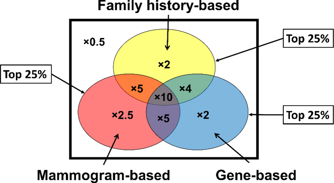
* Screened women in the circles are in the top quartile of risk for mammogram‐based (red), gene‐based (blue), and family history‐based (yellow) risk factors. The numbers inside the regions indicate the risk relative to the mean population risk.
Competing interests
No relevant disclosures.
Acknowledgements
We acknowledge the generous support for our work in this area over many years from the National Breast Cancer Foundation, the Cancer Council Victoria, Cancer Australia, the National Health and Medical Research Council, and the National Institutes of Health (USA).
References
- Noguchi N, Marinovich ML, Wylie EJ, et al. Screening outcomes by risk factor and age: evidence from BreastScreen WA for discussions of risk‐stratified population screening. Med J Aust 2021; 215: 359–365.
- Allweis TM, Hermann N, Bernstein‐Molho R, Guindy M. Personalized screening for breast cancer: rationale, present practices, and future directions. Ann Surg Oncol 2021; 28: 4306–4317.
- Eklund M, Broglio K, Yau C, Connor JT, et al. The WISDOM personalized breast cancer screening trial: simulation study to assess potential bias and analytic approaches. JNCI Cancer Spectr 2018; 2: pky067.
- Hopper JL, Nguyen TL, Schmidt DF, et al. Going beyond conventional mammographic density to discover novel mammogram‐based predictors of breast cancer risk. J Clin Med 2020; 9: 627.
- Boyd NF, Guo H, Martin LJ, Sun L, et al. Mammographic density and the risk and detection of breast cancer. N Engl J Med 2007; 3563: 227–236.
- Cappello NM, Richetelli D, Lee CI. The impact of breast density reporting laws on women’s awareness of density‐associated risks and conversations regarding supplemental screening with providers. J Am Coll Radiol 2019; 16: 139–146.
- Nguyen TL, Aung YK, Evans CF, et al. Mammographic density defined by higher than conventional brightness thresholds better predicts breast cancer risk. Int J Epidemiol 2017; 46: 652–661.
- Schmidt DF, Makalic E, Goudey B, et al. Cirrus: an automated mammography‐based measure of breast cancer risk based on textural features. JNCI Cancer Spectr 2018; 2: pky057.
- Nguyen TL, Schmidt DF, Makalic E, et al. Novel mammogram‐based measures improve breast cancer risk prediction beyond an established mammographic density measure. Int J Cancer 2021; 148: 2193–2202.
- Hopper JL. Genetics for population and public health. Int J Epidemiol 2017; 46: 8–11.
- Freeman K, Geppert J, Stinton C, et al. Use of artificial intelligence for image analysis in breast cancer screening programmes: systematic review of test accuracy. BMJ 2021; 374: n1872.
Provenance: Commissioned; not externally peer reviewed.
From Ambition to Action: Closing Policy, Equity and Implementation Gaps in the Australian Cancer Plan's ‘Enhanced Consumer Experience’
Carla Thamm, Elise Button, Oluwaseyifunmi A. Agbejule, Reegan Knowles, Karen van Gorp, Mark B. Pinkham, Meinir Krishnasamy, Alexandra L. McCarthy, Raymond J. Chan
Sovereignty and Survival: Understanding Lung Cancer Outcomes for Aboriginal and Torres Strait Islander People in Australia, a Case–Control Study
Alice R. T. Bergin, Luc te Marvelde, Roger L. Milne, Benjamin J. Solomon, Kris Ivanova, Peter Savas, Stephen J. Luen, Jay Hamann, Andrea Casey, Nicole Watt, Sue M. Evans, Gail Garvey, Sherene Loi
The Changing Indications for Adult Liver Transplantation in Australia: A Narrative Review
Fadak Mohammadi, Alan J. Wigg, Ken Liu, Simone I. Strasser
Uneven Ground: Survival Differences Among Victorian Lung Cancer Patients by Location of Residence (2011–2023): A Retrospective Cohort Study
Evangeline Samuel, Eldho Paul, Mike Lloyd, Sanuki Tissera, Craig Underhill, Sagun Parakh, Phillip Parente, Inger Olesen, Javier Torres, Katharine See, Gavin M. Wright, David Langton, Thomas John, Matthew Conron, James Bartlett, Nicola Atkin, Nikolajs Zeps, Susan V. Harden, Wasek Faisal, John R. Zalcberg, Rob G. Stirling
Early-Onset Colorectal Cancer With Liver-Only Metastases: A Retrospective Cohort Study Integrating Prospectively Collected Real-World Clinical and Molecular Data From an Australian National Database (2009–2024) to Guide Treatment Planning
Savio G. Barreto, Christos S. Karapetis, Shahid Ullah, Matthew Burge, Susan Caird, Angus Campbell, Azim Jalali, Ross Jennens, Muhammad A. Khattak, Belinda Lee, Stephanie H. Lim, Shehara Mendis, Louise Nott, Timothy J. Price, Jeremy D. Shapiro, Jeanne Tie, Javier Torres, Colin Williams, Rachel Wong, Vanessa Wong, Peter Gibbs
Australian Pathways for Specialist Pain Management and Early Palliative Care for People With Pancreatic Cancer: Developed Using a Community Consensus Approach
Jennifer Philip, Melanie R. Lovell, Kylee Bellingham, Gail Garvey, Gregory B. Crawford, Nicole M. Rankin, Kara Burns, Isabel Young, Vivienne Milch, Dorothy Keefe, Katrina Anderson, James Lawson, Meinir Krishnasamy