




Volume 215 - Issue 6
Interventional bronchoscopy for chronic obstructive pulmonary disease: more than a pipe dream
Authors: Alan M Carew, Jonathan P Williamson, Claude S Farah, Tajalli Saghaie, Martin Phillips and Alvin Ing
Med J Aust 2021; 215 (6): 280-285. || doi: 10.5694/mja2.51218
Published online: 6 September 2021
Published online: 6 September 2021
COPD is the fifth greatest contributor to burden of disease and fifth overall cause of death among Australians
Summary
- Endoscopic lung volume reduction (ELVR) is recognised in both national and international expert guidelines as one of the few additive treatments to benefit patients with advanced chronic obstructive pulmonary disease (COPD) who are otherwise receiving optimal medical and supportive care. Despite these recommendations and a growing evidence base, these procedures are not widely offered across Australia and New Zealand, and general practitioner and physician awareness of this therapy can be improved.
- ELVR aims to mitigate the impact of hyperinflation and gas trapping on dyspnoea and exercise intolerance in COPD. Effective ELVR is of proven benefit in improving symptoms, quality of life, lung function and survival.
- Several endoscopic techniques to achieve ELVR have been developed, with endobronchial valve placement to collapse a single lobe being the most widely studied and commonly practised.
- This review describes the physiological rationale underpinning lung volume reduction, highlights the challenges of patient selection, and provides an overview of the evidence for current and investigational endoscopic interventions for COPD.
Chronic obstructive pulmonary disease (COPD) is the fifth greatest contributor to burden of disease and fifth overall cause of death among Australians.1 It most frequently results from cigarette smoking and leads to progressive small airway narrowing and emphysematous alveolar destruction. Symptoms include progressive dyspnoea, wheeze, and a persistent cough which may be accompanied by chronic sputum production. Patients are also prone to acute exacerbations that can result in hospitalisation, further lung function decline, respiratory failure or death. It is estimated that COPD costs the Australian community $8.8 billion in treatment and lost productivity annually.2
Comprehensive evidence‐based guidelines for the diagnosis and management of COPD in Australia and New Zealand are provided in the Lung Foundation Australia COPD‐X Plan.3 The cornerstones of management are smoking cessation, pulmonary rehabilitation and inhaled pharmacotherapy, with the aims of reducing symptoms, improving lung function and quality of life, and reducing the risk of exacerbations and mortality. For people with advanced disease, additional therapeutic options are limited. Prophylactic macrolide antibiotics may benefit patients with frequent exacerbations but are not widely advocated,4 long term oxygen therapy reduces mortality when COPD is associated with chronic hypoxia,5 and lung transplantation may be appropriate for select patients with limited comorbidities.6 Lung volume reduction is accepted as an additive therapy for certain patients with advanced emphysema refractory to medical treatment,6 but despite support from key international societies, in our experience it is not widely offered in Australia and New Zealand.
In this review, we describe the physiology of dyspnoea in COPD which explains the rationale behind lung volume reduction. We searched PubMed for randomised controlled trials (RCTs) investigating endoscopic lung volume reduction (ELVR) techniques, along with meta‐analyses, case series, subgroup analyses and expert consensus recommendations for the various approaches and their complications. We also review investigational lung volume reduction techniques, and endoscopic therapies which aim to treat chronic bronchitis.
A glossary of key terms is provided in Box 1.
Physiology
The causes of dyspnoea in COPD are numerous, with several processes often intertwined in a given individual. Destruction of elastin and collagen within lung tissue leads to reduced elastic recoil and loss of parenchymal attachments that support small airways. These pathological changes result in a reduced pressure gradient for airflow during expiration, and a propensity for the small airways to narrow and more readily collapse. This small airway narrowing is further compounded by airway inflammation and hyperplasia of mucin‐secreting cells. Together with the obliteration of terminal bronchioles, the overall airway surface area in the lung is reduced.7 Gas trapping within the lungs thereby ensues despite the patient’s expiratory effort. Over time, this results in a gradual increase in lung volume at rest known as hyperinflation.
Hyperinflation and gas trapping impede efficient function of the respiratory muscles, with the overexpanded lung displacing and flattening the diaphragm, impairing its movement during breathing. As a result, significantly more effort is required to expand the hyperinflated lung at higher volumes, which is further magnified during exercise.8 As respiratory demand increases with exercise, acute illness or anxiety, the combination of expiratory airflow limitation and higher respiratory rate leads to dynamic hyperinflation. In this instance, the airspaces fail to empty before the next inhalation begins, causing a progressive increase in end‐expiratory lung volume and compounding the gas trapping. This phenomenon results in increased work of breathing and contributes to the worsening dyspnoea and functional limitation that patients experience.9
Dyspnoea and exercise intolerance are exacerbated by mismatching of ventilation and perfusion, which occurs even in mild COPD and worsens with disease severity as a result of both alveolar destruction and airflow limitation.10 Physical inactivity is also common early in the disease course and predicts hospitalisation and all‐cause mortality, with activity levels tending to deteriorate considerably with disease exacerbations. Deconditioning prompts exercise intolerance, and the consequent muscle weakness leads to a cycle of deteriorating physical activity, frailty and impaired quality of life.11
Lung volume reduction surgery
Despite optimal medical therapy and smoking cessation, many patients remain highly symptomatic with poor quality of life.6 Lung volume reduction surgery (LVRS) was first introduced in 1957, aiming to mitigate the hyperinflation and gas trapping which accompany advanced COPD by resection of the most diseased areas of lung. This has been shown to improve lung elastic recoil, expiratory airflow and respiratory muscle function, while reducing dynamic and static hyperinflation, and potentially decompressing healthier lung tissue.8
In 2003, the seminal National Emphysema Treatment Trial12 found improvements in mortality, exercise capacity, lung function and symptoms following bilateral LVRS in a subgroup of patients with upper lobe predominant emphysema and poor baseline exercise tolerance. These improvements came at the cost of short term mortality of 5.2% in non‐high risk patients, significantly higher than standard of care. This effect was even more pronounced in a high risk subgroup with severely impaired lung function (forced expiratory volume in 1 second [FEV1] ≤ 20% and either diffusion capacity for carbon monoxide ≤ 20% or homogeneous emphysema) who had a 90‐day mortality of 28.6% and were subsequently excluded from the study.
The early mortality seen with bilateral LVRS has been a key driver for development of less invasive, endoscopic means of achieving the same physiological benefits. Although unilateral thoracoscopic LVRS has significantly lower mortality by comparison, the risks of long post‐operative hospital stays, repeat operations for persistent air leaks, and cardiopulmonary morbidity remain.13
Endoscopic lung volume reduction
ELVR aims to achieve the physiological benefits of LVRS with shorter procedures, hospital stays and recovery periods, and lower risks in this vulnerable patient group, without the systemic catabolic state and deconditioning associated with surgery. The inclusion criteria established in the National Emphysema Treatment Trial formed the basis for ELVR candidacy,12 although patient selection remains the most challenging aspect of this field, guided mainly by expert recommendation.
Typical candidates for ELVR are highly symptomatic due to severe COPD despite optimal medical therapy, structured rehabilitation and smoking cessation. They have significant hyperinflation and gas trapping, demonstrated by elevated total lung capacity and residual volume measured during pulmonary function testing, with few additional respiratory or cardiac comorbidities. A general list of characteristics of ELVR candidates are provided in Box 2, based on international expert panel recommendations.14 It is important to note that these eligibility criteria are not absolute, and that certain factors, such as the magnitude of residual volume elevation, may be weighted more heavily depending on the institution. Specific disease characteristics, such as emphysema distribution and the presence or absence of collateral ventilation between lobes, further dictate suitability for ELVR techniques or LVRS. It is therefore recommended that lung volume reduction assessments and procedures are performed in specialist centres, and a multidisciplinary emphysema team involving respiratory physicians, interventional pulmonologists, thoracic surgeons, radiologists, respiratory physiotherapists, transplant physicians and nurse specialists is increasingly needed.15
Endobronchial valves
Lung volume reduction may be achieved through bronchoscopic placement of one‐way valves into segmental airways, which leads to deflation of the target lobe. The Zephyr endobronchial valve (PulmonX) has a silicone duckbill in a nitinol frame, and the Spiration valve system (Olympus) has an umbrella‐shaped polyurethane membrane mounted on a nitinol frame which minimises tissue contact (Box 3). The increasingly robust body of literature for endobronchial valve therapy has led to its recent recognition by the Global Initiative for Chronic Obstructive Lung Disease strategy with level A evidence,6 and the United Kingdom National Institute for Health and Care Excellence guidelines.16 Both valves were approved by the United States Food and Drug Administration in 2018 for use in ELVR.
The first multicentre, international RCT17,18 of Zephyr endobronchial valve placement yielded statistically significant but only modest clinical improvements in lung function, exercise capacity and symptoms following unilateral valve implantation in the most diseased lobe in a phenotypically diverse group of patients with COPD. Post hoc analyses indicated that patients with a heterogeneous distribution of emphysema and complete interlobar fissures had more successful volume reduction and far more meaningful outcomes. This led to the realisation that collateral ventilation, or the presence of channels which bypass the normal airways across interlobar fissures, was a key determinant of effective ELVR by means of airway occlusion.
This principle was tested in the randomised, sham‐controlled BeLieVeR‐HIFi19 study, in which the presence on imaging of intact interlobar fissures adjacent to the target lobe was used as a surrogate for absence of collateral ventilation. Patients also underwent bronchoscopic assessment for collateral ventilation using a dedicated catheter‐based system to measure airflow and pressure in the target airway while under balloon occlusion, although this was not used for inclusion. The STELVIO study20 further refined this process by using both radiologically intact fissures and absence of collateral ventilation by this system for patient selection. These trials alongside the IMPACT21 study, which focused on homogeneous emphysema, confirmed significant improvements in lung function and exercise capacity, with the greatest benefit seen in patients with intact fissures and heterogeneous emphysema (Box 4).
The most recent multicentre studies22,23 have focused on the patient group most likely to benefit from ELVR — individuals with heterogeneous emphysema and no collateral ventilation — and adopted a real world approach by permitting revision or replacement of their valves if lung volume reduction was unsatisfactory on high resolution computed tomography. Both studies confirmed clinically significant improvements in symptoms, exercise capacity, FEV1, and gas trapping.
In a recent meta‐analysis of these trials, endobronchial valve placement in a lobe without collateral ventilation was associated with clinically significant improvements in subjective and objective respiratory outcomes. The St George’s Respiratory Questionnaire is a validated tool which measures the impact of respiratory symptoms on quality of life, producing a score from 0 to 100, with higher values corresponding with quality of life impairment and the minimum clinically important difference being 4 points.3 Scores reduced by a mean of 9.13 points, and distance walked in 6 minutes improved by 49 metres. Improvement in gas trapping was reflected by mean reduction in residual volume of 0.57 L, with airflow improvement indicated by mean increase in FEV1 of 22%.24 Improvements in gas transfer capacity,25 ventilation and perfusion matching,26 and reductions in dynamic hyperinflation during exercise27 have also been reported. Small case series have also suggested the feasibility of endobronchial valve placement in patients with emphysema secondary to α‐1 antitrypsin deficiency,28 and in patients with FEV1 ≤ 20% of predicted29 or with diffusion capacity for carbon monoxide ≤ 20% of predicted30 without excess adverse events.
The most common complication of effective endobronchial valve placement is pneumothorax, owing to a combination of rapid expansion of the adjacent untreated lobe and tearing of fragile lung tissue or adhesions. While this occurs in up to 25% of cases,24 most would consider the risk acceptable given intercostal drains are universally required following LVRS, and rates of prolonged air leak and re‐operation in that cohort are typically high.13 Pneumothorax after endobronchial valve insertion does not influence survival, and in fact appears to be a marker of successful lobar collapse and volume reduction, which in itself is associated with a survival advantage.31 Less frequently encountered complications include infection distal to the valves, haemoptysis, or valve migration or displacement from granulation tissue formation. Further bronchoscopies may be needed to treat complications, or to revise valve placement if the desired effect is not achieved or is lost.
Two RCTs have investigated the Spiration valve system in patients without collateral ventilation to date: REACH32 and EMPROVE.33 These trials reported significant improvements in FEV1 of up to 23% at 12 months and improvement in dyspnoea and quality of life metrics, with the most common complication being pneumothorax, although seemingly at lower rates than in the Zephyr endobronchial valve trials.34 There have not yet been any RCTs directly comparing the two valve types, although a prospective study comparing outcomes of unilateral thoracoscopic LVRS and bronchoscopic endobronchial valve placement is currently underway (ISRCTN19684749).
The published RCTs to date have reported improved outcomes up to 1 year, with the available long term follow‐up data suggesting the benefit gradually declines, likely due to disease progression in the non‐targeted lung. In spite of this, some patients have maintained improvements in symptoms, exercise capacity and lung function at 3 years.35 Longer term data for patients treated with either or both types of valves indicate that treatment responders who develop lobar atelectasis have an improved 5‐year survival of 65%, compared with 44% in those with no lobar volume reduction31 (Box 5).
Biological lung volume reduction
Targeted instillation of a synthetic compound containing glutaraldehyde and polyvinyl alcohol is the basis of biological or polymeric lung volume reduction. This irreversible technique instigates a localised inflammatory response leading to volume loss independent of collateral ventilation, and although the initial prospective study demonstrated effective volume reduction and improvement in lung function and symptoms,36 the larger ASPIRE randomised study raised safety concerns regarding the inflammatory response to the polymer.37 The ASPIRE study was terminated early by the sponsor for financial reasons. This has been revisited with a staged treatment algorithm using lower doses to mitigate the inflammatory response in a completed but as yet unreported trial (NCT02877459).
Our group recently reported combining the use of carefully targeted small doses of the polymer in a patient with collateral ventilation, aiming to seal the leak across the fissure and leading to its cessation without any significant inflammatory response. This was then followed by successful valve placement with collapse of the target lobe.38 Prospective series using this combined approach, and separate trials using autologous blood to terminate collateral ventilation before valve placement (NTR5007; NCT03010449) are underway, aiming ultimately to increase the numbers of patients who may benefit from valve placement.
Bronchial thermal vapour ablation
Bronchial thermal vapour ablation is an irreversible means of segmental ELVR which does not depend on the absence of collateral ventilation for success. Thermal energy is delivered by means of heated water vapour through a balloon‐tipped catheter at bronchoscopy to induce a localised inflammatory reaction in the most diseased lung segments. This then heals with a fibrotic response and subsequent volume loss, with many patients experiencing a localised inflammatory response and systemic features of fatigue, influenza‐like symptoms, or temporary worsening of their respiratory symptoms.39
International multicentre studies have demonstrated improvements in FEV1 of up to 15% at 6 months, with symptomatic improvement and effective volume loss in the target segments.40,41 Post hoc analyses have also indicated that collateral ventilation does not appear to impact clinical outcomes.42 Current recommendations are for treatment of up to three upper lobe segments over two sessions to mitigate the local inflammatory response.39 Research is ongoing in refining this technique, with optimisation of the thermal energy dose required to effect volume loss while reducing the morbidity of a severe inflammatory response through a staged approach, and we await studies on the use of bronchial thermal vapour ablation in patients with homogeneous and lower lobe predominant disease.
Lung volume reduction coils
Endobronchial coils compress hyperinflated lung tissue to restore tension and elastic recoil through deployment of several nitinol rods into target segments, which coil immediately after insertion. This process aims to tether collapsible airways and improve airway resistance, while simultaneously effecting volume reduction independent of collateral ventilation. International RCTs to date have indicated modest improvements in lung function and symptoms, with homogeneous emphysema patients being well represented in these studies.43,44,45 Adverse events include pneumonia, exacerbations of COPD, and pneumothorax. Improved transplant‐free 5‐year survival of 67% in responders with volume loss versus 36% in non‐responders was reported in secondary analysis of RESET trial participants.46 At the time of writing, endobronchial coils are not available in Australia or New Zealand.
Airway bypass
Implantation of artificial airways which bypass the narrowed and collapsible native bronchi has been studied bronchoscopically and surgically. Improvements in lung function in the post‐operative period have been demonstrated but have not proven durable.47,48 These artificial airways appear prone to occlusion, which has limited their efficacy, and they are not in current use.
Targeted therapies for chronic bronchitis
There are now several investigational endoscopic technologies focusing on treatment of chronic bronchitis, rather than emphysema and hyperinflation. These include a system that utilises a liquid nitrogen cryospray catheter to ablate surface epithelium with goblet cell hyperplasia, with the goal of allowing normal epithelium to regenerate (NCT03893370).
Results of the first in‐human study of bronchial rheoplasty for the treatment of chronic bronchitis have recently been reported.49 This endoscopic procedure applies unheated, pulsed electric fields to the airways to reduce the excessive numbers of mucus‐producing cells, based on pre‐clinical studies demonstrating epithelial ablation followed by regeneration of more normal epithelium. The study population with prominent symptoms of chronic bronchitis experienced a significant symptomatic improvement and histological evidence of reduced goblet cell hyperplasia with complications mainly comprising COPD exacerbations.
Targeted lung denervation
Targeted lung denervation uses radiofrequency ablation to disrupt parasympathetic signalling with the aim of decreasing airway resistance and mucus hypersecretion. The AIRFLOW trial prospectively compared targeted lung denervation with sham procedures in a randomised, double‐blind fashion and found fewer respiratory adverse events in a predefined timeframe. Recruitment is underway for a larger randomised, sham‐controlled trial to primarily investigate the rates of exacerbations following targeted lung denervation.50
Conclusion
Lung volume reduction is a therapeutic option for many patients with advanced emphysema who remain highly symptomatic despite maximal medical therapy and for whom few other treatments are available. While inhaled pharmacotherapy, smoking cessation and pulmonary rehabilitation are mandatory evidence‐based therapies for this patient group, a growing array of endoscopic procedures are now available as an alternative to surgery for highly selected patients. Patient selection for lung volume reduction is individualised and complex, and multidisciplinary emphysema teams are increasingly recommended for both assessment and treatment in high volume specialised centres.
The inclusion of ELVR in international expert guidelines and the expanding evidence base highlight the need for awareness of this treatment option. With more research into patient selection, the longer term effects of ELVR, and comparative studies between different endoscopic techniques and surgery, it appears inevitable that these treatments will continue to shift from their position as a niche therapy in Australia and New Zealand towards the mainstream.
Box 1 – Glossary of terms
|
Term |
Definition |
||||||||||||||
|
|
|||||||||||||||
|
Hyperinflation |
Increased lung volume resulting from decreased lung tissue elasticity in COPD. Static hyperinflation is present at rest, indicated by elevated TLC measured during lung function testing. |
||||||||||||||
|
Dynamic hyperinflation |
Progressive rise in end‐expiratory lung volume during periods of increased respiratory demand, such as exercise, illness or anxiety. Results in increased respiratory effort, dyspnoea and exercise limitation. |
||||||||||||||
|
Gas trapping |
Inability to fully exhale to a normal end‐expiratory lung volume, which in COPD results from loss of elastic recoil, airway narrowing and collapsibility. Indicated by elevated RV during lung function testing. |
||||||||||||||
|
Heterogeneous emphysema |
Variable pattern and severity of emphysema distribution within the lung, with separate areas of emphysematous and more preserved lung tissue. Heterogeneity correlates with improved outcomes after lung volume reduction. |
||||||||||||||
|
Homogeneous emphysema |
Minimal difference in distribution and severity of emphysema between different segments or lobes of the lung. |
||||||||||||||
|
Collateral ventilation |
Ventilation of alveoli by pathways other than the normal airways — prevents atelectasis in the presence of bronchial obstruction. If present, prevents clinically meaningful lung volume reduction after endobronchial valve placement. Assessed radiologically by presence of complete interlobar fissures, and bronchoscopically using a catheter system. |
||||||||||||||
|
|
|||||||||||||||
|
COPD = chronic obstructive pulmonary disease; ELVR = endoscopic lung volume reduction; RV = residual volume; TLC = total lung capacity. |
|||||||||||||||
Box 2 – Typical characteristics of lung volume reduction candidates14
- Highly symptomatic; eg, Medical Research Council dyspnoea scale score ≥ 2
- Receiving optimal pharmacological therapy
- Smoking cessation for > 6 months
- Completed or participating in structured rehabilitation program
- FEV1 20–50% of predicted value
- TLC ≥ 100% of predicted value
- RV ≥ 175% of predicted value, or RV:TLC ratio ≥ 0.58
- 6‐minute walk distance of 100–450 metres
- Absence of significant pulmonary hypertension or cardiac disease
- Absence of significant daily sputum production or frequent exacerbations
FEV1 = forced expiratory volume in 1 second; RV = residual volume; RV:TLC = residual volume to total lung capacity; TLC = total lung capacity.
Box 3 – Endobronchial valve systems
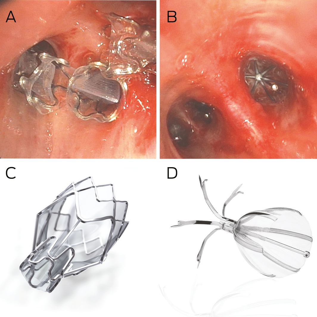
A: Zephyr (PulmonX) endobronchial valves deployed in segmental airways. B: Spiration (Olympus) valve system deployed in segmental airways. C: Zephyr endobronchial valve; image supplied by PulmonX. D: Spiration valve system; image supplied by Olympus.
Box 4 – Heterogeneous and homogeneous emphysema
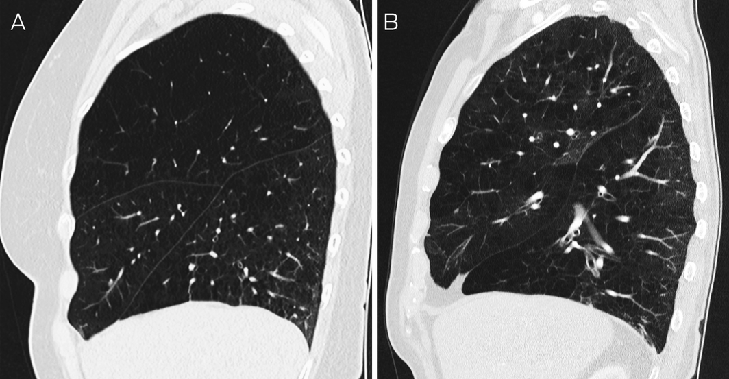
A: Heterogeneous emphysema, more severely affecting the right upper lobe, with complete horizontal and oblique fissures. B: Homogeneous emphysema affecting all right lobes to similar degree, with incomplete horizontal fissure and complete oblique fissure.
Competing interests
Alvin Ing is principal investigator in investigator‐initiated trials sponsored by Olympus Australia, Pulmonx Australia and Gala Therapeutics.
References
- Australian Institute of Health and Welfare. Deaths in Australia (AIHW Cat. No. PHE 229). Canberra: AIHW, 2019. https://www.aihw.gov.au/reports/life‐expectancy‐death/deaths‐in‐australia (viewed July 2020).
- Access Economics. Economic impact of COPD and cost effective solutions. Sydney: Access Economics, 2008. http://www.rnig.org.au/docs/EconomicImpactofCOPDandCostEffectiveSolutions‐226.pdf (viewed Jul 2020).
- Yang IA, Brown JL, George J, et al. The COPD‐X Plan: Australian and New Zealand guidelines for the management of chronic obstructive pulmonary disease 2020. Brisbane: Lung Foundation Australia, 2020. https://copdx.org.au/wp‐content/uploads/2020/04/COPDX‐V2‐61‐Feb‐2020‐FINAL.pdf (viewed Aug 2020).
- Herath SC, Normansell R, Maisey S, Poole P. Prophylactic antibiotic therapy for chronic obstructive pulmonary disease (COPD). Cochrane Database Syst Rev 2018; (10): CD009764.
- Medical Research Council Working Party. Long term domiciliary oxygen therapy in chronic hypoxic cor pulmonale complicating chronic bronchitis and emphysema. Lancet 1981; 1: 681–686.
- Global Initiative for Chronic Obstructive Lung Disease. Global strategy for the diagnosis, management, and prevention of chronic obstructive pulmonary disease: 2020 report. GOLD, 2020. https://goldcopd.org/gold‐reports/ (viewed Aug 2020).
- McDonough JE, Yuan R, Suzuki M, et al. Small‐airway obstruction and emphysema in chronic obstructive pulmonary disease. N Engl J Med 2011; 365: 1567–1575.
- Fessler HE, Scharf SM, Ingenito EP, et al. Physiologic basis for improved pulmonary function after lung volume reduction. Proc Am Thorac Soc 2008; 5: 416–420.
- Gagnon P, Guenette JA, Langer D, et al. Pathogenesis of hyperinflation in chronic obstructive pulmonary disease. Int J Chron Obstruct Pulmon Dis 2014; 9: 187–201.
- Barbera JA, Ramirez J, Roca J, et al. Lung structure and gas exchange in mild chronic obstructive pulmonary disease. Am Rev Respir Dis 1990; 141: 895–901.
- Watz H, Pitta F, Rochester CL, et al. An official European Respiratory Society statement on physical activity in COPD. Eur Respir J 2014; 44: 1521–1537.
- Fishman A, Martinez F, Naunheim K, et al. A randomized trial comparing lung‐volume‐reduction surgery with medical therapy for severe emphysema. N Engl J Med 2003; 348: 2059–2073.
- Clark SJ, Zoumot Z, Bamsey O, et al. Surgical approaches for lung volume reduction in emphysema. Clin Med (Lond) 2014; 14: 122–127.
- Herth FJF, Slebos DJ, Criner GJ, et al. Endoscopic lung volume reduction: an expert panel recommendation ‐ update 2019. Respiration 2019; 97: 548–557.
- Chew J, Mahadeva R. The role of a multidisciplinary severe chronic obstructive pulmonary disease hyperinflation service in patient selection for lung volume reduction. J Thorac Dis 2018; 10: S3335–S3343.
- National Institute for Health and Care Excellence. Chronic obstructive pulmonary disease in over 16s: diagnosis and management (NICE guideline [NG115]). London: NICE, 2019. https://www.nice.org.uk/guidance/ng115/chapter/Recommendations (viewed Aug 2020).
- Sciurba FC, Ernst A, Herth FJ, et al. A randomized study of endobronchial valves for advanced emphysema. N Engl J Med 2010; 363: 1233–1244.
- Herth FJ, Noppen M, Valipour A, et al. Efficacy predictors of lung volume reduction with Zephyr valves in a European cohort. Eur Respir J 2012; 39: 1334–1342.
- Davey C, Zoumot Z, Jordan S, et al. Bronchoscopic lung volume reduction with endobronchial valves for patients with heterogeneous emphysema and intact interlobar fissures (the BeLieVeR‐HIFi study): a randomised controlled trial. Lancet 2015; 386: 1066–1073.
- Klooster K, ten Hacken NH, Hartman JE, et al. Endobronchial valves for emphysema without interlobar collateral ventilation. N Engl J Med 2015; 373: 2325–2335.
- Valipour A, Slebos DJ, Herth F, et al. Endobronchial valve therapy in patients with homogeneous emphysema. results from the IMPACT study. Am J Respir Crit Care Med 2016; 194: 1073–1082.
- Kemp SV, Slebos DJ, Kirk A, et al. A multicenter randomized controlled trial of Zephyr endobronchial valve treatment in heterogeneous emphysema (TRANSFORM). Am J Respir Crit Care Med 2017; 196: 1535–1543.
- Criner GJ, Sue R, Wright S, et al. A multicenter randomized controlled trial of Zephyr endobronchial valve treatment in heterogeneous emphysema (LIBERATE). Am J Respir Crit Care Med 2018; 198: 1151–1164.
- van Geffen WH, Slebos DJ, Herth FJ, et al. Surgical and endoscopic interventions that reduce lung volume for emphysema: a systemic review and meta‐analysis. Lancet Respir Med 2019; 7: 313–324.
- Hsu K, Williamson JP, Peters MJ, Ing AJ. Endoscopic lung volume reduction in COPD: improvements in gas transfer capacity are associated with improvements in ventilation and perfusion matching. J Bronchology Interv Pulmonol 2018; 25: 48–53.
- Yang L, Hsu K, Williamson JP, et al. Changes in ventilation and perfusion following lower lobe endoscopic lung volume reduction (ELVR) with endobronchial valves in severe COPD. Clin Respir J 2019; 13: 453–459.
- Hopkinson NS, Toma TP, Hansell DM, et al. Effect of bronchoscopic lung volume reduction on dynamic hyperinflation and exercise in emphysema. Am J Respir Crit Care Med 2005; 171: 453–460.
- Hillerdal G, Mindus S. One‐ to four‐year follow‐up of endobronchial lung volume reduction in alpha‐1‐antitrypsin deficiency patients: a case series. Respiration 2014; 88: 320–328.
- Darwiche K, Karpf‐Wissel R, Eisenmann S, et al. Bronchoscopic lung volume reduction with endobronchial valves in low‐FEV1 patients. Respiration 2016; 92: 414–419.
- van Dijk M, Hartman JE, Klooster K, et al. Endobronchial valve treatment in emphysema patients with a very low DLCO. Respiration 2020; 99: 163–170.
- Gompelmann D, Benjamin N, Bischoff E, et al. Survival after endoscopic valve therapy in patients with severe emphysema. Respiration 2019; 97: 145–152.
- Li S, Wang G, Wang C, et al. The REACH trial: a randomized controlled trial assessing the safety and effectiveness of the Spiration® valve system in the treatment of severe emphysema. Respiration 2019; 97: 416–427.
- Criner GJ, Delage A, Voelker K, et al. Improving lung function in severe heterogenous emphysema with the Spiration valve system (EMPROVE). A multicenter, open‐label randomized controlled clinical trial. Am J Respir Crit Care Med 2019; 200: 1354–1362.
- Majid A, Labarca G, Uribe JP, et al. Efficacy of the Spiration valve system in patients with severe heterogeneous emphysema: a systematic review and meta‐analysis. Respiration 2020; 99: 62–72.
- Gompelmann D, Heinhold T, Rötting M, et al. Long‐term follow up after endoscopic valve therapy in patients with severe emphysema. Ther Adv Respir Dis 2019; 13: 1753466619866101.
- Herth FJ, Gompelmann D, Stanzel F, et al. Treatment of advanced emphysema with emphysematous lung sealant (AeriSeal®). Respiration 2011; 82: 36–45.
- Come CE, Kramer MR, Dransfield MT, et al. A randomised trial of lung sealant versus medical therapy for advanced emphysema. Eur Respir J 2015; 46: 651–662.
- Ing A, Sullivan C, Hersch N, et al. Reversal of collateral ventilation using endobronchial polymer sealant in a patient with emphysema undergoing endoscopic lung volume reduction (ELVR) with valves: a case report and proof of concept. J Bronchology Interv Pulmonol 2020; 27: e14–e16.
- Gompelmann D, Shah PL, Valipour A, Herth FJF. Bronchoscopic thermal vapor ablation: best practice recommendations from an expert panel on endoscopic lung volume reduction. Respiration 2018; 95: 392–400.
- Snell G, Herth FJ, Hopkins P, et al. Bronchoscopic thermal vapour ablation therapy in the management of heterogeneous emphysema. Eur Respir J 2012; 39: 1326–1333.
- Herth FJ, Valipour A, Shah PL, et al. Segmental volume reduction using thermal vapour ablation in patients with severe emphysema: 6‐month results of the multicentre, parallel‐group, open‐label, randomised controlled STEP‐UP trial. Lancet Respir Med 2016; 4: 185–193.
- Gompelmann D, Eberhardt R, Schuhmann M, et al. Lung volume reduction with vapor ablation in the presence of incomplete fissures: 12‐month results from the STEP‐UP randomized controlled study. Respiration 2016; 92: 397–403.
- Shah PL, Zoumot Z, Singh S, et al. Endobronchial coils for the treatment of severe emphysema with hyperinflation (RESET): a randomised controlled trial. Lancet Respir Med 2013; 1: 233–240.
- Deslée G, Mal H, Dutau H, et al. Lung volume reduction coil treatment vs usual care in patients with severe emphysema: the REVOLENS randomized clinical trial. JAMA 2016; 315: 175–184.
- Sciurba FC, Criner GJ, Strange C, et al. Effect of endobronchial coils vs usual care on exercise tolerance in patients with severe emphysema: The RENEW randomized clinical trial. JAMA 2016; 315: 2178–2189.
- Garner JL, Kemp SV, Srikanthan K, et al. 5‐Year survival after endobronchial coil implantation: secondary analysis of the first randomised controlled trial, RESET. Respiration 2020; 99: 154–162.
- Shah PL, Slebos DJ, Cardoso PF, et al. Bronchoscopic lung‐volume reduction with Exhale airway stents for emphysema (EASE trial): randomised, sham‐controlled, multicentre trial. Lancet 2011; 378: 997–1005.
- Snell GI, Holsworth L, Khorramnia S, et al. Feasibility and safety of a transthoracic pneumostoma airway bypass in severe emphysema patients. Respiration 2017; 93: 236–246.
- Valipour A, Fernandez‐Bussy S, Ing AJ, et al. Bronchial rheoplasty for treatment of chronic bronchitis: 12 month results from a multi‐center study. Am J Respir Crit Care Med 2020; 202: 681–689.
- Slebos DJ, Degano B, Valipour A, et al. Design for a multicenter, randomized, sham‐controlled study to evaluate safety and efficacy after treatment with the Nuvaira® lung denervation system in subjects with chronic obstructive pulmonary disease (AIRFLOW‐3). BMC Pulm Med 2020; 20: 41.
Provenance: Not commissioned; externally peer reviewed.
Barriers and Facilitators to Timely Detection and Optimal Management of Chronic Wet Cough in Aboriginal Children Across Australia From the Perspectives of Carers of Aboriginal Children: A Qualitative Study
Gloria T. Y. Lau, Pam Laird, Robyn Aitken, Melanie Barwick, Liam Bedford, Emily R. Bowden, Jamie Everingham, Angela Fuery, Reece Griffin, Neha Jain, Mina Kinghorn, Renae Knapp, Gabrielle B. McCallum, Peter Morris, Richard Norman, Peter C. Richmond, Aarti Saiganesh, Slade Sibosado, Cameron Taylor, Maree Toombs, Roz Walker, Joan Wilson, Anne B. Chang, André Schultz
Barriers and Facilitators to Timely Detection and Optimal Management of Chronic Wet Cough in Aboriginal Children Across Australia From the Perspective of Health Care Providers: A Qualitative Study
Gloria T. Y. Lau, Pamela Laird, Robyn Aitken, Melanie Barwick, Liam Bedford, Emily R. Bowden, Corey Dalton, Jamie Everingham, Angela Fuery, Reece Griffin, Neha Jain, Mina Kinghorn, Renae Knapp, Gabrielle B. McCallum, Peter Morris, Richard Norman, Peter C. Richmond, Aarti Saiganesh, Slade Sibosado, Maree Toombs, Roz Walker, Joan Wilson, Anne B. Chang, André Schultz
Respiratory Syncytial Virus Disease Burden and Evidence Gaps in Australia Before Immunisation Introduction: A Retrospective Population-Based Study Using 2016–2019 Hospitalisation and 2023 Laboratory Surveillance Data
Chisato Imai-Boulter, Jean Li-Kim-Moy, Clayton Chiu, Sanjay Jayasinghe, Kristine Macartney
Not Gone but Often Forgotten: Action Needed to Address Tuberculosis in Aboriginal and Torres Strait Islander Communities
Nikkita Joshua, Emma L. Smith, Eunice (Cheeky) Love, Christopher P. Lowbridge, Nicole Tukana, Amy Bowden, Megan A. Campbell, Dawn Casey, James Ward
Uneven Ground: Survival Differences Among Victorian Lung Cancer Patients by Location of Residence (2011–2023): A Retrospective Cohort Study
Evangeline Samuel, Eldho Paul, Mike Lloyd, Sanuki Tissera, Craig Underhill, Sagun Parakh, Phillip Parente, Inger Olesen, Javier Torres, Katharine See, Gavin M. Wright, David Langton, Thomas John, Matthew Conron, James Bartlett, Nicola Atkin, Nikolajs Zeps, Susan V. Harden, Wasek Faisal, John R. Zalcberg, Rob G. Stirling
Treatment and Survival Outcomes for Indigenous and Non-Indigenous Australians Within the Victorian Lung Cancer Registry: A Retrospective Cross-Sectional Cohort Study
Melanie Wong, Mike Lloyd, Jessie Zeng, Sanuki Tissera, Kalinda E. Griffiths, Justine Clark, Jonathan Gillies, Lisa Briggs, Jacqueline Lesage, Tom Wood, Craig Underhill, Sagun Parakh, Louis B. Irving, Wasek Faisal, Rob Blum, Gary E. Richardson, Phillip Parente, Michelle Caldecott, Inger Olesen, Javier Torres, Evangeline Samuel, Christopher Lyne, Katharine See, David Langton, Thomas John, Gavin Wright, Matthew Conron, James Bartlett, Golsa Adabi, Maggie Moore, Susan Harden, Zoe K. McQuilten, John R. Zalcberg, Rob Stirling