




Volume 215 - Issue 2
Persistent pathology of the patent foramen ovale: a review of the literature
Authors: Kenneth K Cho, Shaun Khanna, Phillip Lo, Daniel Cheng and David Roy
Med J Aust 2021; 215 (2): 89-93. || doi: 10.5694/mja2.51141
Published online: 5 July 2021
Published online: 5 July 2021
Summary
- A patent foramen ovale (PFO) is an interatrial shunt, with a prevalence of 20–34% in the general population.
- While most people do not have secondary manifestations of a PFO, some reported sequelae include ischaemic stroke, migraine, platypnoea–orthodeoxia syndrome and decompression illness. Furthermore, in some cases, PFO closure should be considered for patients before neurosurgery and for patients with concomitant carcinoid syndrome.
- Recent trials support PFO closure for ischaemic stroke patients with high risk PFOs and absence of other identified stroke mechanisms.
- While PFOs can be associated with migraine with auras, with some patients reporting symptomatic improvement after closure, the evidence from randomised controlled trials is less clear in supporting the use of PFO closure for migraine treatment.
- PFO closure for other indications such as platypnoea–orthodeoxia syndrome, decompression illness and paradoxical embolism are based largely on case series with good clinical outcomes.
- PFO closure can be performed as a day surgical intervention with high procedural success and low risk of complications.
In the fetal circulation, the foramen ovale is a necessary communication allowing shunting of blood from the right to the left atrium.1 At birth, blood returning from the pulmonary circulation increases, causing closure of the foramen ovale.1 However, in 20–34% of the general population, a patent foramen ovale (PFO) persists.1
In most cases, PFOs are not pathogenic, but if the shunt across a PFO is significant, complications can occur.2 We searched the literature for studies exploring the indications, approaches and complications of PFO closure (Supporting Information, appendix 1). For optimal treatment, clinicians should consider the risks and benefits surrounding PFO closure.
Indications for closure
There are multiple indications for PFO closure. The most widely studied are PFO‐associated ischaemic stroke and migraine. Other indications include platypnoea–orthodeoxia syndrome, decompression illness, paradoxical embolism, closure before neurosurgery, carcinoid syndrome and unexplained postoperative hypoxia (Supporting Information, table 1).
PFO‐associated stroke
Stroke is the leading cause of preventable disability worldwide and the fifth most common cause of death in Western societies.3 The term PFO‐associated stroke has been proposed to describe the distinct entity of superficial, large deep, or retinal infarcts causing ischaemic stroke in the presence of a medium to high risk PFO and no other likely cause.4
The Risk of Paradoxical Embolism (RoPE) score is an assessment tool that evaluates the risk of a PFO‐related stroke.5 Scores are based on absence of hypertension, absence of diabetes mellitus, no history of stroke or transient ischaemic attack (TIA), non‐smoking status, presence of cortical infarct on imaging, and age categories.5 It is crucial to exclude atrial fibrillation before PFO‐closure given its high prevalence (~15%). Studies have shown that 24‐hour monitoring should be taken at least 30 days before consideration of PFO closure.6,7 It has been suggested that patients with right‐sided endocardial pacemaker leads should be assessed for PFOs due to increased risk of stroke,8 but other studies have found no significant difference.9,10 Two important factors not covered by the RoPE score are PFO shunt size and presence of an atrial septal aneurysm, with a greater effect of PFO closure preventing stroke recurrence for patients with these anatomical features (relative risk [RR], 0.27; 95% CI, 0.11–0.70; I2 = 42%) compared with patients without these features (RR, 0.80; 95% CI, 0.43–1.47; I2 = 12%).11 In particular, a 2020 meta‐analysis suggested that an atrial septal aneurysm (Box 1) — interatrial septum area greater than 10 mm in width that moves away from the normal plane of the septum by at least 10 mm12 — was a more important predictor for recurrent stroke than shunt size and was independently associated with recurrent PFO‐related stroke (hazard ratio [HR], 3.27; 95% CI, 1.82–5.86).13
Two main mechanisms of PFO‐associated stroke are described in the literature: “atrial fibrillation‐like” pathophysiology and paradoxical embolism,14,15 which are both exacerbated by an atrial septal aneurysm. In patients with PFOs and large atrial septal aneurysms, the left atrium was demonstrated to have an “atrial fibrillation‐like” pathophysiology, which was postulated to cause an increased risk of stroke.15 The same authors found left atrial dysfunction reversed after PFO closure.15 An alternative hypothesis describes PFOs causing transient right‐to‐left shunt leading to paradoxical embolism of venous emboli, causing ischaemic strokes.14 An atrial septal aneurysm can facilitate paradoxical embolism by widening the PFO and redirecting blood flow from the inferior vena cava towards the PFO.13 Box 2 shows a large thrombus from the right atrium extending into the left atrium.
PFO closure for cryptogenic stroke has been studied extensively in the past two decades. Early randomised controlled trials such as CLOSURE I (Evaluation of the STARFlex Closure System in Patients with a Stroke and/or TIA due to Presumed Paradoxical Embolism Through a Patent Foramen Ovale) and the PC Trial (Clinical Trial Comparing Percutaneous Closure of Foramen Ovale Using the Amplatzer PFO Occluder with Medical Treatment in Patients with Cryptogenic Embolism) failed to demonstrate a significant reduction in recurrent stroke risk in patients undergoing PFO closure.5,17,18 The PC Trial was conducted in 29 sites and compared the Amplatzer occluder (Abbot) with medical therapy. The primary endpoint occurred in seven patients of the closure group compared with 11 in the medical therapy group, but this difference was not statistically significant (HR, 0.63; 95% CI, 0.24–1.62).18
There were several limitations identified in both the PC and CLOSURE I trials. For the PC Trial, limitations included the inclusion of TIA and peripheral embolism as endpoints, lack of blinding during recruitment, inclusion of patients receiving anticoagulation, and inclusion of grade 1 shunts. With the CLOSURE I trial, more than one‐quarter of participants had TIAs, and patients at highest risk for paradoxical embolism were excluded, as they would be confounded by long term anticoagulation therapy. Furthermore, CLOSURE I assessed the STARFlex (NMT Medical) system for PFO closure. However, this device is no longer manufactured and had high rates of residual shunt and higher rates of periprocedural complications, which may have contributed to the non‐significant endpoint.17,19
The RESPECT (Randomized Evaluation of Recurrent Stroke Comparing PFO Closure to Established Current Standard of Care Treatment) trial was the first randomised controlled trial that showed PFO closure to be superior to medical therapy (HR, 0.55; 95% CI, 0.31–0.999), and a number‐needed‐to‐treat of 42 to prevent one ischaemic stroke.7 Further meta‐analyses of this patient population group (n = 10 327) have shown that medical therapy was associated with a 6.3‐fold increase in recurrent neurological events.20
The CLOSE (PFO Closure or Anticoagulants v Antiplatelet Therapy to Prevent Stroke Recurrence) trial reported a lower rate of recurrent stroke in the PFO closure group compared with medical therapy (0% v 6.0%; HR, 0.03; 95% CI, 0–0.26).21 Importantly, this trial included all commercially available devices for PFO closure in Europe. Finally, the DEFENSE‐PFO (Device Closure Versus Medical Therapy for Cryptogenic Stroke Patients with High‐Risk Patent Foramen Ovale) trial assessed the Amplatzer PFO occluder versus optimal medical therapy in patients with a high risk PFO (atrial septal aneurysm, hypermobility of the interatrial septum or > 2 mm in size). The primary endpoints only occurred in the medical therapy arm (2‐year event; 12.9%; P = 0.013).22 This trial highlights the importance of PFO closure in this select at‐risk population group.
Persistent PFOs can occur after device insertion. A 2014 study (n = 934) with PFO closure devices found 100% procedural success rates, with 86% of patients having no residual shunt at 6‐month follow‐up.23 Residual shunt at 6 months was higher in the patients with the Cardia device (31% of patients) compared with the Amplatzer device (9% of patients; P < 0.001), but without significant differences in the clinical endpoints of stroke, TIA and peripheral embolism, and no significant differences in procedural complications rates.23 A residual shunt is also more common in the presence of an atrial septal aneurysm (odds ratio [OR], 7.6; 95% CI, 1.38–42.35).24 Post‐closure monitoring with a transthoracic echocardiogram is crucial, specifically the day after device implantation and with a follow‐up contrast transthoracic echocardiogram or transoesophageal echocardiogram at 6 months to exclude residual shunt.25
Migraine
It has been proposed that migraines are associated with PFOs with right‐to‐left cardiac shunts.26 The presence of auras may identify a subset of patients with migraine who are more likely to benefit from intervention.26 Although unclear mechanistically, theories include paradoxical embolism causing cerebral ischaemia and deliverance of venoactive substrates.27
Three major randomised controlled trials have investigated the effectiveness of PFO closure for migraines: the MIST (Migraine Intervention with STARFlex Technology) trial, the PRIMA (Percutaneous Closure of PFO in Migraine With Aura) trial, and the PREMIUM (Prospective, Randomized Investigation to Evaluate Incidence of Headache Reduction in Subjects With Migraine and PFO Using the AMPLATZER PFO Occluder to Medical Management) trial.
The MIST trial found no significant impact of PFO closure on its primary outcome of migraine cessation (n = 147) with moderate or large PFOs.28 However, its investigators were criticised for methodological shortcomings and trial misconduct.29,30 The PRIMA trial (n = 107) used optimal medical therapy as the control group and did not find a significant difference in the reduction in number of migraine‐days per‐month.31 The PREMIUM trial also did not find a significant difference for its primary outcome of 50% reduction in the monthly number of migraine attacks.32 The current evidence to support PFO closure for symptomatic relief of migraines is unclear.33,34
Other indications
Platypnoea–orthodeoxia syndrome
Platypnoea–orthodeoxia syndrome is characterised by dyspnoea and hypoxaemia in an upright position that resolves when supine.35 Causes of platypnoea–orthodeoxia syndrome include PFOs, atrial septal defects, lung parenchymal diseases and intrapulmonary arteriovenous dysfunction or hepatopulmonary syndrome.35 In PFO‐associated platypnoea–orthodeoxia syndrome the theorised mechanism is interatrial septal distortion on upright position which changes its relationship to the inferior vena cava and eustachian valve, leading to increased blood flow from the inferior vena cava through the septal defect. Confirmation requires a transoesophageal echocardiogram with bubble study, in both supine and sitting/upright position, and should demonstrate right‐to‐left shunting at rest. A case series (n = 54) concluded that percutaneous PFO closure led to improvement in oxygen saturations (mean oxygen saturation on room air pre‐procedure, 81% ± 8%; mean post‐procedure, 95.1% ± 0.5%).36
Decompression illness
Symptoms of decompression illness occur when gas bubbles enter the arterial circulation by effectively bypassing the pulmonary filtration mechanism through a PFO and right‐to‐left shunt.37 An observational study demonstrated that PFO closure led to the elimination of arterial bubbles after simulated dives.37 This also manifests clinically, as a prospective non‐randomised controlled trial of 104 divers with a history of decompression illness (including 26 divers with PFO who had PFO closure and 39 with PFO who did not have PFO closure) found that PFO closure prevented neurological symptoms of decompression illness.38
Paradoxical embolism
Paradoxical embolism occurs when a dislodged venous thrombus enters the systemic circulation via an intracardiac or pulmonary shunt, potentially resulting in multi‐organ ischaemia and infarction.39 In patients with pulmonary embolism, PFOs (> 4 mm) increase the risk of systemic embolism by fivefold and the risk of death by tenfold compared with patients without PFOs.40 Paradoxical embolisms can occur in the non‐cerebral circulation, including the coronary arteries, visceral organs and limbs. Iatrogenic material, such as in sclerotherapy, can also cause paradoxical embolism.41 PFO closure in these circumstances should be assessed case by case, especially when some patients may require lifelong anticoagulation if PFO closure is deemed not beneficial.
Before neurosurgery
A PFO is traditionally considered a contraindication for neurosurgery performed in the semi‐sitting position, which is primarily used for procedures of the posterior fossa or cervical spine. As there is an increased chance of venous air embolism perioperatively due to the patient’s position, a PFO may lead to paradoxical embolism to the heart or brain through the right‐to‐left shunt.42 A systematic review of studies (four studies, n = 977) found that air embolism occurred in 40.2% of patients with PFO during posterior fossa or cervical spine neurosurgery in the semi‐sitting position. The authors suggest preventive PFO closure may be indicated in patients with a large PFO, permanent shunt, or history of paradoxical embolism.42
Postoperative unexplained hypoxaemia
There have been multiple case reports of hypoxaemia due to acquired right‐to‐left shunts in patients with a PFO following thoracic or laparoscopic abdominal surgery.43,44 While not completely understood, patients who develop unexplained hypoxaemia after the operation should be considered for a transthoracic echocardiogram and bubble study to assess for PFOs.
Associated carcinoid syndrome
Carcinoid tumours can induce valvular heart disease known as carcinoid heart disease through a fibrotic process.45 While right‐sided carcinoid heart disease is more common due to the pulmonary inactivation of serotonin, a PFO and right‐to‐left shunt may lead to left‐sided carcinoid heart disease. A case series (n = 3) suggested percutaneous PFO closure should be considered in patients experiencing systemic deoxygenation in the setting of carcinoid heart disease.45 After PFO closure, patients had improved New York Heart Association functional class (mean class pre‐procedure, 3.67 ± 0.58; post‐procedure, 2.33 ± 0.58) and 6‐minute walk test results (mean walking distance pre‐procedure, 377 ± 45 m; post‐procedure, 460 ± 72 m; P = 0.03), despite participants having residual right‐to‐left shunts.45
Approach to therapy
The initial investigation when suspecting a PFO is a transthoracic echocardiogram with a positive bubble study during rest or Valsalva manoeuvre. A transoesophageal echocardiogram is the gold standard for PFO assessment to confirm the defect, assess size, confirm presence of an atrial septal aneurysm and determine the most appropriate closure method.46 Alternatively, PFO diagnosis can be made through invasive guidewire evidence where the interatrial septum is crossed at the level of the PFO.22 In some circumstances, such as a complicated (associated with other cardiac abnormalities) or very large (> 3 cm) defect associated with extreme atrial septal aneurysm, surgical referral is occasionally indicated.46
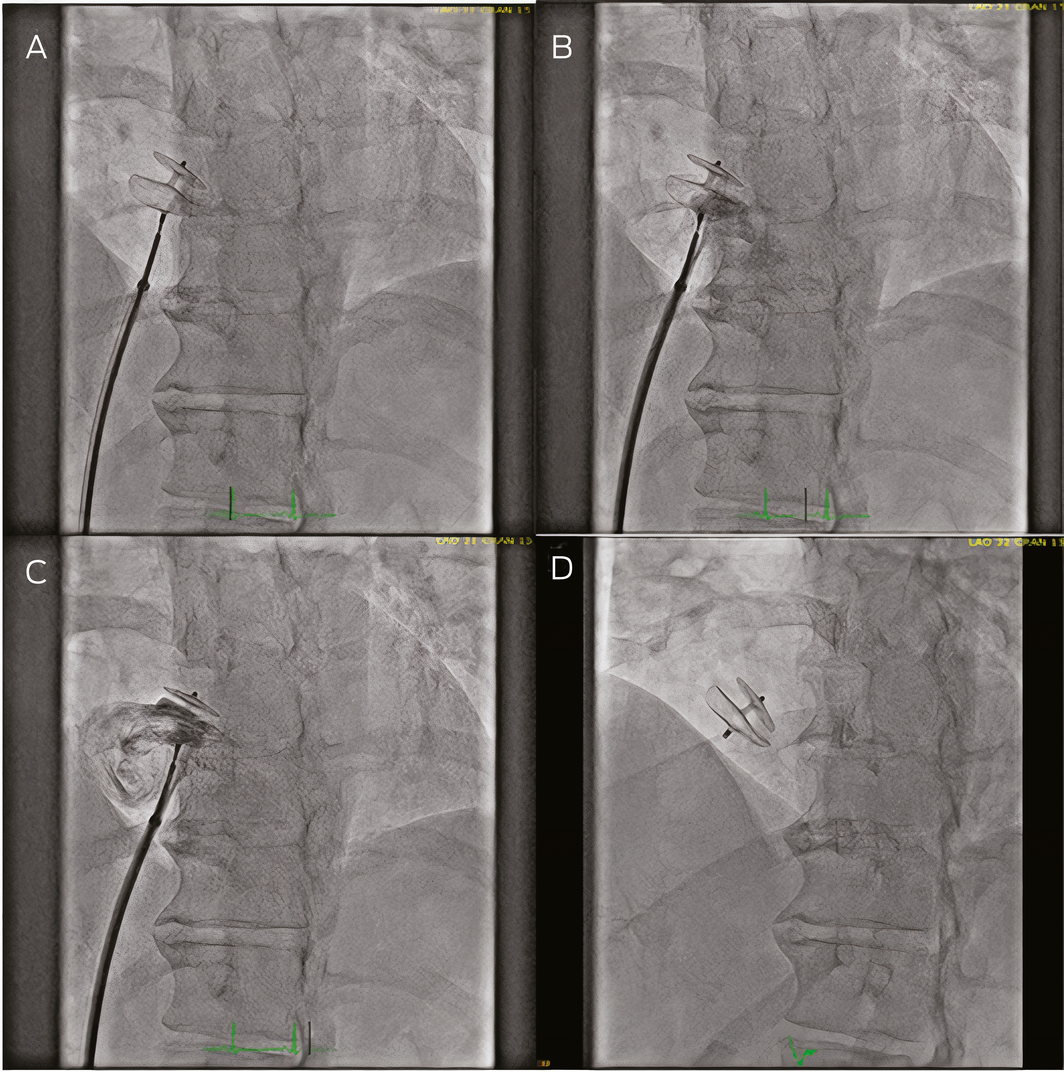
Percutaneous PFO closure commonly occurs as a day procedure in an experienced centre under conscious sedation with fluoroscopic guidance.46 In some cases, a general anaesthetic may be necessary especially if a transoesophageal echocardiogram is used where septal anatomy is challenging. A multipurpose catheter is inserted via the femoral vein with or without a 0.0035 inch guide wire support and advanced until the PFO is crossed. Once the appropriate closure device is selected, it is inserted within the delivery sheath through the access site, and the left atrial disc is expanded followed by retraction of the delivery system and then deployment of the right atrial disc (Box 3). In some cases intraprocedural echocardiogram is required to confirm the positioning of the closure device.47 If the position is satisfactory, the closure device is released from the delivery sheath. Post‐procedural care involves femoral vein compression, and often patients can resume normal activities within 24–48 hours.48 Dual antiplatelet therapy is recommended until the device is covered by endothelium. While there are no formal guidelines on duration, patients generally continue dual antiplatelet therapy for 3–6 months, and patients with PFO‐associated stroke should receive aspirin lifelong.1
Complications of percutaneous PFO closure
Several companies have created PFO closure devices, all with high rates of effective closure and low rates of complication. In Australia, three devices have Therapeutic Goods Administration approval: the GORE CARDIOFORM Septal Occluder (WL GORE and Associates), the Figulla Flex II PFO Occluder (Occlutech) and the Amplatzer PFO Occluder. One per cent of patients experience procedural complications, including arteriovenous fistula, branch occlusion of a retinal artery, atrial fibrillation, transient leg paraesthesia, postoperative minor bleeding at the access site, and periprocedural catheter thrombosis.23 A meta‐analysis of five randomised controlled trials exploring percutaneous closure for PFO‐associated stroke (n = 3630) found a significant increase in atrial arrhythmias (RR, 4.8; 95% CI, 2.2–10.7) but no significantly increased risk of mortality or bleeding.49
Conclusions
PFOs are common in the general population and, although rare, can lead to several pathologies. Recent studies have shown PFO closure to be an effective treatment for PFO‐associated stroke. While the evidence for routine closure for other indications such as migraine is less robust, smaller studies suggest patients can benefit from PFO closure in select situations. PFO closure is generally well tolerated with very high rates of closure success and low complications rates. Further research is required to identify subgroups who may further benefit from PFO closure.
Box 1 – Transoesophageal echocardiogram image of atrial septal aneurysm
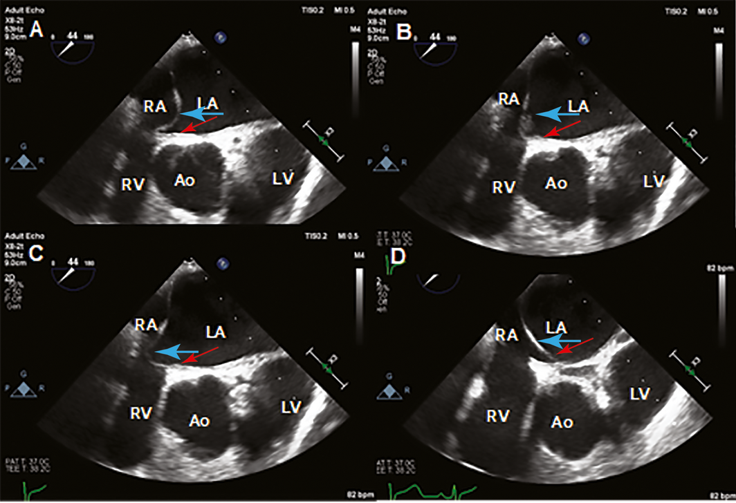
Ao = aorta; LA = left atrium; LV = left ventricle; RA = right atrium; RV = right ventricle. Images of transoesophageal echocardiogram illustrating the anatomical location of a PFO (red arrows) and an atrial septal aneurysm (blue arrows). An atrial septal aneurysm is defined as an interatrial septum area of at least 10 mm in width that moves away from the normal plane of the septum by at least 10 mm.
Box 2 – Transoesophageal echocardiogram showing thrombus in right atrium extending through a patent foramen ovale (PFO) into the left atrium
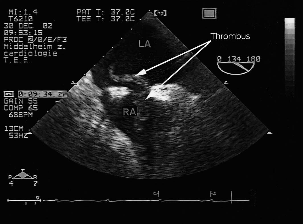
LA = left atrium; RA = right atrium. Thrombus (white arrow) in right atrium extending into left atrium through a PFO. Reprinted with permission from Delvigne et al.16
Competing interests
David Roy receives consultancy fees from Abbott Vascular, Medtronic, Boston Scientific and Edwards Lifesciences; he is a Proctor for Abbott Vascular, Medtronic and Edwards Lifesciences.
References
- Calvert PA, Rana BS, Kydd AC, Shapiro LM. Patent foramen ovale: anatomy, outcomes, and closure. Nat Rev Cardiol 2011; 8: 148.
- Roy D, Sharma R, Bunce N, et al. Selecting the optimal closure device in patients with atrial septal defects and patent foramen ovale. Interv Cardiol 2012; 4: 85–100.
- Collado FMS, Poulin MF, Murphy JJ, et al. Patent foramen ovale closure for stroke prevention and other disorders. J Am Heart Assoc 2018; 7: e007146.
- Elgendy AY, Saver JL, Amin Z, et al. Proposal for updated nomenclature and classification of potential causative mechanism in patent foramen ovale‐associated stroke. JAMA Neurol 2020; 77: 878–886.
- Mojadidi MK, Zaman MO, Elgendy IY, et al. Cryptogenic stroke and patent foramen ovale. J Am Coll Cardiol 2018; 71: 1035–1043.
- Poulin MF, Kavinsky CJ. The approval of PFO closure in the United States. Pract Neurol 2018; 1: 13–38.
- Saver JL, Carroll JD, Thaler DE, et al. Long‐term outcomes of patent foramen ovale closure or medical therapy after stroke. N Engl J Med 2017; 377: 1022–1032.
- DeSimone CV, Friedman PA, Noheria A, et al. Stroke or transient ischemic attack in patients with transvenous pacemaker or defibrillator and echocardiographically detected patent foramen ovale. Circulation 2013; 128: 1433–1441.
- Poddar KL, Nagarajan V, Krishnaswamy A, et al. Risk of cerebrovascular events in patients with patent foramen ovale and intracardiac devices. JACC Cardiovasc Interv 2014; 7: 1221–1226.
- Agboola KM, Lee JM, Liu X, et al. Interaction of cardiac implantable electronic device and patent foramen ovale in ischemic stroke: a case‐only study. Pacing Clin Electrophysiol 2019; 42: 341–348.
- Turc G, Calvet D, Guérin P, et al. Closure, anticoagulation, or antiplatelet therapy for cryptogenic stroke with patent foramen ovale: systematic review of randomized trials, sequential meta‐analysis, and new insights from the CLOSE study. J Am Heart Assoc 2018; 7: e008356.
- Mittal S. Atrial septal aneurysm. J Heart Stroke 2018; 3: 1050.
- Turc G, Lee J‐Y, Brochet E, et al. Atrial septal aneurysm, shunt size, and recurrent stroke risk in patients with patent foramen ovale. J Am Coll Cardiol. 2020; 75: 2312–2320.
- Belkin RN, Kisslo J. Atrial septal aneurysm: recognition and clinical relevance. Am Heart J 1990; 120: 948–957.
- Rigatelli G, Aggio S, Cardaioli P, et al. Left atrial dysfunction in patients with patent foramen ovale and atrial septal aneurysm: an alternative concurrent mechanism for arterial embolism? JACC Cardiovasc Interv 2009; 2: 655–662.
- Delvigne M, Vermeersch P, Van Den Heuvel P. Thrombus‐in-transit causing paradoxical embolism in cerebral and coronary arterial circulation. Acta Cardiol 2004; 59: 669–672.
- Furlan AJ, Reisman M, Massaro J, et al. Closure or medical therapy for cryptogenic stroke with patent foramen ovale. N Engl J Med 2012; 366: 991–999.
- Meier B, Kalesan B, Mattle HP, et al. Percutaneous closure of patent foramen ovale in cryptogenic embolism. N Engl J Med 2013; 368: 1083–1091.
- Thaler DE, Wahl A. Critique of closure or medical therapy for cryptogenic stroke with patent foramen ovale: the hole truth? Stroke 2012; 43: 3147–3149.
- Agarwal S, Bajaj NS, Kumbhani DJ, et al. Meta‐analysis of transcatheter closure versus medical therapy for patent foramen ovale in prevention of recurrent neurological events after presumed paradoxical embolism. JACC Cardiovasc Interv 2012; 5: 777–789.
- Mas J‐L, Derumeaux G, Guillon B, et al. Patent foramen ovale closure or anticoagulation vs. antiplatelets after stroke. N Engl J Med 2017; 377: 1011–1021.
- Mojadidi MK, Bogush N, Caceres JD, et al. Diagnostic accuracy of transesophageal echocardiogram for the detection of patent foramen ovale: a meta‐analysis. Echocardiography 2014; 31: 752–758.
- Puricel S‐G, Arroyo DA, Goy J‐J, et al. A propensity score‐matched comparison between Cardia and Amplatzer PFO closure devices‐insights from the SOLUTION registry (Swiss percutaneOus patent foramen ovale cLosUre in recurrent clinical events prevenTION). EuroIntervention 2014; 11: 230–237.
- Marchese N, Pacilli MA, Inchingolo V, et al. Residual shunt after percutaneous closure of patent foramen ovale with AMPLATZER occluder devices‐influence of anatomic features: a transcranial Doppler and intracardiac echocardiography study. EuroIntervention 2013; 9: 382–388.
- Davison P, Clift PF, Steeds RP. The role of echocardiography in diagnosis, monitoring closure and post‐procedural assessment of patent foramen ovale. Eur J Echocardiogr 2010; 11: i27–i34.
- Shi YJ, Lv J, Han XT, Luo GG. Migraine and percutaneous patent foramen ovale closure: a systematic review and meta‐analysis. BMC Cardiovasc Disord 2017; 17: 203.
- Meier B, Lock JE. Contemporary management of patent foramen ovale. Circulation 2003; 107: 5–9.
- Dowson A, Mullen MJ, Peatfield R, et al. Migraine Intervention With STARFlex Technology (MIST) trial: a prospective, multicenter, double‐blind, sham‐controlled trial to evaluate the effectiveness of patent foramen ovale closure with STARFlex septal repair implant to resolve refractory migraine headache. Circulation 2008; 117: 1397–1404.
- Dyer C. Migraine doctor is suspended for serious breach of professional standards. BMJ 2015; 350: h982.
- Carroll JD. Migraine Intervention With STARFlex Technology trial: a controversial trial of migraine and patent foramen ovale closure. Circulation 2008; 117: 1358–1360.
- Mattle HP, Evers S, Hildick‐Smith D, et al. Percutaneous closure of patent foramen ovale in migraine with aura, a randomized controlled trial. Eur Heart J 2016; 37: 2029–2036.
- Tobis JM, Charles A, Silberstein SD, et al. Percutaneous closure of patent foramen ovale in patients with migraine: the PREMIUM trial. J Am Coll Cardiol 2017; 70: 2766–2774.
- Takagi H, Umemoto T. A meta‐analysis of case‐control studies of the association of migraine and patent foramen ovale. J Cardiol 2016; 67: 493–503.
- Tariq N, Tepper SJ, Kriegler JS. Patent foramen ovale and migraine: closing the debate — a review. Headache 2016; 56: 462–478.
- Agrawal A, Palkar A, Talwar A. The multiple dimensions of platypnea–orthodeoxia syndrome: a review. Respir Med 2017; 129: 31–38.
- Shah AH, Osten M, Leventhal A, et al. Percutaneous intervention to treat platypnea–orthodeoxia syndrome: the Toronto experience. JACC Cardiovasc Interv 2016; 9: 1928–1938.
- Honěk J, Šrámek M, Šefc L, et al. Effect of catheter‐based patent foramen ovale closure on the occurrence of arterial bubbles in scuba divers. JACC Cardiovasc Interv 2014; 7: 403–408.
- Billinger M, Zbinden R, Mordasini R, et al. Patent foramen ovale closure in recreational divers: effect on decompression illness and ischaemic brain lesions during long‐term follow‐up. Heart 2011; 97: 1932–1937.
- Windecker S, Stortecky S, Meier B. Paradoxical embolism. J Am Coll Cardiol 2014; 64: 403–415.
- Serra W, De Iaco G, Reverberi C, Gherli T. Pulmonary embolism and patent foramen ovale thrombosis: the key role of TEE. Cardiovasc Ultrasound 2007; 5: 26.
- IStephens R, Dunn S. Non‐ST-elevation myocardial infarction following foam ultrasound‐guided sclerotherapy. Phlebology 2014; 29: 488–490.
- Klein J, Juratli TA, Weise M, Schackert G. A systematic review of the semi‐sitting position in neurosurgical patients with patent foramen ovale: how frequent is paradoxical embolism? World Neurosurg 2018; 115: 196–200.
- Brugts J, Liesting C, Kofflard M, van den Bos E. Right-to‐left atrial shunting with normal intracardiac pressures following cardiac surgery: pathophysiology and management. J Card Surg 2012; 27: 335–337.
- Riddles T, Brown J, Judge D. A case of platypnoea orthodeoxia following laparoscopic cholecystectomy. BMJ Case Rep 2018; 2018: bcr‐2017221714.
- Mansencal N, Mitry E, Pillière R, et al. Prevalence of patent foramen ovale and usefulness of percutaneous closure device in carcinoid heart disease. Am J Cardiol 2008; 101: 1035–1038.
- Calvert PA, Klein AA. Anaesthesia for percutaneous closure of atrial septal defects. BJA Educ 2008; 8: 16–20.
- Meier B. Closure of patent foramen ovale: technique, pitfalls, complications, and follow up. Heart 2005; 91: 444–448.
- Meier B. Patent foramen ovale and closure technique with the amplatzer occluder. Scientifica (Cairo) 2014; 2014: 129196.
- Riaz H, Khan MS, Schenone AL, et al. Transcatheter closure of patent foramen ovale following cryptogenic stroke: an updated meta‐analysis of randomized controlled trials. Am Heart J 2018; 199: 44–50.
Provenance: Not commissioned; externally peer reviewed.
Fatty Liver Disease in Australia: A Narrative Review on the Epidemiology, Natural History, Prognostication and Management in People With Metabolic Dysfunction
Karl Vaz, Daniel Clayton-Chubb, William W. Kemp, Stuart K. Roberts, Ammar Majeed
Sex Differences in Management and Outcomes of People With ST-Elevation Myocardial Infarction, New South Wales, 2011–2020: A Retrospective Cohort Study
Adam C. Livori, Jedidiah I. Morton, Dianna J. Magliano, Jonathan E. Shaw
Rethinking Rheumatic Fever Diagnosis: Progress, Pitfalls and the Path Towards a Definitive Test
Anna P. Ralph, Rachel Webb, Tom Parks, Nicole J. Moreland, Asha C. Bowen, Benjamin Jones, Emma Ndagire, Andrea Z. Beaton
Sex Differences in Management and Outcomes of People With ST-Elevation Myocardial Infarction, New South Wales, 2011–2020: A Retrospective Cohort Study
Samia Kazi, Simone Marschner, Clara K. Chow
Distribution of Cardiovascular Disease Risk Based on the Updated 2023 Guideline-Recommended Australian Cardiovascular Disease Risk Algorithm and Comparison With the 2012 Algorithm: An Observational Study
Nina Lazarevic, Meghana Bhat, Grace Joshy, Danielle C. Butler, Mark Woodward, Anushka Patel, Rod T. Jackson, Garry Jennings, Rosemary Wyber, Ellie Paige, Emily Banks
They Brought the Hospital to Me
Hugh N. Burrill