




Volume 214 - Issue 9
Management of temporomandibular disorders in the primary care setting
Authors: Samuel V Thambar, Sachin Kulkarni, Joshua Tesar and Scott Armstrong
Med J Aust 2021; 214 (9): 412-414.e1. || doi: 10.5694/mja2.51044
Published online: 17 May 2021
Published online: 17 May 2021
Temporomandibular disorders are a major cause of orofacial pain resulting in significant
morbidity and reduction in an individual’s perceived quality of life
Temporomandibular disorders (TMDs) are a major cause of orofacial pain resulting in significant morbidity and reduction in an individual’s perceived quality of life. They are a group of disorders affecting the temporomandibular joint (TMJ), masticatory muscles and surrounding structures. Up to 70% of the general population have detectable signs of TMDs, of whom only 15% are significantly affected and only 5% seek treatment, with women being twice as likely to be affected.1 Most acute articular inflammation is transient, resulting in patients not seeking treatment, and those with chronic pain tend to have acute exacerbations managed with simple conservative measures.
The TMJ is a ginglymoarthrodial joint between the mandibular condyle and temporal bone of the skull, with the external auditory canal as its posterior border. It is the most frequently used joint in the body and comprises two joint spaces separated by an articular disc, allowing it to function as both a sliding and hinge joint. The joint has proximity to numerous other structures and is intrinsically related to the masticatory muscles, with the lateral pterygoid having anterior attachments to both the condylar head and the disc. Branches of the third division of the trigeminal nerve provide sensory innervation to the TMJ via the auriculotemporal and masseteric branches of the mandibular nerve.2
Presentation and clinical assessment
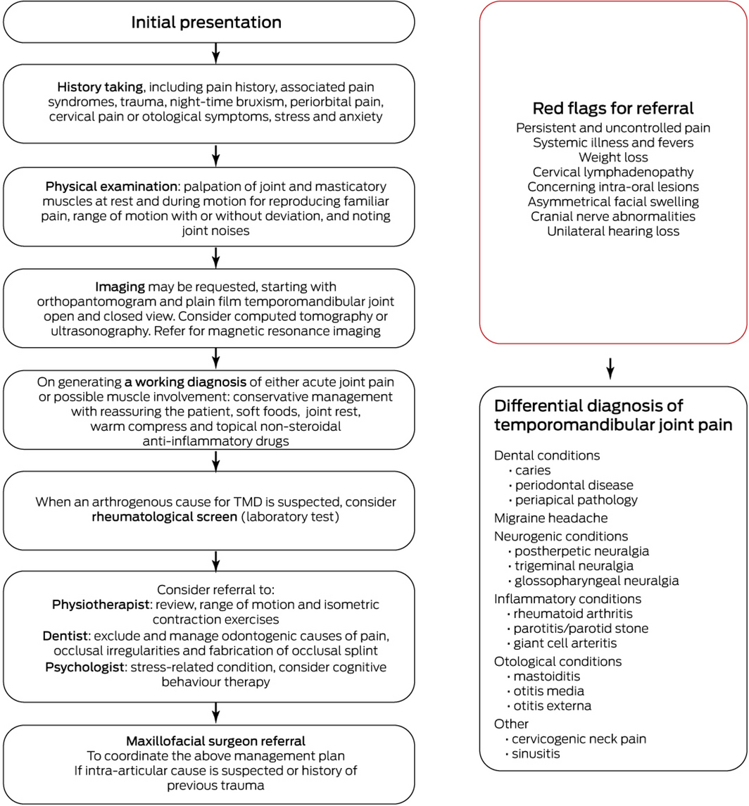
Given the multifactorial aetiological nature of TMDs, diagnosis is often complex and based on specific history and examination findings (Box). Patients typically describe pain in and around the TMJ on mouth opening and closing, with or without clicking or crepitus, especially during speaking or mastication, with trismus (restriction of mouth opening) and decreased lateral excursions of the jaw being common problems.3 Patients may also describe radiating periorbital pain, cervical neck pain and temporal headaches.3 Given its anatomical location and shared innervation with the external ear and tympanic membrane, patients may report otological symptoms such as otalgia, aural fullness, tinnitus and subjective hearing loss.4
Assessment should begin with a history of the pain, and any jaw movements that elucidate the pain. History should be taken for other pain conditions such as fibromyalgia, sleep apnoea and psychiatric illnesses such as anxiety or depression, as they are consistently associated with TMDs, as well as previous orthodontic treatment, trauma and posterior dentition loss.5 Specific questions should be asked for night‐time bruxism, which results in ongoing degenerative destruction of the TMJ and morning temporal and jaw ache.3
Physical examination should include palpation of the TMJ and masticatory muscles, both static and during movement, including intra‐oral palpation of the lateral pterygoid muscle where possible. Mouth opening should be assessed, with the mean being 51.3 mm in men and 44.3 mm in women, correlating roughly with a width of three fingers.6 Mouth opening less than 25 mm suggests dysfunction.6 Clicking, crepitus and locking of the jaw should also be assessed. Crepitus may occur in patients with osteoarthritis and is a sign of articular surface disruption.5 Clicking or locking of the jaw is often caused by disc displacement, most frequently anteriorly. Painless clicking of the joint during opening is often untreated and monitored for becoming symptomatic, upon which disc displacement should be evaluated with a magnetic resonance imaging (MRI) scan. A potential cause of locking could also be oromandibular dystonia, characterised by sustained or rhythmic involuntary contractions of the tongue, facial and masticatory musculature.7 A distinct pattern for oromandibular dystonia is that involuntary movements that present as bruxism cease on sleeping, whereas TMD‐related bruxism continues during sleep.7 Otoscopic examination should also be performed to exclude a primary otological cause, along with thorough examination of the oral cavity, palpation for cervical lymphadenopathy, and cranial nerve examination.
Imaging that may assist in diagnosis includes plain film orthopantamography and plain film open and closed views of the TMJ. These allow for an initial assessment of condylar integrity as well as potential odontogenic causes of pain. Following this, computed tomography may aid in assessment of condylar pathologies and other bony irregularities. MRI assesses the integrity of the joint, the articular disc, its surrounding structures, and the presence of a joint effusion. Given their high cost, MRI scans are usually reserved for patients with persistent TMD symptoms where an intracapsular pathology of the TMJ is suspected, and for pre‐operative planning purposes organised by specialist maxillofacial surgeons. When MRI is unavailable, ultrasonography is a non‐invasive investigation to confirm internal derangement of the joint. A rheumatological laboratory screen (including antinuclear antibody, C‐reactive protein, erythrocyte sedimentation rate, rheumatoid factor, and anti‐cyclic citrullinated peptide tests) should be ordered when an arthrogenous cause for TMDs is suspected.
Most common diagnoses tend to be acute arthralgia presenting as joint pain modified by mandibular movement, as well as masticatory myalgia. Masticatory myalgia is best managed by a multidisciplinary team with dentists and physiotherapists. Diagnoses that are more complex or chronic may warrant referral to a maxillofacial surgeon. A pathway for managing TMDs, along with a differential diagnosis of temporomandibular pain and a list of red flag symptoms which may require urgent investigation, is presented in the Box.
Conservative management options
There are various treatment options for TMDs, ranging from conservative interventions for myofascial TMDs to surgical management options reserved for patients with arthrogenous TMDs. In 40% of patients, the condition is self‐limiting without treatment. Alternatively, conservative treatment measures, including jaw rest, soft diet, warm compress over the region, non‐steroidal anti‐inflammatory drugs (NSAIDs) and occlusal splints, provide adequate pain relief in 50–90% of patients when given concurrently.8 NSAIDs are indicated for joint pain secondary to inflammation and, if not contraindicated, are the main pharmacological agent used for TMDs.9 Topical formulations have been shown to be as beneficial as systemic medications.9 Soft diet for joint rest and avoidance of triggers are beneficial, along with warm compress over the TMJ region. Cognitive behaviour therapy is commonly used, given the high association of psychological factors affecting patients with TMDs. TMJ physiotherapy has been proven to be effective, through range of movement and isometric contraction exercises.10 There is also increasing evidence for the role of acupuncture as an adjunctive treatment for short term analgesia.11
Occlusal splints
Occlusal splint therapy is a conservative management option for TMDs that reduces joint loading, relaxes masticatory muscles and prevents the ill effects of bruxism.12 An occlusal splint is a removable device fabricated by a general dentist, fitting over the occlusal surfaces of the teeth. Occlusal splints are effective in managing TMD especially when the pathophysiology is related to sleep‐related bruxism and masticatory muscle pain.12 They are worn throughout the night by the patient for a duration of at least 3 months, followed by review to assess patient response. There are two major types: muscle relaxation splints and anterior repositioning splints. Referral to a general dentist should be made for determination, manufacturing and reviewing of the best occlusal splint for the patient. A dental referral is also important to exclude and manage odontogenic causes of orofacial pain, and for management of occlusal irregularities.
Botulinum toxin A
Recently, botulinum toxin has been increasingly used in the management of bruxism and myogenic orofacial pain. Botulinum toxin A, an exotoxin produced by Clostridium botulinum, decreases muscle contraction by inhibiting acetylcholine release at the neuromuscular junction, lasting between 3 and 6 months.13 Botulinum toxin A also has a direct effect on peripheral nociceptors. A systematic review concluded that despite showing benefits, clear consensus was lacking on the therapeutic benefit of botulinum toxin A in the management of myofascial TMDs.13 Nevertheless, the review found that botulinum toxin A can improve outcomes in patients with myofascial TMDs who have had at least 3 months of appropriate conservative management, and for these patients, referral can be made to a maxillofacial surgeon for consideration and administration of this therapy.13 If a patient is unresponsive to botulinum toxin A, other management options should be considered.
Surgical management options and referral
There are various surgical management options performed by maxillofacial surgeons for patients with arthrogenous TMD, with pain localised to the TMJ and/or internal derangement of the joint. Arthrocentesis is utilised to wash out the superior joint space and may be supplemented with injection of various medicaments including anti‐inflammatory agents, hyaluronic acid and analgesics. It may also be combined with arthroscopy as a diagnostic aid in select patients for internal derangement of the joint. Eminoplasty is used for pain management, joint restriction and crepitus, and involves altering the articular eminence without significantly altering the bony architecture to allow the condyle to move freely back into its normal position.10 Other surgical options include condylar shave and condylar neck osteotomy, discectomy and discopexy, with total joint replacement being the final stage, reserved for those with significant pathology or end‐stage joint disease.14 Surgical options, however, involve substantially increased risk and recovery periods for patients. A careful and considered approach should therefore be employed, with a preference for conservative strategies where possible, as the first step in management.14 Any patient with prior TMJ trauma, joint internal derangement, pain not responsive to at least 3 months of conservative management options, or suspicion of any other intra‐articular cause should be referred to a maxillofacial surgeon.15 Additionally, early referral to maxillofacial surgeons and early arthrocentesis results in better outcomes for patients with internal derangement of the TMJ.
Conclusion
TMDs encompass a broad range of disorders. Clarification regarding assessment, diagnosis and management among primary care providers is instrumental in improving the patient pathway, and alleviating pain and frustration at delay to diagnosis. We have summarised the latest evidence to improve understanding of the condition’s multifactorial aetiological nature and guide initial management techniques. These techniques rely on a multidisciplinary approach, and include dental review, occlusal splint fabrication, soft diet, NSAIDs, physiotherapy, psychological assessment, plain film imaging, and referral to a maxillofacial surgeon for further investigation where required. These management techniques are in line with those for other chronic musculoskeletal conditions, where patient education and motivation play a key role, along with a patient‐centred approach to reduce their experience of pain and improve their quality of life.
Competing interests
No relevant disclosures.
References
- Carlsson GE. Epidemiology and treatment need for temporomandibular disorders. J Orofac Pain 1999; 13: 232–237.
- Rodriguez-Lopez MJ, Fernandez-Baena M, Aldaya-Valverde C. Management of pain secondary to temporomandibular joint syndrome with peripheral nerve stimulation. Pain Physician 2015; 18: E229–E236.
- Guarda-Nardini L, Manfredini D, Salamone M, at al. Efficacy of botulinum toxin in treating myofascial pain in bruxers: a controlled placebo pilot study. Cranio 2008; 26: 126–135.
- Ramirez LM, Ballesteros LE, Sandoval GP. Topical review: temporomandibular disorders in an integral otic symptom model. Int J Audiol 2008; 47: 215–227.
- Scrivani SJ, Keith DA, Kaban LB. Temporomandibular disorders. N Engl J Med 2008; 359: 2693–2705.
- Nagi R, Sahu S, Gahwai D, Supreet J. Study on evaluation of normal range of maximum mouth opening among Indian adults using three finger index: a descriptive study. J Indian Acad Oral Med Radiol 2017; 29: 186–190.
- Britton D, Alty JE, Mannion CJ. Oromandibular dystonia: a diagnosis not to miss. Br J Oral Maxillofac Surg 2020; 58: 520–524.
- Fonseca RJ, Marciani RD, Turvey TA. Oral and maxillofacial surgery. 2nd ed. St Louis: Elsevier, 2009: 881–897.
- Kulkarni S, Thambar S, Arora H. Evaluating the effectiveness of nonsteroidal anti-inflammatory drug(s) for relief of pain associated with temporomandibular joint disorders: a systematic review. Clin Exp Dent Res 2020; 6: 134–146.
- Rajapakse S, Ahmed N, Sidebottom AJ. Current thinking about the management of dysfunction of the temporomandibular joint: a review. Br J Oral Maxillofac Surg 2017; 55: 351–356.
- Cho SH, Whang WW. Acupuncture for temporomandibular disorders: a systematic review. J Orofac Pain 2010; 24: 152–162.
- Okeson J. Management of temporomandibular disorders and occlusion. 7th ed. St Louis: Elsevier, 2012.
- Thambar S, Kulkarni S, Armstrong S, Nikolarakos D. Botulinum toxin in the management of temporomandibular disorders: a systematic review. Br J Oral Maxillofac Surg 2020; 58: 508–519.
- Dimitroulis G. The role of surgery in the management of disorders of the temporomandibular joint: a critical review of the literature. Part 1. Int J Oral Maxillofac Surg 2005; 34: 107–113.
- Gauer RL, Semidey MJ. Diagnosis and treatment of temporomandibular disorders. Am Fam Physician 2015; 91: 378–386.
Provenance: Not commissioned; externally peer reviewed.
Non-Technical Errors Associated With Deaths in Surgical Care, Australia, 2012–2019, by Surgical Specialty (Australian and New Zealand Audit of Surgical Mortality)
Jesse Ey, Guy J. Maddern
Localised Herpes Simplex Following Midline Laparotomy
Jessica S. Bulluss, Paul Chee, Matthew J. Verheyden
Non‐technical errors associated with deaths in surgical care, Australia, 2012–2019, by surgical specialty (Australian and New Zealand Audit of Surgical Mortality): a retrospective cohort study
Jesse Ey, Victoria Kollias, Octavia Lee, Kelly Hou, Matheesha Herath, John B North, Ellie Treloar, Suzanne Edwards, Martin Bruening, Adam J Wells, Guy J Maddern
Changes in patient management after preoperative MRI for newly diagnosed breast cancer: a multicentre prospective observational study
Michael L Marinovich, Nehmat Houssami, Andrew Spillane, Gregory B Mann, Donna Taylor, Michelle Reintals, Nadine Phillips, Max K Bulsara, Patsy Siok Hwa Soon, Tracey Dickens, Christobel M Saunders
The first Australian evidence‐based guidelines on male infertility
Darren J Katz, Liza O’Donnell, Robert I McLachlan, Tim J Moss, Clare V Boothroyd, Veena Jayadev, Sarah R Catford
The first live term birth following uterus transplantation in Australia
Rebecca Deans, Brigitte Gerstl, Antonia W Shand, Sarah Lyons, Aaron Budden, Helen L Barrett, Grant Luxton, Mangalee Fernando, Kenneth Yong, Karen Keung, Kaushalya Arulpragasam, Henry Pleass, King Man Wan, Eva Kehag, Jana‐Emily Pittman, Mianna Lotz, Maria Fenn, Erin Nesbitt‐Hawes, Lily Byun, Katrina Tang, Mats Brannstrom, Jason Abbott