




Medical education
Lessons from practice
Volume 214 - Issue 11
Ureaplasma urealyticum septic oligoarthritis in a patient with severe secondary hypogammaglobulinaemia
Authors: Peter Bradhurst, Carlos El‐Haddad, John Ng and Nicolás Urriola
Med J Aust 2021; 214 (11): 506-507.e1. || doi: 10.5694/mja2.51107
Published online: 21 June 2021
Published online: 21 June 2021
A 64-year-old woman presented to the emergency department with 7 days of right ankle pain
Clinical record
A 64‐year‐old woman presented to the emergency department with 7 days of right ankle pain. In the preceding 48 hours, she had developed worsening erythema over the right ankle and was unable to bear weight, associated with non‐specific constitutional symptoms including poor appetite and lethargy. She did not have a history of fevers, rigors, night sweats, or antecedent trauma.
Previous history was significant for follicular stage 3A non‐Hodgkin lymphoma treated 5 years before presentation, for which she received six cycles of rituximab, cyclophosphamide, doxorubicin, vincristine and prednisolone and remained in clinical remission. The patient was also in remission from transitional cell carcinoma of the bladder after undergoing cystectomy and ileal conduit formation 2 years before this presentation.
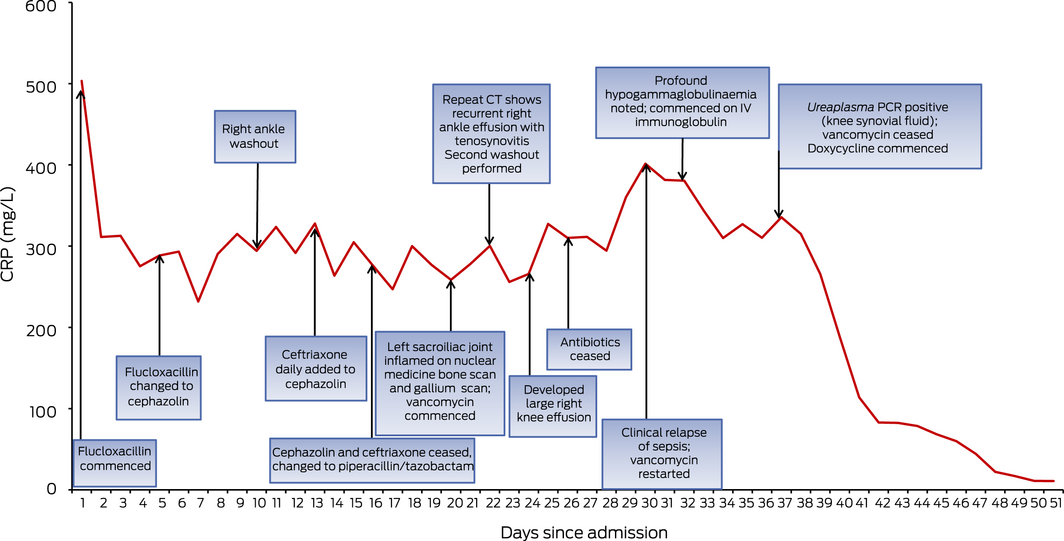
C‐reactive protein (CRP) on admission was 503.4 mg/L (reference interval [RI], ≤ 5 mg/L), with an erythrocyte sedimentation rate of 125 mm/h (RI, < 20 mm/h). Despite initiation of intravenous flucloxacillin (2 g every 6 hours), symptoms progressed. On day 10 of admission, an ultrasound of the right ankle showed a small joint effusion. Aspiration revealed turbid synovial fluid with a cell count of 138 600 × 106 cells/mL, comprised of 100% neutrophils. Polarised light microscopy, gram stain and culture were negative for crystals and organisms respectively, as were bacterial and fungal cultures from synovial fluid and biopsy obtained from joint washout. Despite intravenous flucloxacillin and washout, the patient developed fevers over 38°C. Antimicrobial cover was expanded with the addition of intravenous piperacillin/tazobactam (4 g/0.5 g every 8 hours) without clinical improvement.
After 20 days with no improvement, nuclear medicine bone scan and gallium scan revealed significantly increased uptake in the left sacroiliac joint on both studies, with left‐sided sacroiliitis confirmed on magnetic resonance imaging. Percutaneous sampling of this area revealed normal histology with negative cultures and polymerase chain reaction (PCR) test for tuberculosis. Whole body computed tomography (CT) scan and transoesophageal echocardiogram were performed but did not demonstrate any deep foci of infection. Vancomycin was added to cover for multiresistant Staphylococcus aureus but no response was observed. By day 21, the patient developed recurrent right ankle effusion, with CT imaging revealing thick rim enhancing effusions of the tibialis posterior and peroneus longus tendons (Box 1). A second washout of the right ankle was required and full cultures were negative. On day 24, a large right knee effusion developed, with negative bacterial and fungal cultures of the aspirated samples.
On day 33 of admission, peripheral assessment of the patient’s immune system was performed to further characterise the patient’s risk of infection with atypical organisms. This demonstrated an absence of B cells and an IgG level of 1.56 g/L (RI, 6.39–15.60 g/L). A synovial fluid Ureaplasma urealyticum nucleic acid detection (NAD) test was ordered on all synovial fluid samples to date, because of known susceptibility to U. urealyticum septic arthritis in patients with hypogammaglobulinaemia, and returned positive. A congruent 16S ribosomal RNA (rRNA) subunit panbacterial PCR ordered concurrently also demonstrated evidence of U. urealyticum infection. The patient was commenced on oral doxycycline 100 mg twice a day, along with intravenous immunoglobulin (0.4g/kg of body weight, monthly) with a rapid clinical improvement (Box 2).
Discussion
Septic arthritis caused by Mycoplasmataceae, including the genera Mycoplasma and Ureaplasma, in the setting of primary immunodeficiency with hypogammaglobulinaemia was first reported in 1978.1 U. urealyticum, U. parvum, M. hominis, M. salivarium and M. pneumoniae have been previously implicated. Mycoplasmataceae are defined by the absence of a cell wall, conferring a natural immunity against β‐lactam antibiotics and making them undetectable on gram stain.2 The genus Ureaplasma is further differentiated by an ability to hydrolyse urea enabling inhabitation of the urogenital tract.2 Diagnosis of extra‐urogenital Ureaplasma infection relies on a high index of clinical suspicion. Mycoplasmataceae are fastidious organisms, requiring specialised media and incubation conditions for culture.2 NAD is theoretically more sensitive because it may detect DNA of a single cell, exceeding the threshold of culture that requires 100–1000 organisms, making it the diagnostic modality of choice in this case.2
There have been increasing reports of Mycoplasmataceae septic arthritis due to iatrogenic immunodeficiency due to the emergence of B cell‐depleting agents such as rituximab.3 Hypogammaglobulinaemia is the major risk factor for disseminated Mycoplasmataceae infection.2 Profound hypogammaglobulinaemia is a well documented complication of B cell depletion therapy, with 4.2% of patients requiring intravenous immunoglobulin post‐treatment in a large case series.4
The ribosomal 16S rRNA panbacterial PCR is an adjunct microbiological investigation that targets the highly conserved region of the bacterial 16S rRNA subunit present in all bacterial ribosomal genes.5 The broad nature of this investigation allows for the non‐specific detection of bacterial components, potentially eliminating immune‐mediated causes of arthritis from consideration.5 A 16S rRNA panbacterial PCR can detect non‐viable organisms that may allow for organism identification in cases where broad spectrum antibiotics have already been administered.5 In similar diagnostic dilemmas where an infectious aetiology is considered, early use of 16S rRNA panbacterial PCR may facilitate earlier diagnosis with reduced morbidity and time of hospital admission.
Lessons from practice
- Mycoplasmataceae are a rare but well recognised cause of septic polyarthritis and should be considered early when in association with hypogammaglobulinaemia.
- Rituximab is a commonly used monoclonal antibody specific for CD20. It is known to be an iatrogenic cause of secondary hypogammaglobulinaemia, and patients who have received rituximab are at increased risk for infections for years after treatment.
- An assessment of immune function with immunoglobulin levels and lymphocyte counts is essential in patients previously treated with rituximab with unusual or recurrent infection.
- Ribosomal 16S panbacterial polymerase chain reaction (PCR) is a molecular assay that may reveal evidence of bacterial infection in cases where the aetiology of inflammatory arthritis remains uncertain.
Box 1 – Computed tomography imaging of the ankle showing thick rim enhancing effusions of the tibialis posterior and peroneus longus tendons*
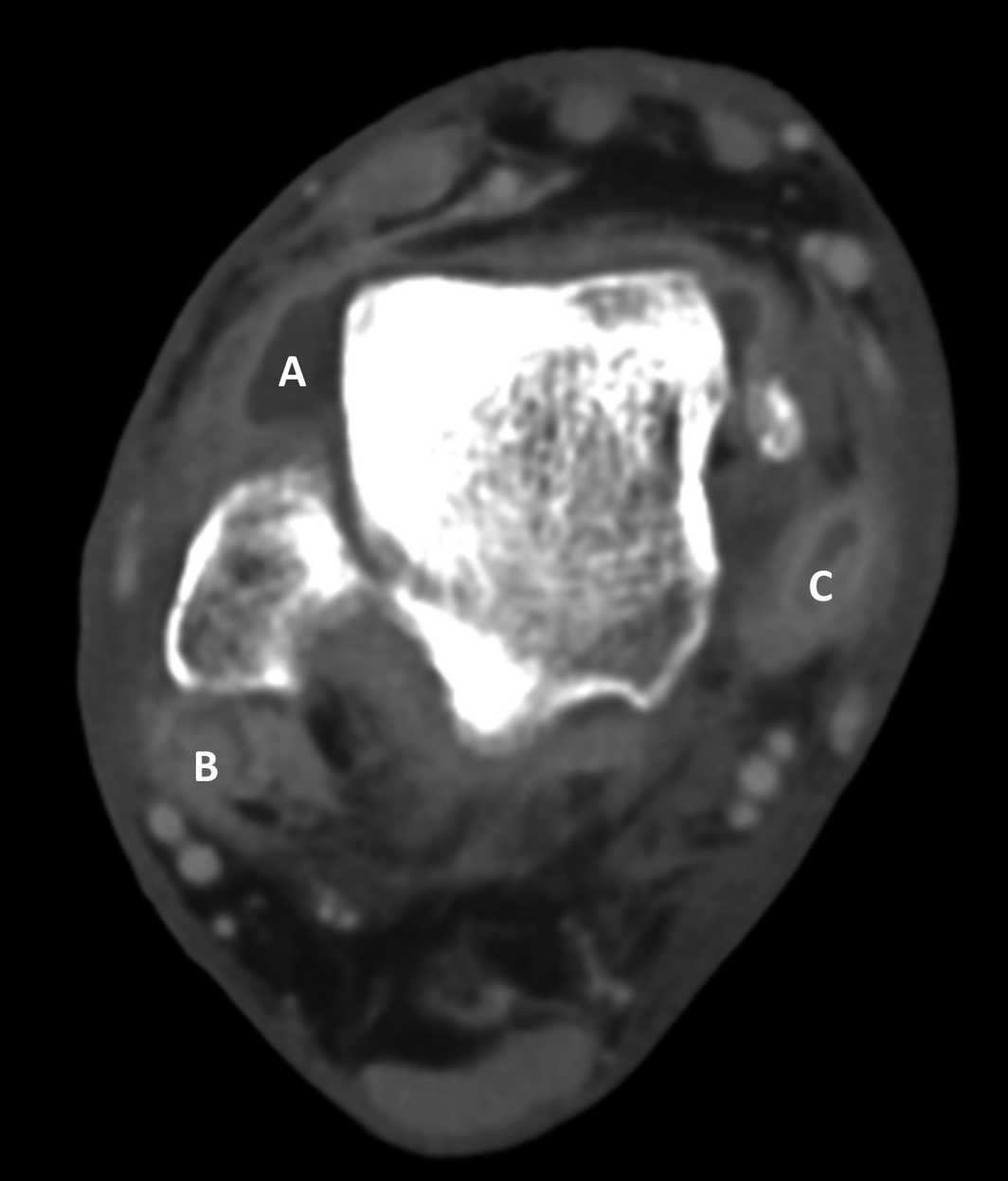
* A: An irregular, thick rimmed, loculated effusion of the ankle joint with synovial contrast enhancement. B: Loculated effusion within the peroneus longus tendon sheath causing marked tendon thickening. C: Rim‐enhancing loculated effusion within the posterior tibialis tendon with marked tendon thickening.
Competing interests
No relevant disclosures.
References
- Webster AD, Taylor‐Robinson D, Furr PM, Asherson GL. Mycoplasmal (ureaplasma) septic arthritis in hypogammaglobulinaemia. Br Med J 1978; 1: 478–479.
- Waites KB, Xiao L, Paralanov V, et al. Mycoplasma and ureaplasma. In: de Filippis I, McKee ML. Molecular typing in bacterial infections. Totowa: Humana Press, 2013: 229–281.
- George MD, Cardenas AM, Birnbaum BK, Gluckman SJ. Ureaplasma septic arthritis in an immunosuppressed patient with juvenile idiopathic arthritis. J Clin Rheumatol 2015; 21: 221–224.
- Roberts DM, Jones RB, Smith RM, et al. Rituximab‐associated hypogammaglobulinemia: incidence, predictors and outcomes in patients with multi‐system autoimmune disease. J Autoimmun 2015; 57: 60–65.
- Sontakke S, Cadenas MB, Maggi RG, et al. Use of broad range16S rDNA PCR in clinical microbiology. J Microbiol Methods 2009; 76: 217–225.
Provenance: Not commissioned; externally peer reviewed.
Rhabdomyolysis After COVID-19
Navya Kataria, Abel Devadass, Lana Sundac, Laura De Souza, Nabeel Sheikh, Samantha Ng, Scott Campbell, Yishen Wang, Ross S. Francis
Technological advances in the search for a CURE for asthma
Sara Quon, Timothy M Johanson, Ridhima Wadhwa, Alen Faiz, Anthony Flynn, Gary P Anderson, Amanda J Cox, Nicholas P West, Michael P Menden, Rhys S Allan
The CURE Asthma roadmap
Gary P Anderson, Anthony Flynn, Phil G Bardin, John D Blakey, Shyamali C Dharmage, Paul Foster, Peter G Gibson, Adam Jaffe, Alan James, Christine R Jenkins, Sundram Sivamalai, Peter D Sly, Guy B Marks, Vanessa M McDonald, Judy Wetttenhall
The first live term birth following uterus transplantation in Australia
Rebecca Deans, Brigitte Gerstl, Antonia W Shand, Sarah Lyons, Aaron Budden, Helen L Barrett, Grant Luxton, Mangalee Fernando, Kenneth Yong, Karen Keung, Kaushalya Arulpragasam, Henry Pleass, King Man Wan, Eva Kehag, Jana‐Emily Pittman, Mianna Lotz, Maria Fenn, Erin Nesbitt‐Hawes, Lily Byun, Katrina Tang, Mats Brannstrom, Jason Abbott
The public health impacts of mining in Australia
Javier Cortes‐Ramirez, Ruby N Michael, Leisa‐Maree Toms, Melissa Haswell
Lingual Raynaud phenomenon
Michael Taggart, Mina John