




Medical education
Lessons from practice
Volume 213 - Issue 2
A case of toxigenic, pharyngeal diphtheria in Australia
Authors: Sarah Grigg, David Hogan, F Shaun Hosein, Dean Johns, Amy Jennison and Shradha Subedi
Med J Aust 2020; 213 (2): 64-65.e1. || doi: 10.5694/mja2.50566
Published online: 4 May 2020
Published online: 4 May 2020
Clinical record
A 42‐year‐old woman presented to the Sunshine Coast University Hospital, Queensland, with a 5‐day history of odynophagia, orthopnoea and rapid onset of neck swelling over 12 hours. She had returned one week prior from a year‐long trip to Central America, Sri Lanka and Indonesia. Relevant past medical history included nephrotic syndrome due to minimal change disease, use of prednisolone 2.5 mg daily and previous treatment with rituximab. Childhood vaccinations were reported, but she had no booster travel vaccinations.
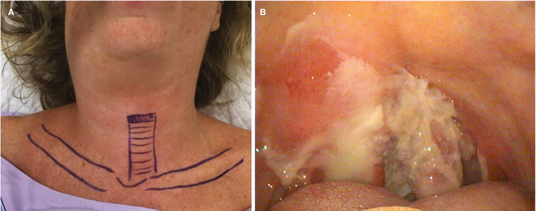
On examination, she had right‐sided neck swelling, consistent with “bull neck” (Box, A), and an exudative right tonsil with a haemorrhagic component (Box, B). The patient had several healed skin lesions and a 2 cm non‐healing ulcer on her buttock.
Flexible nasendoscopy showed supraglottic oedema with a patent airway. The diagnosis of diphtheria was strongly considered, with differentials including peritonsillar abscess and tonsillitis. Computed tomography scan of the neck demonstrated peritonsillar phlegmon and oedema in the parapharyngeal space, pre‐vertebral fat and subcutaneous neck tissues. She was commenced on intravenous benzylpenicillin, lincomycin and dexamethasone, was placed on contact and droplet precautions, and was admitted to the intensive care unit. Multiple tissue and swab samples were taken from the pharyngeal membrane and the buttock wound and urgently sent to the laboratory for culture into selective media.
She underwent elective intubation 24 hours later due to worsening laryngeal oedema. Tissue and swabs from the pharyngeal membrane and sacral wound grew Corynebacterium diphtheriae. Diphtheria antitoxin (DAT) 100 000 IU was administered 36 hours into her admission. The isolates were confirmed to be toxigenic by polymerase chain reaction.1,2 The patient was discharged from the intensive care unit on Day 6. On Day 7, she developed anterior T wave inversions on her electrocardiogram, with an elevated troponin value (0.39 μg/L; reference range, < 0.040 μg/L). Her cardiac enzymes showed serial improvement. She developed a moderate glossopharyngeal and vagal palsy, which resolved after 3 weeks, and peripheral neuropathy, which resolved after 4 months. The cardiac and neurological sequelae were thought to be complications of pharyngeal diphtheria.
The local Public Health Unit and the infection management service identified 12 staff and seven close community contacts. All contacts had nasal and throat swabs taken, were treated with oral erythromycin and were vaccinated where appropriate.3 Staff were excluded from work until returning negative throat and nasal cultures at 48–72 hours.
Discussion
Diphtheria is an acute pharyngeal or cutaneous infection caused by toxigenic strains of C. diphtheriae — a gram‐positive, non‐motile, non‐encapsulated bacillus.4 The infection spreads by respiratory droplets or direct contact with nasopharyngeal secretions or skin lesions. The incubation period of diphtheria is commonly 2–5 days. Data from the World Health Organization show that diphtheria is endemic to South‐East Asia, including Indonesia, Malaysia and the Philippines.5 Our case illustrates the need for a thorough travel history and administration of timely antitoxin therapy in suspected diphtheria cases to limit diphtheria‐related neurological and cardiovascular consequences.
Diphtheria is rare in Australia after the widespread use of the effective vaccine following World War II, with most cases associated with sporadic importations. There have been seven cases of diphtheria reported since 2001, including one that was fatal in 2011.6 Diphtheria affects the upper respiratory tract, presenting with sore throat and cervical lymphadenopathy; a coating membrane forms in about a third of cases. Simultaneous infection of the skin and respiratory tract is uncommon. Accumulation of the C. diphtheriae organism within the membrane along with fibrin debris result in the appearance of a white pseudomembrane.4 The pathognomonic bull neck is caused by superficial oedema of neck tissues and is associated with a more severe course and higher mortality. The diphtheria toxin is produced by toxigenic strains of the bacterium and affects the cardiovascular, renal and nervous systems via haematogenous spread. The toxin is bound on cell surface receptors and acts to arrest protein synthesis.7 Toxin‐producing infections have a mortality rate between 5% and 10%.8
Diphtheritic myocarditis occurs in 10–20% of patients with pharyngeal diphtheria manifesting as ST disturbance, corrected QT interval (QTc) prolongation, or heart block.9 Cardiac abnormalities are associated with extensive respiratory tract involvement and bull neck appearance as well as neurological sequelae, which occur in 75% of patients with severe respiratory disease.4 Cranial nerve neuropathy develops first; often presenting as swallowing difficulties and resulting in aspiration.
DAT and antibiotics should be administered promptly upon clinical suspicion. Early administration of DAT reduces circulating toxin load and reduces clinical sequelae.3 Our patient received DAT at 36 hours, yet significant neurological sequelae were observed up to 4 months later. Penicillin and/or erythromycin are the antimicrobials of choice; however, resistance has been described.10
Lessons from practice
- Diphtheria should be suspected in patients presenting with pseudomembranous tonsillitis, significant neck swelling and relevant travel history.
- It is important for clinicians to liaise with their local laboratory and Public Health Unit in suspicious cases so appropriate investigations and follow‐up can be established.
- Timely administration of diphtheria antitoxin is imperative and should not be delayed awaiting laboratory confirmation.
- Booster vaccinations should be considered before travel, particularly in patients who may have waning immunity.
Competing interests
No relevant disclosures.
References
- Nakao H, Pruckler JM, Mazurova IK, et al. Heterogeneity of diphtheria toxin gene, tox, and its regulatory element, dtxR, in Corynebacterium diphtheriae strains causing epidemic diphtheria in Russia and Ukraine. J Clin Microbiol 1996; 34: 1711–1716.
- Sing A, Berger A, Schneider‐Brachert W, et al. Rapid detection and molecular differentiation of toxigenic Corynebacterium diphtheriae and Corynebacterium ulcerans strains by LightCycler PCR. J Clin Microbiol 2011; 49: 2485.
- Queensland Government, Queensland Health. Diphtheria [website]. Queensland Health, 2012. https://www.health.qld.gov.au/cdcg/index/diphtheria (viewed Mar 2020).
- Hadfield TL, McEvoy P, Polotsky Y, et al. The pathology of diphtheria. J Infect Dis 2000; 181 (Suppl): S116–S120.
- World Health Organization. Diphtheria [website]. WHO, 2017. http://www.who.int/immunization/monitoring_surveillance/burden/diphtheria/en/ (viewed Mar 2020).
- NNDSS Annual Report Working Group. Australia's notifiable disease status, 2014: annual report of the National Notifiable Diseases Surveillance System. Commun Dis Intell Q Rep 2016; 40: E48–E145.
- Lubran MM. Bacterial toxins. Ann Clin Lab Sci 1988; 18: 58–71.
- Adler NR, Mahony A, Friedman ND. Diphtheria: forgotten, but not gone. Intern Med J 2013; 43: 206–210.
- Lumio JT, Groundstroem KW, Melnick OB, et al. Electrocardiographic abnormalities in patients with diphtheria: a prospective study. Am J Med 2004; 116: 78–83.
- Tiwari TSP, Wharton M. Diphtheria toxoid. In: Plotkin SA, Orenstein WA, Offit PA, Edwards KM, editors. Plotkin's vaccines, 7th ed. Elsevier, 2018: 261–275.
Provenance: Not commissioned; externally peer reviewed.
Designing Housing to Reduce Overcrowding-Related Harms: Rheumatic Heart Disease as the Canary in the Coal Mine
Simon Quilty, Veronica Matthews, Angus Baumann, James Marangou, Bo Remenyi, Gavin Wheaton, Serena Morton Nabanunga, Norman Frank Jupurrurla, Simon Robinson, Steve Mintern, Cary Duffield, Joshua R. Francis, Paul C. Memmott
Australian Climate Leadership in 2026: COP-Out or Step-Up for Health?
Catherine G. A. Pendrey, Angie Bone, Aletha Ward, Francis Nona, Michelle Isles, Paul M. Kelly, Nicholas J. Talley
The impact of the Breast Screen NSW transition from film to digital mammography, 2002–2016: a linked population health data analysis
Rachel Farber, Nehmat Houssami, Katy J L Bell
Shifting focus to adolescent wellbeing and inclusive participation in the digital age
Allyson R Todd, Elena Wang, Stephanie R Partridge
The CURE Asthma roadmap
Gary P Anderson, Anthony Flynn, Phil G Bardin, John D Blakey, Shyamali C Dharmage, Paul Foster, Peter G Gibson, Adam Jaffe, Alan James, Christine R Jenkins, Sundram Sivamalai, Peter D Sly, Guy B Marks, Vanessa M McDonald, Judy Wetttenhall
Protecting public interest journalism as a public health good
Bronwyn Fredericks, Neha Lalchandani, Melissa A Sweet, Alex Cramb, Carmel Williams