




Medical education
Lessons from practice
Volume 212 - Issue 9
May–Thurner syndrome: an overlooked cause of venous thromboembolism
Authors: Farooq Akram and Roshni G Sadashiv
Med J Aust 2020; 212 (9): 402-403.e1. || doi: 10.5694/mja2.50548
Published online: 18 May 2020
Published online: 18 May 2020
Correction(s) for this article:
Erratum | Published online: 7 September 2020
A 48-year-old Chinese woman with obesity and no significant past medical history presented to the emergency department with acute onset pain and swelling in the entire left leg
Clinical record
A 48‐year‐old Chinese woman with obesity and with no significant past medical history presented to the emergency department with acute onset pain and swelling in the entire left leg. She had been on a long‐haul flight from New York to Singapore 3 weeks before presentation. There was no family history of unprovoked venous thromboembolism (VTE) and no symptoms suggestive of malignancy.
Doppler ultrasound assessment showed extensive deep vein thrombosis (DVT) of the left common iliac vein, external iliac vein, common femoral vein, superficial femoral vein and popliteal vein. VTE risk factors included obesity, recent air travel, and use of a combined oral contraceptive pill for endometriosis, which was discontinued on admission. The VTE was deemed to be a provoked event and thrombophilia work‐up was considered unwarranted. Anticoagulation with rivaroxaban was commenced and was discontinued after 3 months of planned therapy.
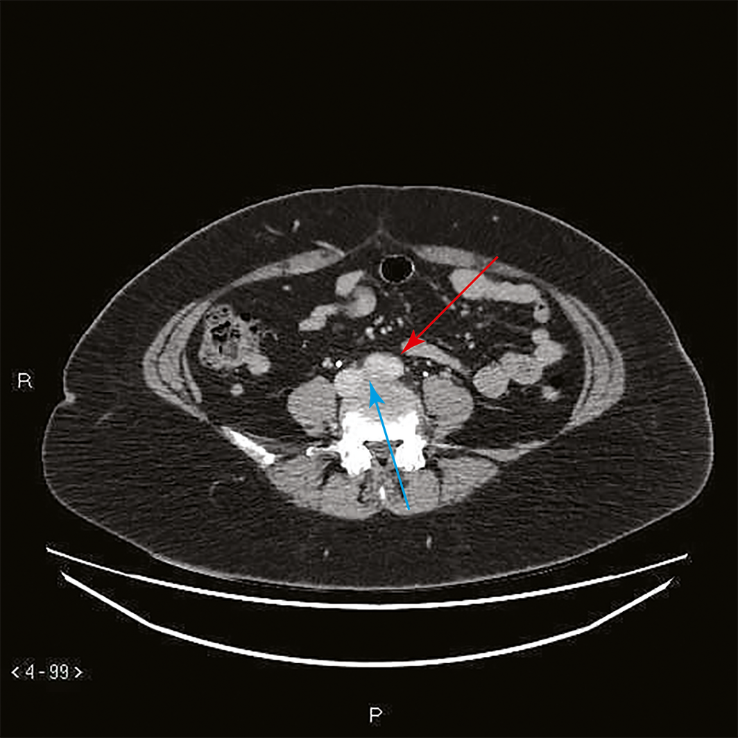
Three months after completing the planned course of rivaroxaban, the patient re‐presented with acute left leg pain and swelling. Ultrasound evaluation of the leg showed extensive thrombosis in the same proximal veins. Inherited thrombophilia work‐up and tests for antiphospholipid antibodies were unremarkable. A computed tomography (CT) venogram showed a thrombus in the left common and external iliac veins with significant narrowing of the thrombus as it passed posterior to the proximal right common iliac artery, suggestive of May–Thurner syndrome (MTS) (Box).
Anticoagulation was re‐initiated and the patient was referred to vascular surgeons. The surgeons offered catheter‐directed thrombolysis, with a view to balloon angioplasty and stent placement into the common iliac vein, but the patient declined endovascular intervention.
Discussion
MTS is defined as an extrinsic compression of the left common iliac vein by the right common iliac artery against the lumbar vertebra. It classically presents as an unprovoked left‐sided acute iliofemoral DVT among women aged in their 30s to 50s (women to men ratio of 2:1).1
In our patient, the first DVT was considered provoked from transient risk factors and anticoagulation was limited to 3 months, as per current VTE management guidelines.2
The VTE recurrence may be considered unprovoked; however, a hidden anatomical defect (MTS) along with obesity contributed to this recurrence. Even though the first DVT episode was provoked, early detection and definitive therapy of MTS in this patient might have prevented VTE recurrence.3,4,5,6,7
Demonstration of venous occlusion by extrinsic compression is required to diagnose MTS. This is usually done by abdominal CT scan or by CT or magnetic resonance imaging venography. Nevertheless, despite appropriate imaging, MTS may occasionally remain undiagnosed, as the extensive iliac vein thrombus may not show extrinsic venous compression. In such cases, when the index of suspicion remains high, thrombectomy followed by intravascular ultrasound (gold standard) confirms the venous occlusion.3
A meta‐analysis of fifteen retrospective studies4 and case series on the efficacy of combining endovascular interventions, involving catheter‐directed thrombolysis and iliac vein stenting with anticoagulation compared with anticoagulation alone, have shown a reduction in post‐thrombotic syndrome and a high iliac vein patency rate, ranging from 87.5% to 92% at one and 3 years of follow‐up.5,6,7
As reported previously,8 MTS is not an uncommon cause of a common diagnosis. Clinicians routinely carry out thrombophilia work‐up, while MTS is often overlooked in patients with unprovoked, and even provoked, extensive iliofemoral DVT, as many patients with MTS have transient provoking risk factors. This current practice may be attributed to MTS not being mentioned in international guidelines and, hence, a lack of awareness of this condition among physicians. In the authors’ experience, multiple cases of extensive left iliofemoral DVT remained undiagnosed until they were investigated specifically for MTS. Some patients had VTE recurrence after cessation of anticoagulation, and some cases of MTS were detected at follow‐up.
It remains unclear if MTS qualifies as a persistent provoking risk factor. VTE classifications have not taken into account anatomical causes of VTE, nor do guidelines provide insight as to when such causes should be investigated. Some authors have described MTS as an anatomical variant, which does not qualify the event as provoked. If endovascular intervention in MTS reduces the risk of recurrence, then it may be considered as a persistent provoking factor.3,4,5,6,7 The real‐life questions that remain unanswered are the duration of anticoagulation in provoked and unprovoked VTE with underlying MTS without endovascular intervention (declined or not suitable), and in unprovoked VTE after endovascular intervention.
In summary, MTS remains an underdiagnosed condition. Undiagnosed and untreated patients with MTS may experience recurrent VTE and higher incidence of post‐thrombotic syndrome. Early diagnosis and endovascular intervention may improve clinical outcomes.
We suggest physicians think beyond thrombophilia screening when encountering unprovoked and even provoked acute extensive left iliofemoral cases of DVT.
Lessons from practice
- The true incidence of symptomatic May–Thurner syndrome (MTS) remains unknown, as it has likely been underestimated and underdiagnosed in clinical practice.
- MTS should be strongly considered among cases of acute or recurrent extensive left‐sided iliofemoral deep vein thrombosis.
- MTS may occasionally be difficult to diagnose despite appropriate imaging; a high index of suspicion should trigger intervention with intravascular ultrasound (gold standard) to confirm the venous occlusion.
- Early identification and early management with endovascular intervention, including iliac vein balloon angioplasty and stent placement in selected patients, may be considered.
Box – Computed tomography venogram showing a thrombus in the left common and external iliac veins, with significant narrowing of the thrombus as it passes posterior to the proximal right common iliac artery, suggestive of May–Thurner syndrome. The blue arrow shows the compressed left common iliac vein, while the red arrow shows the distal abdominal aorta just before it bifurcates
Competing interests
No relevant disclosures.
References
- Thijs W, Rabe KF, Rosendaal FR, Middeldorp S. Predominance of left‐sided deep vein thrombosis and body weight. J Thromb Haemost 2010; 8: 2083–2084.
- Kearon C, Akl EA, Comerota AJ, et al. Antithrombotic therapy for VTE disease: antithrombotic therapy and prevention of thrombosis, 9th ed: American College of Chest Physicians Evidence‐Based Clinical Practice Guidelines. Chest 2012; 141 (Suppl): e419S–e496S.
- Lee AI, Ochoa Chaar CI. The hidden lesion. N Engl J Med 2016; 375: 1198–1199.
- Casey ET, Murad MH, Zumaeta‐Garcia M, et al. Treatment of acute iliofemoral deep vein thrombosis. J Vasc Surg 2012; 55: 1463–1473.
- Hager ES, Yuo T, Tahara R, et al. Outcomes of endovascular intervention for May–Thurner syndrome. J Vasc Surg Venous Lymphat Disord 2013; 1: 270–275.
- DeRubertis BG, Alktaifi A, Jimenez JC, et al. Endovascular management of non‐malignant iliocaval venous lesions. Ann Vasc Surg 2013; 27: 577–586.
- O'Sullivan GJ, Semba CP, Bittner CA, et al. Endovascular management of iliac vein compression (May–Thurner) syndrome. J Vasc Interv Radiol 2000; 11: 823–826.
- Peters M, Syed RK, Katz M, et al. May–Thurner syndrome: a not so uncommon cause of a common condition. Proc (Bayl Univ Med Cent) 2012; 25: 231–233.
Linked content
-
MJA Letter: May–Thurner syndrome: an overlooked cause of venous thromboembolism
Provenance: Not commissioned; externally peer reviewed.
Splenic injury in severe cases of the zoonoses Q fever and rickettsial infection: diagnostic challenges
Ashleigh Drury, Philippa Harrison, Aiveen Bannan
Health and economic benefits of improving pre‐hospital identification of stroke in Australian women: a modelling study
Thomas Gadsden, Lei Si, Emily R Atkins, Cheryl Carcel, Xia Wang, Stephen Jan, Mark Woodward, Laura E Downey
Updating the diagnosis and management of elevated serum ferritin levels in the era of routine ferritin testing of blood donors by Australian Red Cross Lifeblood
Gary D Zhang, James Chen, Daniel M Johnstone, Martin B Delatycki, Katie Allen, John K Olynyk
Early cardiovascular collapse after envenoming by snakes in Australia, 2005–2020: an observational study (ASP‐31)
Geoffrey K Isbister, Katherine Z Isoardi, Angela L Chiew, Shane Jenkins, Nicholas A Buckley
Updated recommendations for warfarin reversal in the setting of four‐factor prothrombin complex concentrate
Danielle Robinson, James McFadyen, Eileen Merriman, Chee Wee Tan, Ross Baker, Huyen Tran
Updating the diagnosis and management of iron deficiency in the era of routine ferritin testing of blood donors by Australian Red Cross Lifeblood
Gary D Zhang, Daniel Johnstone, Michael F Leahy, John K Olynyk