




Volume 211 - Issue 9
The management of diverticulitis: a review of the guidelines
Authors: Hayley You, Amy Sweeny, Michelle L Cooper, Michael Von Papen and James Innes
Med J Aust 2019; 211 (9): 421-427. || doi: 10.5694/mja2.50276
Published online: 29 July 2019
Published online: 29 July 2019
Diverticular disease is a common gastrointestinal disorder with significant health burden; recent evidence is changing practice
Summary
- Radiological evidence of inflammation, using computed tomography (CT), is needed to diagnose the first occurrence of diverticulitis. CT is also warranted when the severity of symptoms suggests that perforation or abscesses have occurred.
- Diverticulitis is classified as complicated or uncomplicated based on CT scan, severity of symptoms and patient history; this classification is used to direct management.
- Outpatient treatment is recommended in afebrile, clinically stable patients with uncomplicated diverticulitis.
- For patients with uncomplicated diverticulitis, antibiotics have no proven benefit in reducing the duration of the disease or preventing recurrence, and should only be used selectively.
- For complicated diverticulitis, non‐operative management, including bowel rest and intravenous antibiotics, is indicated for small abscesses; larger abscesses of 3–5 cm should be drained percutaneously. Patients with peritonitis and sepsis should receive fluid resuscitation, rapid antibiotic administration and urgent surgery.
- Surgical intervention with either Hartmann procedure or primary anastomosis, with or without diverting loop ileostomy, is indicated for peritonitis or in failure of non‐operative management.
- Colonoscopy is recommended for all patients with complicated diverticulitis 6 weeks after CT diagnosis of inflammation, and for patients with uncomplicated diverticulitis who have suspicious features on CT scan or who otherwise meet national bowel cancer screening criteria.
Diverticular disease is one of the most common gastrointestinal disorders and carries a significant health burden in industrialised countries. The disease is characterised by diverticulosis: the presence of mucosal and submucosal herniations or “pockets” known as diverticula.1 Although diverticulosis is largely asymptomatic, 4% of individuals with diverticula develop diverticulitis throughout their lifetime.2 Recent evidence concerning diverticular disease is changing methods of practice. The aim of this review is to analyse the current literature and guidelines on the assessment and management of diverticulitis.
Pathogenesis
Aetiology
The pathogenic aetiology of diverticulosis is poorly understood. Diverticular development, however, is thought to involve three principal contributors: colonic wall structural abnormalities, increased intraluminal pressure, and dietary fibre deficiency.
It is thought that diverticula develop from age‐related mucosal wall degeneration and localised increases in colonic pressure, resulting in herniations at points of weakness.3 The sigmoid colon, which has the highest intraluminal pressures and narrowest calibre, is the most common site for diverticula formation. Some cases involve the descending colon or, more rarely, the whole colon.
Unlike the small intestine and rectum, the colon is comprised of only one complete inner circular muscular layer. Diverticula typically form in rows parallel to the taeniae coli of the outer longitudinal layer.4 This site is where the vasa recta blood vessels penetrate the muscle to supply the mucosal and submucosal bowel layers. Compared with normal controls, the colonic characteristics involved include microscopic muscular atrophy; abnormal elastin deposition in the taeniae coli, resulting in shortening of the muscle layer; and increased collagen cross‐linking, which mimics natural ageing patterns.5 These features lead to loss of compliance of the bowel wall and muscular weakness, predisposing to diverticula formation, particularly when the intraluminal pressure is also increased. Chronic inflammation of the mucosa leads to muscular hypertrophy of the affected area and enteric nerve remodelling resulting in altered motility.
Progression
Diverticulosis is largely asymptomatic, although it can be accompanied by changes in bowel habits such as constipation or diarrhoea and may or may not progress to diverticulitis (Box 1). The dominant theory explaining progression to diverticulitis is that faecal stasis causes an obstructive faecolith to be trapped in the neck of a diverticulum,1 which may eventually lead to localised inflammation.4 Uncomplicated diverticulitis is diagnosed by evidence of inflammation without abscess, perforation or peritonitis. Complicated diverticulitis occurs when the inflammation leads to abscess, perforation and/or peritonitis.
Method
We undertook a systematic search for guidelines on assessment, diagnosis, classification, imaging, management and prevention of diverticulitis and diverticular disease. National Guideline Clearinghouse, EMBASE, MEDLINE and PubMed were searched initially in May 2017. Updates to the guidelines were sought in September 2018.
National and international guidelines published between 2007 and 2017 with full text English versions were included.6,7,8,9,10,11,12,13 Guideline recommendations were analysed and summarised based on topic to find areas of consensus or controversy. In areas of controversy, the references in guideline documents and journal articles that were systematic reviews, meta‐analyses or randomised controlled trials from the past 10 years were reviewed for supplemental and up‐to‐date evidence.
Clinical assessment
Diverticulitis is a severe episode of lower abdominal pain that is usually left‐sided, accompanied by a low grade fever, leucocytosis and change in bowel movements.12 Guidelines classify diverticulitis as complicated and uncomplicated based on computed tomography (CT) images (Box 2).
Initial examination of suspected diverticulitis
The American Society of Colon and Rectal Surgeons (ASCRS) strongly recommends an initial examination consisting of a specific history of presenting complaint, physical examination, complete blood count, and urinalysis.6
Commencing treatment without imaging is reasonable in symptomatic patients with a previous history of diverticulitis, after completing a detailed history and physical examination.14 The guidelines from the Netherlands Society of Surgery (NSS) and Danish Surgical Society (DSS) support using a clinical scoring system to improve diagnostic accuracy, foregoing the need for imaging for patients with prior episodes of diverticulitis.7,8 Three presentation characteristics: lower left quadrant abdominal pain only, C‐reactive protein (CRP) above 50 mg/L, and absence of vomiting, when all present, are reported to correctly diagnose diverticulitis for 97% of cases.7
Yet despite improved clinical scoring systems, misdiagnosis is common for patients presenting with the first episode of diverticulitis, due to the plethora of differentials, including irritable bowel syndrome, appendicitis, urinary tract infections, kidney stones, neoplasia and bowel obstruction.9 Because of this, all guidelines recommend CT scanning to support the first diverticulitis diagnosis and to evaluate the extent of disease by detection of any complications.
In summary, clinical evaluation alone is often insufficient in the first diagnosis of diverticulitis and can lead to misdiagnosis. Radiological evidence of inflammation is needed for definitive diagnosis of diverticulitis.
Imaging for acute diverticulitis
Imaging plays an essential role in both diagnosis and staging of diverticulitis.
Computed tomography
CT is considered the best imaging choice for initial evaluation of patients with suspected diverticulitis6,9,13 because of its high sensitivity and specificity (94% and 99%, respectively) and its ability to detect other causes of left lower quadrant pain.15 CT serves four major functions: it confirms the diagnosis of diverticulitis, evaluates the severity and extent of the disease, guides management plans for the treatment of abscesses, and detects other causes of abdominal pain. All major guidelines agree on its high predictive accuracy in diagnosing diverticulitis.
In the authors’ clinical opinion, CT is not indicated routinely as a means to assess resolution of a bout of diverticulitis. However, when an outpatient's symptoms are not improving or have worsened after 5 days, a repeat CT would be recommended to assure that disease has not progressed.
There is generally a lack of consensus regarding the use of contrast in CT.16 Some guidelines and studies classify CT using oral, intravenous or colonic contrast as optimal.7,8,9,12,17 Other guidelines do not offer discussion or recommend contrast‐enhanced over unenhanced CT.10,11
Barium enema
In the past, barium enema was the first‐line imaging examination for diverticulitis. It is now surpassed by CT, mostly due to evidence showing superior diagnostic accuracy with CT.18 Barium enema is now discouraged following case studies reporting diverticular perforations. Nevertheless, water‐soluble iodinated contrast enema is still used in some centres to evaluate suspected perforation.9 In any case, CT is now so specific that contrast enema is rarely required to confirm perforation.
In summary, CT has replaced barium enema as the primary imaging choice.
Ultrasound
Transabdominal, high resolution ultrasound — generally used in Europe — is considered an alternative imaging modality for suspected diverticulitis. The reported summary sensitivity is 92% and specificity is 90%.16 The guidelines from the Association of Coloproctology of Great Britain and Ireland (ACPGBI) recommend that CT or ultrasound should be used depending on local expertise.10 This was agreed upon by expert physicians from six countries at the 2nd International Symposium of Diverticular Disease of the Colon in 2016.12 Most other guidelines6,7,8,9,13 also recognise that ultrasound may be useful in patients in whom CT scanning is contraindicated (eg, pregnancy, contrast allergy, renal insufficiency). Two European guidelines recommend a conditional CT scan after a negative or inconclusive ultrasound.7,13
Ultrasound has acknowledged limitations compared with CT.15,16,17,18 It is highly operator‐dependant and requires sonographer expertise. It is also dependent on body habitus as it cannot penetrate extensive soft tissue or air‐filled structures, resulting in poor image quality in patients with obesity or in those with overlying gas. In addition, probing may cause discomfort in patients with abdominal tenderness.
In summary, ultrasound can be used to diagnose diverticulitis if carried out by an expert sonographer and is preferred in select patients where CT scanning is contraindicated.
Magnetic resonance imaging
The use of magnetic resonance imaging (MRI) in the diagnosis of diverticulitis is an area of controversy. Preliminary data show potential for MRI in assessing diverticulitis, with one of its major advantages being its lack of ionising radiation. Moreover, it is less operator‐dependant than ultrasound. Two small studies19,20 also suggest that MRI provides a better image to distinguish colonic carcinoma from inflammation; however, these preliminary data require confirmation by larger studies. The sensitivity and specificity of MRI are reported to be as high as 94% and 92%, respectively.20
The ASCRS recommends MRI as a useful alternative to CT to limit the patient's radiation exposure.6 The American College of Radiology, ACPGBI, NSS and DSS acknowledge the potential of MRI, but fall short of recommendation due to lack of systematic analysis and consensual data. The clinical applicability in Australia is further questioned, as Medicare rebates do not apply to MRI scans for this indication.
In summary, MRI use is not currently widely recommended for diagnosis of diverticulitis.
Biochemical tests
All guidelines recommend that clinical diagnosis should consider presenting complaint, physical examination and biochemistry. However, there is controversy in whether biochemical tests can confirm diagnosis. All guidelines recognise that biochemistry alone is insufficient.
CRP is recognised as the most powerful independent factor in the differentiation of diverticulitis from other abdominal conditions,6,7,8 but it only has diagnostic power if considered in combination with other factors, including white cell count. Other guidelines do not discuss biochemical markers for diverticulitis. The negative predictive value of CRP below 50 mg/L is 79% for perforation in acute sigmoid diverticulitis.21
In summary, biochemical markers are recommended in routine evaluation. A high CRP and a high white cell count may help determine severity of disease. Biochemical tests, however, do not confirm diagnosis.
Colonoscopy
Perforated colon cancer mimics both clinical evaluation and CT findings of diverticulitis. Because of this, in the past, all major guidelines recommended a routine colonoscopy after CT‐diagnosed diverticulitis to avoid misdiagnosis of a colonic neoplasm.
However, systematic reviews of the literature have now specified that the evidence base supports routine colonoscopy only for cases of complicated diverticulitis.22,23 There are insufficient data to support the recommendation of routine colonoscopy for uncomplicated diverticulitis; its value has been further rebuked by large studies showing that the incidence of colorectal cancers after uncomplicated diverticulitis was not different to that observed in the general population.23,24 A 2014 systematic review and meta‐analysis23 reported that routine colonoscopy in this group of patients yielded the same cancer incidence (around 0.7%) as that of the general population undergoing asymptomatic screening.
Nonetheless, colonoscopy is still indicated for some uncomplicated diverticulitis cases, such as patients in whom CT scan has identified short segments of disease with several diverticula (suggesting a more malign pathology) and patients who would otherwise fulfil the criteria for routine national screening.25
In Australia, the need for colonoscopy is based on the results of faecal occult blood testing, age, and other risk factors, including family history,26 set out in the National Bowel Cancer Screening Program.25 Similar recommendations are shared by the American Gastroenterological Association, which adds that colonoscopy should only be performed if a high quality colonoscopy has not been done recently (within 12 months). This decision is based on timing and quality of previous colonoscopy, comorbidities, persistent symptoms, and patient preference.
For complicated cases of diverticulitis, a follow‐up colonoscopy is still considered warranted to rule out a colonic neoplasm.6,7,9,10
When colonoscopy is indicated, the NSS does not recommend colonoscopy in the acute phase, as air insufflation and scope manipulation may cause a full perforation.7 A 6‐week waiting period after diagnosis is recommended by several guidelines,6,7,10 to allow time for resolution of inflammation.
In summary, colonoscopy is recommended for all cases of complicated diverticulitis 6 weeks after CT‐diagnosed inflammation, and in uncomplicated diverticulitis where there are concerning findings on CT or where the patient otherwise meets national screening criteria.
Management of uncomplicated diverticulitis
Outpatient management
In the past 5–10 years, the management of diverticulitis has changed depending on whether diverticulitis is complicated or uncomplicated. Some major guidelines6,7,12 have been updated and now recommend outpatient treatment for afebrile clinically stable cases of uncomplicated diverticulitis with no other reason for admission. Outpatient management is safe, with high success rates.27,28,29
A retrospective study found that among a cohort of 693 patients, outpatient treatment was successful in 94% of patients.30 In another retrospective cohort study, 91.5% of patients with uncomplicated diverticulitis were successfully treated as outpatients without developing complications or subsequent hospital admission for a period of 48 months.31
Uncomplicated diverticulitis has historically been managed with antibiotic therapy,32 but most international guidelines6,7,8,11,13 now recommend a clear liquid diet, and antibiotic use on a selective case‐by‐case basis only. Although the use of antibiotics in outpatient management of diverticulitis remains the norm in the United States, a recent review notes that there is insufficient evidence to defend routine antibiotic use in this setting.33 Furthermore, antibiotics may cause adverse effects, such as allergic reactions and pseudomembranous colitis. This, coupled with the ever‐present concern of antibiotic resistance, makes the decision to use antibiotics more difficult to defend. The revised recommendations are based on recent randomised controlled trials and Cochrane systematic reviews finding that antibiotic therapy has no clear benefit in reducing complications, shortening recovery or preventing recurrence of episodes.34,35 Selective antibiotic use on an outpatient basis should be limited to patients who are immunocompromised.
When outpatient antibiotics are warranted, oral antibiotics are typically given for 7–10 days. Antibiotics should include complete coverage against gram‐positive, gram‐negative and both anaerobic and aerobic strains.28
A comprehensive outpatient management strategy for uncomplicated diverticulitis was set out in 2018:36
- clear liquid diet for 2–3 days;
- low fibre diet until pain improves;
- acetaminophen plus antispasmodics for pain; and
- use of antibiotics on a case‐by‐case basis.
Although we were unable to locate evidence in support of the clear liquid diet, a clinical rationale supporting this type of management is that resting the bowel will ease abdominal pain through the prevention of hard stool formation. Furthermore, if there is concern that an operative approach may become necessary, clear liquid diets offer a shorter period of fasting before the operation.
In summary, outpatient treatment is safe and is recommended for afebrile patients with uncomplicated diverticulitis who can tolerate oral hydration and have adequate family support. Antibiotics are to be used selectively for patients presenting with uncomplicated diverticulitis.
Management of complicated diverticulitis
All guidelines currently recommend the use of intravenous broad spectrum antibiotics and bowel rest for patients with complicated diverticulitis.6,7,8,9,10,11,12,13
Non‐operative treatment
Due to the lack of quality trials to provide evidence of the optimal treatment strategy, there is no universal practice for the management of complicated diverticulitis.
It is generally agreed that bowel rest and intravenous antibiotics are sufficient for small abscesses of less than 3 cm. Larger abscesses of 3–5 cm should be percutaneously drained if accessible.13 Inpatient non‐operative treatment, including broad spectrum antibiotics, bowel rest and percutaneous drainage (followed by oral antibiotics and liquid or low residue diet as outpatient) are successful in 91% of all patients with complicated diverticulitis and in 95% of patients deemed appropriate for non‐operative treatment (ie, with perforation with or without abscess without peritonitis).38 There is no recommended method for drainage.
In summary, smaller pericolic abscesses can be conservatively managed with bowel rest and antibiotics; larger abscesses of 3–5 cm should be percutaneously drained.
Surgical intervention
Although most patients can be managed without surgical intervention, diverticulitis that is not resolved by non‐operative management should be treated with surgery. In some patients, recurrent bouts of inflammation may lead to luminal stenosis, also warranting intervention. Stenosis not amenable to ballooning procedures will require surgery. When surgery is indicated, a laparoscopic approach confers more favourable patient outcomes.33
For recurrent cases of diverticulitis that could be managed without surgery, elective surgery is offered on a case‐by‐case basis. This depends on the patient's wishes, anaesthetic risk, and consideration of the toll recurrent disease is taking on the patient's activities of daily living and/or ability to work.39
Peritonitis is a life‐threatening complication of acute diverticulitis. Perforation due to inflammatory wall damage results in either purulent or faecal peritonitis, in which the patient experiences organ dysfunction from septicaemia. Major guidelines currently recommend that patients with peritonitis and sepsis receive fluid resuscitation, rapid antibiotic administration and urgent surgery.
Even though all guidelines recommend urgent surgical intervention in emergency settings, there is controversy over the preferred methods. The options include simple colostomy formation, colonic resection with construction of end colostomy (Hartmann procedure), and colonic resection with primary anastomosis with or without diverting loop ileostomy.40,41
A systematic review published in 2014 demonstrates a minor preference for primary colorectal anastomosis compared with Hartmann procedure but only when performed by experienced surgeons.42 Some randomised controlled trials demonstrate a small improvement in the stoma reversal rate for patients undergoing anastomosis but only for those subsets in which operator experience was high.42 American guidelines recommend a two‐stage procedure: Hartmann or colonic resection with primary anastomosis and diverting protective ostomy,11,43 which is almost always accompanied by loop ileostomy.
In view of these findings, the ASCRS recommends that the decision for anastomosis with or without an ileostomy following resection of the colon should be decided on a case‐by‐case basis. In general, primary anastomosis with proximal diversion is recommended by ASCRS and NSS for patients with peritonitis; however, it should be based on factors such as haemodynamic instability, acidosis, acute organ failure and comorbidities, in conjunction with surgeon expertise. It is generally accepted that Hartmann procedure may be more appropriate and may have clinical value in patients who are haemodynamically unstable, high risk, older or have multiple comorbidities.6,13,44
Laparoscopic lavage
There is lack of clinical evidence to support the safety and efficacy of laparoscopic lavage as an alternative to colonic resection. All guidelines discourage the use of lavage in purulent or faecal peritonitis.6,7,8,9,10,11,12,13
When surgical intervention is required, Hartmann procedure or primary anastomosis with our without diversion, such as diverting loop ileostomy, is indicated, with the latter only recommended in experienced surgeons’ hands.
Prevention
Several reviews make conditional recommendations for a range of protective factors32,33,35 that may prevent the development of diverticular disease and some of its sequelae. However, only some general lifestyle recommendations (eg, body mass index < 30, vigorous exercise, avoiding smoking, and limiting red meat consumption) are backed by sufficient evidence45,46,47,48,49,50 showing reduction in the incidence of diverticular disease (Box 3).
Conclusion
In conclusion, the most recent evidence available and international guidelines recommend changing some age‐old practices in the diagnosis and management of diverticulitis. The most significant changes pertain to patients with uncomplicated diverticulitis: clinical diagnosis for patients with a history of diverticulitis and mild symptoms, the increased use of outpatient management, use of antibiotics on a selective case‐by‐case basis, and avoidance of routine colonoscopy unless another clear indication exists.
Box 1 – Diverticulosis and its manifestations
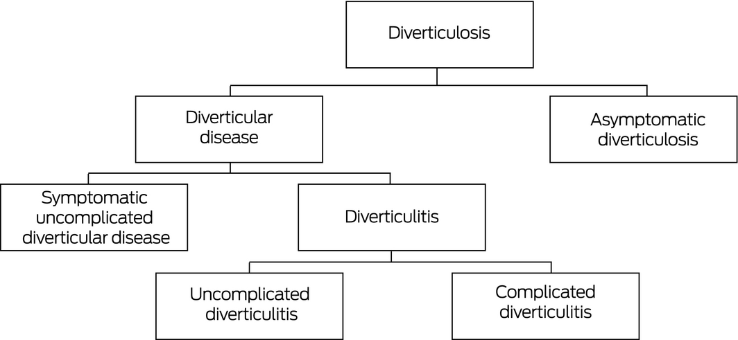
Diverticulosis: asymptomatic presence of mucosal and submucosal herniations due to defects in weaker areas of the muscular wall of the colon.
Diverticular disease: a wide spectrum of disease including diverticular bleeding and diverticulitis.
Symptomatic uncomplicated diverticular disease (SUDD): subtype of diverticular disease in which there are persistent, recurrent abdominal symptoms without signs of overt diverticulitis.
Diverticulitis: diverticula become acutely inflamed, most likely due to obstruction of the neck by faecal matter leading to bacterial overgrowth.
Uncomplicated diverticulitis: diverticulitis without perforation, abscess, bleeding, fistula, peritonitis or stenosis.
Complicated diverticulitis: diverticulitis with complicating features such as perforation, abscess, bleeding, fistula, peritonitis or stenosis; it may be localised or lead to infection of the peritoneal cavity; stricture, obstruction or bleeding may be evident. Glossary adapted from Hong et al.1
Box 2 – Computed tomography (CT) scans showing uncomplicated (A and B)* and complicated diverticulitis (C and D)†
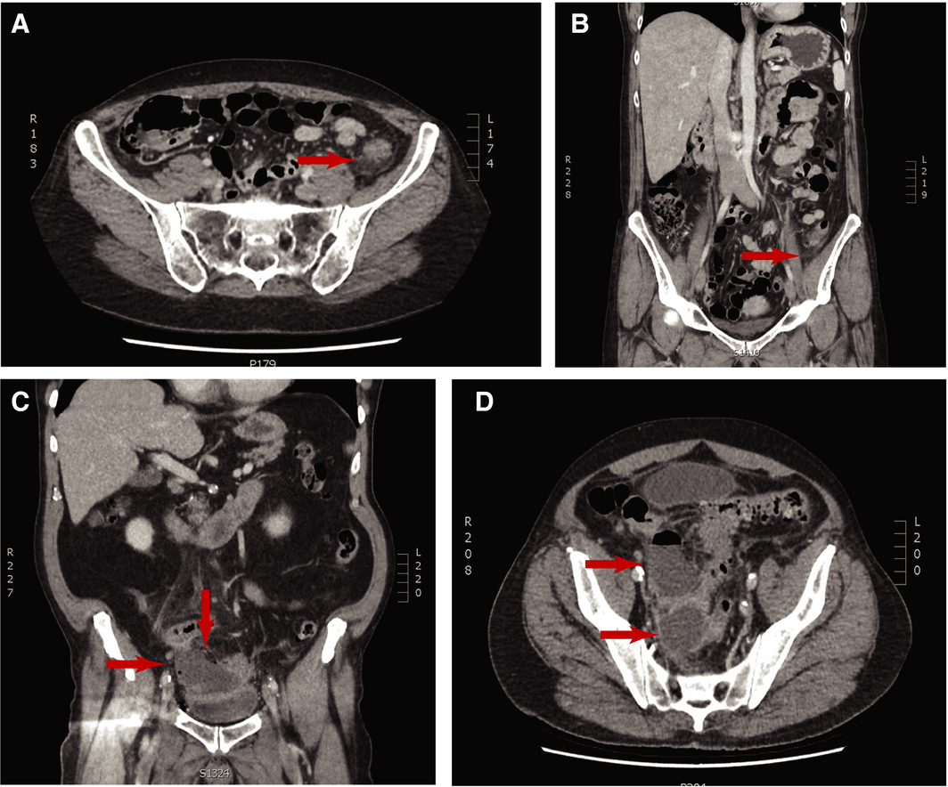
* Uncomplicated diverticulitis (A and B): two views of a CT scan of the colon showing fat stranding, indicative of local inflammation, but no abscess or perforation. †Complicated diverticulitis (C and D): two views of a CT scan of the colon showing a collection of pus or gas, suggesting an abscess.
Box 3 – Summary of current evidence on diagnosis, management and prevention of diverticular disease
|
Topic |
Conclusion of recommendations |
Evidence |
NHMRC level of evidence* |
||||||||||||
|
|
|||||||||||||||
|
Clinical assessment |
|||||||||||||||
|
Definition |
A uniform clinical definition is lacking |
na |
na |
||||||||||||
|
Initial examination of suspected diverticulitis |
Clinical evaluation alone is insufficient for initial diagnosis. Require radiological imaging for diagnostic support |
Studies suggest that clinical scoring systems have potential in diagnosis, but still require imaging for diagnostic support6,7,8,9,10,11,12,13,14 |
III‐3 |
||||||||||||
|
Imaging |
|||||||||||||||
|
Barium |
No longer indicated: CT is superior |
II |
|||||||||||||
|
Evidence of increased perforation risks with barium are based on case studies and expert opinion |
|
||||||||||||||
|
CT |
Recommended as primary imaging choice (highly sensitive and specific) |
CT is the gold standard for diagnosis and staging of acute diverticulitis6,7,8,9,10,11,12,13,14,15,16,17 |
I |
||||||||||||
|
Ultrasound |
Use is recommended as an alternative to CT for contraindicated patients or pregnant or fertile women |
Ultrasound and CT are comparable; however, CT is superior due to its higher specificity and advantage in identifying alternative diagnoses15,16,17,18 |
I |
||||||||||||
|
MRI |
Not widely used |
IV |
|||||||||||||
|
Other |
|||||||||||||||
|
Biochemical tests |
Recommended in routine evaluation but not for diagnosis |
Lack of evidence to show high diagnostic power of biochemical examination in isolation21 |
na |
||||||||||||
|
Colonoscopy |
Colonoscopy after resolution of CT‐diagnosed complicated diverticulitis is recommended in appropriate patients |
Insufficient data to support recommendation of routine colonoscopy for uncomplicated diverticulitis22,23,24,25 |
III |
||||||||||||
|
Management of uncomplicated diverticulitis |
|||||||||||||||
|
Uncomplicated diverticulitis |
Outpatient management is safe and recommended in patients without complications, comorbidities, fever and adequate family support |
High level of support for mild cases of uncomplicated diverticulitis27,28,29,30,31 |
I |
||||||||||||
|
Antibiotic therapy |
Guidelines are yet to implement antibiotic‐free strategies but recommend selective use |
Growing body of evidence demonstrating no benefit of antibiotic therapy in patients with uncomplicated diverticulitis33,34,35,36 |
I |
||||||||||||
|
Management of complicated diverticulitis |
|||||||||||||||
|
Non‐operative treatment |
Smaller abscesses (< 5 cm) can be conservatively managed with antibiotics, while larger ones also require percutaneous drainage |
Increasing evidence in success of non‐operative therapy for management of abscesses13,38 |
I |
||||||||||||
|
Urgent operative therapy |
Recommended in emergency cases of purulent or faecal peritonitis, or when non‐operative management fails |
III |
|||||||||||||
|
Preferred surgical procedure |
Both Hartmann procedure and primary anastomosis with or without diversion is indicated. Physician is to determine procedure on case‐by‐case basis |
Lack of significant clinical evidence; however, RCTs lean towards primary anastomosis42,43,44 |
III‐1 |
||||||||||||
|
Laparoscopic lavage |
Not recommended as an alternative surgical procedure due to conflicting evidence |
Some RCTs concluded that laparoscopic lavage was feasible and safe in patients with perforated diverticulitis; however, others do not recommend it over colectomy6,7,8,9,10,11,12,13 |
I |
||||||||||||
|
Prevention |
|||||||||||||||
|
Vigorous exercise |
Vigorous physical activity is recommended to reduce the risk of diverticulitis |
Systematic review and meta‐analyses have shown an inverse relationship between vigorous physical activity and incidence of diverticulitis45 |
I |
||||||||||||
|
BMI < 30 |
Obesity increases the risk of diverticular disease |
Systematic reviews and meta‐analyses show small increases in BMI result in a higher risk of diverticular disease and complications45 |
I |
||||||||||||
|
Avoiding smoking |
Avoiding smoking reduces the risk of diverticulitis |
Systematic reviews and meta‐analyses show that tobacco smoking is associated with increased incidence of diverticulitis and complications46 |
I |
||||||||||||
|
Limiting red meat consumption |
Limited intake of red meat is recommended to reduce the development of diverticular disease |
Large cohort study suggests that red meat, especially processed red meat, was associated with increased incidence of diverticular disease47 |
III‐1 |
||||||||||||
|
High fibre diet |
High fibre diet is recommended in combination with healthy lifestyle factors as above |
Systematic reviews are inconclusive50 |
na |
||||||||||||
|
|
|||||||||||||||
|
BMI = body mass index; CT = computed tomography; MRI = magnetic resonance imaging; na = not applicable; NHMRC = National Health and Medical Research Council; RCT = randomised controlled trial. *If applicable.51 |
|||||||||||||||
Competing interests
No relevant disclosures.
Acknowledgements
Amy Sweeny receives funding from the Emergency Medicine Foundation as a member of the Research Support Network.
References
- Hong MK, Skandarajah AR, Hayes IP. Diverticulosis, diverticular disease and diverticulitis — definitions and differences. Journal of Stomal Therapy Australia 2015; 35: 8–10.
- Shahedi K, Fuller G, Bolus R, et al. Long‐term risk of acute diverticulitis among patients with incidental diverticulosis found during colonoscopy. Clin Gastroenterol Hepatol 2013; 11: 1609–1613.
- Gaglia A, Probert C. Diverticular disease. Medicine 2015; 43: 320–323.
- Matrana M, Margolin D. Epidemiology and pathophysiology of diverticular disease. Clin Colon Rectal Surg 2009; 22: 141–146.
- Jeyarajah S, Papagrigoriadis S. Review article: the pathogenesis of diverticular disease — current perspectives on motility and neurotransmitters. Aliment Pharmacol Ther 2011; 33: 789–800.
- Feingold D, Steele S, Lee S, et al. Practice parameters for the treatment of sigmoid diverticulitis. Dis Colon Rectum 2014; 57: 284–294.
- Andeweg C, Mulder I, Felt‐Bersma R, et al. Guidelines of diagnostics and treatment of acute left‐sided colonic diverticulitis. Dig Surg 2013; 30: 278–292.
- Andersen J, Bundgaard L, Elbrønd H, et al. Danish national guidelines for treatment of diverticular disease. Dan Med J 2012; 59: 4453.
- McNamara M, Lalani T, Camacho M, et al. ACR appropriateness criteria: left lower quadrant pain — suspected diverticulitis. J Am Coll Radiol 2014; 7.
- Royal College of Surgeons. 2014 Commissioning guide: colonic diverticular disease. London: RCS, 2017. https://www.acpgbi.org.uk/content/uploads/2017/02/Commissioning-guide-colonic-diverticular-disease-RCS-2014.pdf (viewed Sept 2018).
- Stollman N, Smalley W, Hirano I; AGA Institute Clinical Guidelines Committee. American Gastroenterological Association Institute guideline on the management of acute diverticulitis. Gastroenterology 2015; 149: 1944–1949.
- Tursi A, Picchio M, Elisei W, et al. Management of patients with diverticulosis and diverticular disease: consensus statements from the 2nd International symposium on diverticular disease. J Clin Gastroenterol 2016; 50: S101–S107.
- Binda GA, Cuomo R, Laghi A, et al. Practice parameters for the treatment of colonic diverticular disease: Italian Society of Colon and Rectal Surgery (SICCR) guidelines. Techn Coloproctol 2015; 19: 615–626.
- Vennix S, Morton DG, Hahnloser D, et al; Research Committee of the European Society of Coloproctology. Systematic review of evidence and consensus on diverticulitis: an analysis of national and international guidelines. Colorectal Dis 2014; 16: 866–878.
- Laméris W, van Randen A, Bipat S, et al. Graded compression ultrasonography and computed tomography in acute colonic diverticulitis: meta‐analysis of test accuracy. Eur Radiol 2008; 18: 2498.
- Liljegren G, Chabok A, Wickbom M, et al. Acute colonic diverticulitis: a systematic review of diagnostic accuracy. Colorectal Dis 2007; 9: 480–488.
- Sarma D, Longo W. Diagnostic imaging for diverticulitis. J Clin Gastroenterol 2008; 42: 1139–1141.
- Destigter K, Keating D. Imaging update: acute colonic diverticulitis. Clin Colon Rectal Surg 2009; 22: 147–155.
- Öistämö E, Hjern F, Blomgvist L, et al. Cancer and diverticulitis of the sigmoid colon. Differentiation with computed tomography versus magnetic resonance imaging: preliminary experiences. Acta Radiol 2013; 54: 237–241.
- Heverhagen J, Sitter H, Zielke A, Klose K. Prospective evaluation of the value of magnetic resonance imaging in suspected acute sigmoidal diverticulitis. Dis Colon Rectum 2008; 51: 1810–1815.
- Käser SA, Fankhauser G, Glauser PM, et al. Diagnostic value of inflammation markers in predicting perforation in acute sigmoid diverticulitis. World J Surg 2010; 34: 2717–2722.
- Sai V, Velayos F, Neuhaus J, Westphalen A. Colonoscopy after CT diagnosis of diverticulitis to exclude colon cancer: a systematic literature review. Radiol 2012; 263: 383–390.
- Sharma P, Eglinton T, Hider P, Frizelle F. Systematic review and meta‐analysis of the role of routine colonic evaluation after radiologically confirmed acute diverticulitis. Ann Surg 2014; 259: 263–272.
- Andrade P, Ribeiro A, Ramalho R, et al. Routine colonoscopy after acute uncomplicated diverticulitis — challenging a putative indication. Dig Surg 2017; 34: 197–202.
- Cancer Council Australia: Cancer guidelines wiki: clinical practice guidelines for the prevention, early detection and management of colorectal cancer [website]. https://wiki.cancer.org.au/australia/Guidelines:Colorectal_cancer (viewed Oct 2017).
- Jenkins MA, Quakrim DA, Boussioutas A, et al. Revised Australian national guidelines for colorectal cancer screening: family history. Med J Aust 2018; 209: 455–460. https://www.mja.com.au/journal/2018/209/10/revised-australian-national-guidelines-colorectal-cancer-screening-family
- Alonso S, Pera M, Parés D, et al. Outpatient treatment of patients with uncomplicated acute diverticulitis. Colorectal Dis 2010; 12: 278–282.
- Tursi A. Efficacy, safety, and applicability of outpatient treatment for diverticulitis. Drug Healthc Patient Saf 2014; 6: 29–36.
- van Dijk S, Bos K, de Boer M, et al. A systematic review and meta‐analysis of outpatient treatment for acute diverticulitis. Int J Colorectal Dis 2018; 33: 505–512.
- Etzioni D, Chiu V, Cnnom R, et al. Outpatient treatment of acute diverticulitis: rates and predictors of failure. Dis Colon Rectum 2010; 53: 861–865.
- Ünlü C, Gunadi P, Gerhards M, et al. Outpatient treatment for acute uncomplicated diverticulitis. Euro J Gastroenterol Hepatol 2013; 25: 1038–1043.
- Carabotti M, Annibale B. Treatment of diverticular disease: an update on latest evidence and clinical implications. Drugs Context 2018; 7: 212526.
- Young‐Fadock TM, Solomon CG; editors. Diverticulitis. N Engl J Med 2018; 379: 1635–1642.
- Chabok A, Påhlman L, Hjern F, et al. Randomised clinical trial of antibiotics in acute uncomplicated diverticulitis. Br J Surg 2012; 99: 532–539.
- Shabanzadeh D, Wille‐Jørgensen P. Antibiotics for uncomplicated diverticulitis. Cochrane Database Syst Rev 2012; 11: CD009092.
- Swanson S, Strate LL. Acute colonic diverticulitis. Ann Intern Med 2018; 168: 65–80.
- Paolillo C, Sartelli M, Miele V, et al. A three‐step approach to patients with suspected acute diverticulitis in the emergency department: an interdisciplinary algorithm proposal. Emerg Care J 2018; 14: 46–50.
- Dharmarajan S, Hunt S, Birnbaum E, et al. The efficacy of nonoperative management of acute complicated diverticulitis. Dis Colon Rectum 2011; 54: 663–671.
- Fozard J, Armitage N, Schofield J, Jones O. ACPGBI position statement on elective resection for diverticulitis. Colorectal Dis 2011; 13: 1–11.
- Abbas S. Resection and primary anastomosis in acute complicated diverticulitis: a systematic review of the literature. Int J Colorectal Dis 2007; 22: 351–357.
- Morris A, Regenbogen S, Hardiman K, Hendren S. Sigmoid diverticulitis: a systematic review. J Am Med Assoc 2014; 311: 287–297.
- Regenbogen S, Hardman K, Hendren S, Morris A. Surgery for diverticulitis in the 21st century: a systematic review. J Am Med Assoc 2014; 149: 292–302.
- Floch M, Longo W. United States guidelines for diverticulitis treatment. J Clin Gastroenterol 2016; 50: 53–56.
- Zingg U, Pasternak I, Dietrich M, et al. Primary anastomosis vs Hartmann's procedure in patients undergoing emergency left colectomy for perforated diverticulitis. Colorectal Dis 2010; 12: 54–60.
- Aune D, Sen A, Leitzmann M, et al. Body mass index and physical activity and the risk of diverticular disease: a systematic review and meta‐analysis of prospective studies. Eur J Nutr 2017; 56: 2423–2438.
- Aune D, Sen A, Leitzmann M, et al. Tobacco smoking and the risk of diverticular disease — a systematic review and meta‐analysis of prospective studies. Colorectal Dis 2017; 19: 621–623.
- Cao Y, Strate L, Keeley B, et al. Meat intake and risk of diverticulitis among men. Gut 2018; 67: 466–472.
- Strate L, Liu Y, Aldoori W, Giovannucci E. Physical activity decreases diverticular complications. Am J Gastroenterol 2009; 104: 1221–1230.
- Strate L, Liu Y, Syngal S, et al. Nut, corn, and popcorn consumption and the incidence of diverticular disease. JAMA 2008; 300: 907–914.
- Strate L. Lifestyle factors and the course of diverticular disease. Dig Dis 2012; 30: 35–45.
- National Health and Medical Research Council. A guide to the development, implementation and evaluation of clinical practice guidelines. Canberra: NHMRC, 1999. https://nhmrc.gov.au/about-us/publications/guide-development-evaluation-and-implementation-clinical-practice-guidelines; (viewed June 2019).
Linked content
-
MJA Podcast: Ms Amy Sweeny
-
InSight+: Uncomplicated diverticulitis: putting antibiotics on hold
Provenance: Not commissioned; externally peer reviewed.
Early-Onset Colorectal Cancer With Liver-Only Metastases: A Retrospective Cohort Study Integrating Prospectively Collected Real-World Clinical and Molecular Data From an Australian National Database (2009–2024) to Guide Treatment Planning
Savio G. Barreto, Christos S. Karapetis, Shahid Ullah, Matthew Burge, Susan Caird, Angus Campbell, Azim Jalali, Ross Jennens, Muhammad A. Khattak, Belinda Lee, Stephanie H. Lim, Shehara Mendis, Louise Nott, Timothy J. Price, Jeremy D. Shapiro, Jeanne Tie, Javier Torres, Colin Williams, Rachel Wong, Vanessa Wong, Peter Gibbs
Australian Pathways for Specialist Pain Management and Early Palliative Care for People With Pancreatic Cancer: Developed Using a Community Consensus Approach
Jennifer Philip, Melanie R. Lovell, Kylee Bellingham, Gail Garvey, Gregory B. Crawford, Nicole M. Rankin, Kara Burns, Isabel Young, Vivienne Milch, Dorothy Keefe, Katrina Anderson, James Lawson, Meinir Krishnasamy
Cause‐specific mortality among Queensland people with cirrhosis, by cirrhosis aetiology and decompensation status, 2007–22: a retrospective cohort study
Vikas Bhasker, Jessica R Fong, Paul J Clark, Gunter F Hartel, Richard Skoien, James O’Beirne, Elizabeth E Powell, Patricia C Valery
Clinical practice guidelines for hepatocellular carcinoma surveillance for people at high risk in Australia: summary of recommendations
Jacob George, Nicole L Allard, Stuart K Roberts, Leon A Adams, Jane Davies, Behzad Hajarizadeh, Jennifer H MacLachlan, Suzanne E Mahady, Rosalie Altus, Catherine Brown, David C Fry, Belinda Greenwood‐Smith, Natali Smud, Patricia C Valery, Nafisa Yussf, Kate Broun, Denise Campbell, Karen Canfell, Chelsea Carle Harrison, Victoria Freeman, Paul Grogan, Catherine Holliday, Suzanne Hughes, Anna Kelly, Cathelijne Kemenade, Claire Latumahina, Amanda McAtamney, Megan Varlow, Joachim Worthington, Susan Yuill, Eleonora Feletto
Assessment of metabolic dysfunction‐associated fatty liver disease in primary care: a consensus statement summary
Leon A Adams, William W Kemp, Kate R Muller, Elizabeth E Powell, Stuart K Roberts, Luis Calzadilla Bertot, Stephanie Best, Gary Deed, Jon D Emery, Samantha L Hocking, Graham R Jones, John S Lubel, Sinead Sheils, Stephen M Twigg, Gerald F Watts, Jacob George
Treatment outcomes for people with hepatitis C referred to tertiary care in Victoria, 2021–22: a retrospective observational study
Elly Layton, Nicole Matthews, Brendan Quinn, Nasra Higgins, Gabrielle Lindeman, Mielle Abbott, Jennifer MacLachlan, Elizabeth Birbilis, Margaret E Hellard, Joseph Doyle, Benjamin C Cowie, Mark Stoové