




Volume 211 - Issue 4
Antiphospholipid syndrome: a clinical review
Authors: Veronica Mezhov, Julian D Segan, Huyen Tran and Flavia M Cicuttini
Med J Aust 2019; 211 (4): 184-188. || doi: 10.5694/mja2.50262
Published online: 5 August 2019
Published online: 5 August 2019
Antithrombotic treatment is gold standard and effective
Summary
- Antiphospholipid syndrome is characterised by recurrent thrombosis (arterial, venous, microvascular) and/or pregnancy complications in the presence of persistent antiphospholipid antibodies (lupus anticoagulant, anti‐β2‐glycoprotein 1 and anticardiolipin).
- It can be a primary disease or associated with another autoimmune disease (especially systemic lupus erythematosis).
- Testing for antiphospholipid antibodies should be considered in patients < 50 years of age with unprovoked venous or arterial thromboembolism, thrombosis at unusual sites or pregnancy complications.
- The mainstay of treatment is antithrombotic therapy and recommendations vary based on arterial, venous or pregnancy complications.
- If associated with systemic lupus erythematosis, hydroxychloroquine is recommended both as primary and secondary prophylaxis.
- Antithrombotic treatment is gold standard and effective.
Antiphospholipid syndrome (APS) is a systemic autoimmune disease characterised by vascular thrombosis or pregnancy complications associated with persistent antiphospholipid antibodies.1 The prevalence of APS in the general population is estimated to be 40–50 per 100 000.2 Although the association with individual antiphospholipid antibodies is controversial,3 antiphospholipid antibodies are thought to account for 10–15% of recurrent pregnancy loss.4
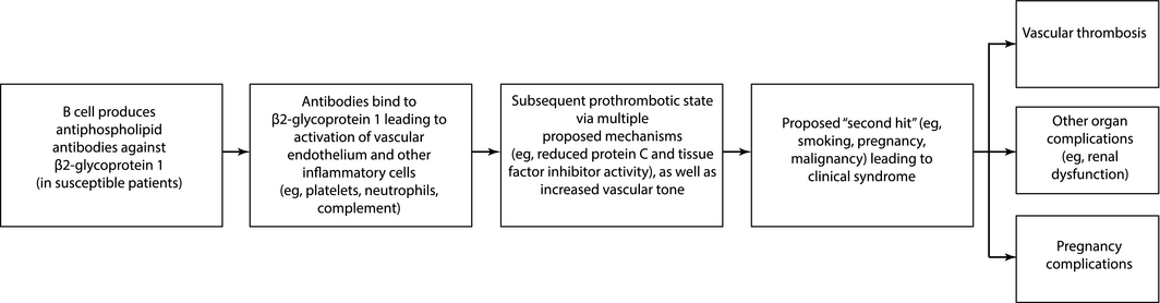
APS is frequently associated with systemic lupus erythematosis (SLE) and other autoimmune diseases, but occurs in the absence of other autoimmune disease in many cases (primary APS). In autoimmune disease, particularly SLE, the prevalence is as high as 30%.5 The pathogenesis and key clinical effects of APS are shown schematically in Box 1.
The prothrombotic state in APS is in large part due to the three characteristic antibodies: lupus anticoagulant, anticardiolipin and anti‐β2‐glycoprotein 1.6
The risk of thrombosis is increased with:
- lupus anticoagulant or antibodies against β2‐glycoprotein 1 occurring alone (higher risk of thrombosis than with anticardiolipin alone);
- high antibody titres (particularly IgG);7
- positivity for multiple antibodies7 (associated with the highest risk of thrombosis); or
- additional risk factors for thrombosis at the time of diagnosis (eg, hypertension, smoking and diabetes mellitus for arterial thrombosis,8 and hyperlipidaemia for venous thrombosis9).
The antibodies have variable associations with the specific clinical manifestations in APS, but the magnitude of these vary widely based on method of testing, cut‐off values and populations studied. In a population with SLE, the reported sensitivities of IgG anticardiolipin and anti‐β2‐glycoprotein 1 for prior thrombosis were 5.5–22.0% and 3.7–11.0% while the specificities were 86.9–95.0% and 93.7–97.7%, respectively.10 The clinical utility of these tests improves with typical disease features, multiple positive antibodies and persistence of antibodies when tested more than 12 weeks apart.
Although the exact pathogenesis of thrombosis and other complications in APS is complex and poorly understood, it is thought that these result from multiple interconnected mechanisms resulting in inflammation, vasculopathy and thrombosis.6
Clinical features
The hallmark features of APS are recurrent thrombotic events and pregnancy complications.1 Common types of venous thrombosis include deep vein thrombosis and pulmonary embolism. Stroke and transient ischaemic attack are the most common types of arterial thrombosis,11 with APS accounting for 10% of strokes in patients aged under 50 years.12 Recurrent thrombotic events are common, with an estimated annual recurrence rate of 5–12% with anticoagulation.13
Recurrent pregnancy complications are the other characteristic feature of APS. Complications include fetal death after 10 weeks’ gestation, premature birth due to severe pre‐eclampsia or placental insufficiency, and recurrent embryonic loss before 10 weeks’ duration.
Haematological abnormalities, including a prolonged activated partial thromboplastin time that fails to correct with mixing with normal plasma (owing to the presence of a lupus anticoagulant) and mild to moderate thrombocytopenia are commonly seen in APS. Less common haematological abnormalities include haemolytic anaemia and thrombotic microangiopathies (eg, thrombotic thrombocytopenia purpura).14 Other features of APS include cognitive dysfunction (even in the absence of strokes), renal disease, cardiac valvular disease, and cutaneous manifestations such as severe skin ulceration and necrosis.
Rarely, patients with APS may present with simultaneous thromboses affecting multiple organs, termed catastrophic APS, which is fatal in up to 50% of patients if not treated promptly.15
Diagnosis
There are no diagnostic criteria for APS, and caution is needed in extrapolating classification criteria developed for research purposes (Box 2)1 to clinical practice, as they have not been validated for clinical use. Less common manifestations of the disease also do not meet the research criteria.
Consider a diagnosis of APS in patients with recurrent or unexplained thrombotic events or pregnancy complications (Box 2), particularly in those who are young or have an autoimmune disease (eg, SLE). Other clinical features (eg, livedo reticularis, or an otherwise unexplained prolonged activated partial thromboplastin time) (Box 3) may support the diagnosis. However, other causes of thrombosis should be considered, including malignancy, heparin‐induced thrombocytopenia and thrombophilias.
The diagnosis of APS is established by the presence of antiphospholipid antibodies (lupus anticoagulant, anticardiolipin and anti‐β2‐glycoprotein 1) and the appropriate clinical presentation. Antibodies should be present on repeat testing at least 12 weeks apart given they can appear transiently in other conditions (eg, infection). Testing for other antiphospholipid antibodies directed at other antigens (eg, antiphosphatidylserine/prothrombin antibodies) remains controversial, and their routine use is not recommended.
False positive results may occur. Antiphospholipid antibodies may be seen in up to 12% of the general population, with the prevalence increasing with age.16 In the absence of APS, antiphospholipid antibodies may be seen with infections, medications and malignancy. Although the antiphospholipid antibodies are thought to be key to the development of thrombosis in APS, the majority of otherwise healthy individuals with antiphospholipid antibodies will not develop APS, particularly when the antibodies are seen in isolation or in low titres, or are absent on repeat testing.17
Treatment
APS is managed in conjunction with a haematologist. If associated with an autoimmune disease (such as SLE), it may also be managed by a rheumatologist. Additionally, an obstetrician is involved in the management of pregnancy‐related complications of APS.
The management of APS includes primary prophylaxis for first thrombotic event and obstetric event, secondary prophylaxis for venous and arterial thrombotic events, management of recurrent thromboses, and management of obstetric complications. Recent treatment guidelines have been proposed by international taskforces including the Haemostasis and Thrombosis Task Force of the British Committee for Standards in Haematology and the International Congress on Antiphospholipid Antibodies. In this review, we consider these as well as significant new data (Box 4).18,19,20,21,22,23,24,25,26,27,28,29,30,31,32,33,34
Primary prophylaxis
The use of aspirin to prevent a first thrombotic event in the presence of antiphospholipid antibodies remains controversial.35 However, it may be considered in patients with high risk antiphospholipid antibodies (ie, triple or multiple positivity, lupus anticoagulant, persistent medium to high titre antibodies) and if other thrombotic risk factors (eg, hypertension, smoking, diabetes, hyperlipidaemia, recent surgery) are present.18 For patients with APS associated with SLE, hydroxychloroquine has been shown to be of benefit as primary prophylaxis leading to a reduction in thromboembolic events and is thus recommended.18 However, the use of hydroxychloroquine in primary APS is currently not recommended.36
Prevention of venous thrombosis
In patients with APS who developed unprovoked venous thrombosis, anticoagulation with unfractionated heparin or low molecular weight heparin followed by a vitamin K antagonist (warfarin) aiming for an international normalised ratio (INR) of 2–3 is recommended.18 Higher intensity warfarin (INR, 3–4) has not been shown to reduce the risk of recurrent venous thrombotic events in two randomised controlled trials (RCTs)19,20 and was associated with a higher rate of bleeding.20 Anticoagulation should continue long term as the risk of recurrent thrombosis is high if it is stopped.22 Patients undergoing long distance air travel may consider adopting other general measures for venous thromboembolism prevention (eg, compression stockings).37
Prevention of arterial thrombosis
There is no consensus due to lack of high quality evidence for the optimal management of APS with arterial thrombosis. Owing to the higher rates of recurrent arterial thrombosis in APS,37 experts recommend anticoagulation with warfarin, aiming for an INR > 3.0, or combination aspirin and warfarin with an INR target of 2–3.18 Although cohort studies suggest a lower rate of recurrent thrombosis with warfarin at INR > 3,38 two RCTs did not show a difference in recurrence rate with warfarin with a higher INR target.19,20 A small RCT and retrospective review suggested a lower rate of recurrent arterial thrombosis on combination aspirin and warfarin.21,39 Data from a prospective cohort study, the Antiphospholipid Antibody and Stroke Study, suggested that warfarin or aspirin monotherapy were equally effective in preventing ischaemic stroke in patients with a prior history of stroke and a single positive antiphospholipid antibody test result.40 Therefore, some experts have suggested that such patients could be managed with aspirin alone, provided there are no other indications for anticoagulation.22
Recurrent thrombosis on anticoagulation
A recurrent thrombotic event despite therapeutic anticoagulation is a well recognised but relatively uncommon scenario.41 There is no high quality evidence to support a particular management strategy. Potential options include intensifying warfarin therapy to INR 3–4, adding aspirin (although this is associated with a higher risk of bleeding), adding hydroxychloroquine, adding a statin, using a different anticoagulant such as low molecular weight heparin, or a combination of these.23 Statins have pleiotropic immunomodulatory, anti‐inflammatory and antithrombotic properties but clinical trials are lacking.30
Obstetric APS
The current recommended treatment is low dose aspirin and prophylactic dose low molecular weight heparin.22 For patients with obstetric and thrombotic complications, treatment should include low dose aspirin and therapeutic dose low molecular weight heparin. However, up to 20% of pregnancies are unsuccessful despite treatment.42 Risk factors for an unsuccessful pregnancy include triple antiphospholipid antibody positivity, associated autoimmune disease and thrombotic manifestations.43 Treatments for refractory obstetric APS include hydroxychloroquine,24 low dose prednisolone until 14 weeks’ gestation, immunoglobulin, plasma exchange and immunoadsorption.25
Asymptomatic antiphospholipid antibody carriers should be considered for post partum thromboprophylaxis given the increased risk of thrombosis in this period.22
Low dose aspirin has been used as primary prophylaxis in patients with antiphospholipid antibodies, but there are no clear data showing a benefit44 and further studies are needed.
Catastrophic APS
Catastrophic APS is characterised by multiple thrombi with a systemic inflammatory response and has a high mortality rate.45 Due to the rarity of this condition and the high mortality rate, there are no controlled trials evaluating optimal treatment. A retrospective review from the international registry of patients with catastrophic APS46 found that anticoagulation, high dose steroids, plasma exchange and/or immunoglobulin (triple therapy) had the highest rate of survival27 and is recommended, albeit with low certainty, as the treatment for catastrophic APS.26
Novel therapies
Direct oral anticoagulants
There is insufficient evidence to make recommendations on the use of direct oral anticoagulants in APS.47 There have been two RCTs evaluating their use in APS. The first trial compared rivaroxaban and warfarin and had as its primary outcome measure the mean percentage change of thrombin potential, a marker of hypercoagulability. It found rivaroxaban to be inferior to warfarin in inhibiting thrombin generation. It examined rates of venous thromboembolism as a secondary outcome and found no difference between the two interventions as well as no difference in adverse events.48 The second trial, which examined thrombosis as the primary outcome in a high risk population (triple antiphospholipid antibody positivity) was terminated early due to a higher rate of arterial thrombotic events in the rivaroxaban arm.29 Other trials evaluating direct oral anticoagulants such as apixaban in APS are ongoing.
Immunomodulatory therapy
Immunomodulatory therapy has been increasingly investigated due to the immune‐based mechanisms involved in APS. B cell inhibition has been suggested to have a role in mouse models.49 A pilot open‐label phase 2 case series of 19 participants given rituximab suggested that it may have a role in the treatment of non‐thrombotic manifestations (eg, skin ulcers, cognitive dysfunction).31 Another case series review of 24 patients who received rituximab also noted variable improvements in skin ulcers, thrombocytopenia, cardiac valve dysfunction and cognitive dysfunction, and in two patients with thrombosis.32 The main adverse events included infectious complications. Complement activation has also been shown to initiate and amplify APS.50 There have been case reports describing the use of eculizumab, a monoclonal antibody to C5, in the treatment of APS. It was successfully used as treatment for thrombotic microangiopathy after renal transplant for patients with a prior history of systemic lupus erythrematosus and renal thrombotic microangiopathy,34 and as prophylaxis against recurrent thrombotic microangiopathy following renal transplantation.33 The main risk is infection with encapsulated organisms and patients must therefore be immunised against Neisseria meningitidis.
Practice points
A clinical vignette and tips for clinicians are provided in Box 5.
Box 2 – Revised classification criteria for antiphospholipid syndrome
Antiphospholipid syndrome is present if at least one of the clinical criteria and one of the laboratory criteria that follow are met.
Clinical criteria
- Vascular thrombosis:
- ▶ One or more clinical episodes of arterial, venous or small vessel thrombosis, in any tissue or organ. Thrombosis must be confirmed by objective validated criteria. For histopathological confirmation, thrombosis should be present without significant evidence of inflammation in the vessel wall.
- Pregnancy morbidity:
- ▶ One or more unexplained deaths of a morphologically normal fetus at or beyond the 10th week of gestation, with normal fetal morphology documented by ultrasound or by direct examination of the fetus.
- ▶ One or more premature births of a morphologically normal neonate before the 34th week of gestation because of (i) eclampsia or severe pre‐eclampsia defined according to standard definitions, or (ii) recognised features of placental insufficiency.
- ▶ Three or more unexplained consecutive spontaneous abortions before the 10th week of gestation, with maternal anatomical or hormonal abnormalities and paternal and maternal chromosomal causes excluded.
- Lupus anticoagulant present in plasma, on two or more occasions at least 12 weeks apart.
- Anticardiolipin antibody of IgG and/or IgM isotype in serum or plasma, present in medium or high titre (ie, > 40 GPL or MPL or > 99th percentile), on two or more occasions, at least 12 weeks apart.
- Anti‐β2‐glycoprotein 1 antibody of IgG and/or IgM isotype in serum or plasma (in titre > 99th percentile), present on two or more occasions, at least 12 weeks apart.
Box 3 – Non‐criteria manifestations of antiphospholipid syndrome
|
System |
Manifestation |
||||||||||||||
|
|
|||||||||||||||
|
Haematological |
|
||||||||||||||
|
Neurological |
|
||||||||||||||
|
Cardiac |
|
||||||||||||||
|
Renal |
|
||||||||||||||
|
Cutaneous |
|
||||||||||||||
|
|
|||||||||||||||
|
|
|||||||||||||||
Box 4 – Guideline‐based treatment recommendations in antiphospholipid syndrome (APS)
|
Treatment |
Strength of recommendation and quality of evidence |
||||||||||||||
|
|
|||||||||||||||
|
Thromboprophylaxis in SLE |
|||||||||||||||
|
Hydroxychloroquine18 |
Strong recommendation, moderate quality evidence |
||||||||||||||
|
Low dose aspirin18 |
Weak recommendation, moderate quality evidence |
||||||||||||||
|
Thromboprophylaxis without SLE with high risk profile |
|||||||||||||||
|
Low dose aspirin18 |
Weak recommendation, low or very low quality evidence |
||||||||||||||
|
Secondary thromboprophylaxis after venous thrombosis |
|||||||||||||||
|
Warfarin to target INR 2–318 |
Strong recommendation, moderate quality evidence |
||||||||||||||
|
Secondary thromboprophylaxis after arterial thrombosis |
|||||||||||||||
|
Warfarin to target INR > 3;18 not supported by two RCTs19,20 |
Non‐graded |
||||||||||||||
|
OR |
|||||||||||||||
|
Combined oral anticoagulant and antiplatelet;18 single RCT21 |
|||||||||||||||
|
Single‐agent antiplatelet or warfarin in patients with a single positive aPL test result22 |
Strong recommendation, moderate quality evidence |
||||||||||||||
|
Recurrent thrombosis on anticoagulation |
|||||||||||||||
|
Low molecular weight heparin, hydroxychloroquine, statin;18 intensify warfarin to INR 3–4 or add aspirin;23 lack of evidence‐based data |
Non‐graded |
||||||||||||||
|
Obstetric APS |
|||||||||||||||
|
Low dose aspirin and low molecular weight heparin22 |
Strong recommendation, moderate quality evidence |
||||||||||||||
|
Refractory obstetric APS |
|||||||||||||||
|
Add hydroxychloroquine24 |
Observational data only |
||||||||||||||
|
Add low dose prednisolone 10 mg/day up to 14th week of gestation25 |
Observational study |
||||||||||||||
|
Add intravenous immunoglobulin25 |
Pilot RCT and observational studies |
||||||||||||||
|
Add plasmapharesis25 |
Observational studies |
||||||||||||||
|
Catastrophic APS |
|||||||||||||||
|
Combination high dose steroids, heparin, plasma exchange and/or immunoglobulin;26 retrospective cohort study27 |
Conditional recommendation, low certainty of evidence |
||||||||||||||
|
Novel treatments |
|||||||||||||||
|
Direct oral anticoagulant use not routinely recommended;28 recent RCT not supportive29 |
Insufficient evidence to make recommendations |
||||||||||||||
|
Rituximab (B cell inhibition);30 based on small case series31,32 |
May have a role in difficult‐to‐treat patients, particularly those with haematological and microthrombotic manifestations |
||||||||||||||
|
Eculizumab (complement inhibition);28 based on case reports33,34 |
May have a role in life‐threatening resistant APS as salvage therapy |
||||||||||||||
|
|
|||||||||||||||
|
aPL = antiphospholipid antibodies; INR = international normalised ratio; RCT = randomised controlled trial; SLE = systemic lupus erythematosis. |
|||||||||||||||
Box 5 – Clinical vignette and tips for clinicians
Clinical vignette
- A 62‐year‐old man with antiphospholipid syndrome with previous deep vein thrombosis and pulmonary embolism complicated by chronic thromboembolic pulmonary hypertension was referred to a tertiary centre for a pulmonary endarterectomy. He was positive only for lupus anticoagulant. He was receiving long term warfarin aiming for an international normalised ratio of 2–3.
- He underwent a successful pulmonary endarterectomy but re‐presented within a few weeks after discharge with abdominal skin ulceration and necrosis, and skin biopsy showed necrosis of epidermal and dermal connective tissues with microthrombosis in small and medium sized vessels with surrounding dermal panniculitis. In case this was due to warfarin‐induced skin necrosis, he was transitioned to danaparoid but he had progressively worse skin necrosis as well as progressive thrombocytopenia with a platelet nadir of 18 × 109/L (reference interval, 150–396 × 109/L). His provisional diagnosis was a flare of antiphospholipid syndrome precipitated by surgery with non‐criteria manifestations of skin necrosis and thrombocytopenia. He was treated with a trial of immunosuppression with pulsed methylprednisolone 1 g daily for 3 days, intravenous immunoglobulin, three doses of rituximab 500 mg weekly, as well as hyperbaric oxygen therapy. Following treatment, his platelet count improved to 50 × 109/L and abdominal wounds also improved. He was safely discharged home after a 4‐week stay in hospital.
Explaining antiphospholipid syndrome to patients
- Antiphospholipid syndrome is a condition caused by the immune system which may result in clotting and pregnancy complications.
- Clots can occur anywhere in the body but particularly the heart, brain, legs and lungs.
- Problems in pregnancy include recurrent miscarriages and early pre-term birth.
- It is managed most commonly by a haematologist together with an obstetrician (if associated with pregnancy) and a rheumatologist (if associated with another disease of the immune system).
- The condition is readily treatable with blood-thinning medications that are usually given long term.
Competing interests
Huyen Tran has received grants from Bayer Health and Pfizer and speaker honoraria from Bayer Health and Boehringer Ingelheim.
Acknowledgements
We thank James McFadyen, Indi Rasaratnam, Juan Aw and Shom Bhattacharjee of Alfred Health for contributing to the content and review of this article.
References
- Miyakis S, Lockshin MD, Atsumi T, et al. International consensus statement on an update of the classification criteria for definite antiphospholipid syndrome (APS). J Thromb Haemost 2006; 4: 295–306.
- Gomez‐Puerta JA, Cervera R. Diagnosis and classification of the antiphospholipid syndrome. J Autoimmun 2014; 48–49: 20–25.
- Lockshin MD, Kim M, Laskin CA, et al. Prediction of adverse pregnancy outcome by the presence of lupus anticoagulant, but not anticardiolipin antibody, in patients with antiphospholipid antibodies. Arthritis Rheum 2012; 64: 2311–2318.
- Rai RS, Regan L, Clifford K, et al. Antiphospholipid antibodies and beta 2‐glycoprotein‐I in 500 women with recurrent miscarriage: results of a comprehensive screening approach. Hum Reprod 1995; 10: 2001–2005.
- Ünlü O, Zuily S, Erkan D. The clinical significance of antiphospholipid antibodies in systemic lupus erythematosus. Eur J Rheumatol 2016; 3: 75–84.
- Giannakopoulos B, Krilis SA. The pathogenesis of the antiphospholipid syndrome. N Engl J Med 2013; 368: 1033–1044.
- Kelchtermans H, Pelkmans L, de Laat B, Devreese KM. IgG/IgM antiphospholipid antibodies present in the classification criteria for the antiphospholipid syndrome: a critical review of their association with thrombosis. J Thromb Haemost 2016; 14: 1530–1548.
- Zuo Y, Fan J, Sarode R, et al. Identifying additional risk factors for thrombosis and pregnancy morbidities among antiphospholipid antibodies carriers. Clin Appl Thromb Hemost 2018; 24: 980–985.
- Danowski A, de Azevedo MN, de Souza Papi JA, Petri M. Determinants of risk for venous and arterial thrombosis in primary antiphospholipid syndrome and in antiphospholipid syndrome with systemic lupus erythematosus. J Rheumatol 2009; 36: 1195–1199.
- Vikerfors A, Johansson A‐B, Gustafsson JT, et al. Clinical manifestations and anti‐phospholipid antibodies in 712 patients with systemic lupus erythematosus: evaluation of two diagnostic assays. Rheumatology 2013; 52: 501–509.
- Cervera R, Piette JC, Font J, et al. Antiphospholipid syndrome: clinical and immunologic manifestations and patterns of disease expression in a cohort of 1,000 patients. Arthritis Rheum 2002; 46: 1019–1027.
- Sciascia S, Sanna G, Khamashta MA, et al. The estimated frequency of antiphospholipid antibodies in young adults with cerebrovascular events: a systematic review. Ann Rheum Dis 2015; 74: 2028–2033.
- Cervera R, Serrano R, Pons‐Estel GJ, et al. Morbidity and mortality in the antiphospholipid syndrome during a 10‐year period: a multicentre prospective study of 1000 patients. Ann Rheum Dis 2015; 74: 1011–1018.
- Uthman I, Godeau B, Taher A, Khamashta M. The hematologic manifestations of the antiphospholipid syndrome. Blood Rev 2008; 22: 187–194.
- Bucciarelli S, Espinosa G, Cervera R, et al. Mortality in the catastrophic antiphospholipid syndrome: causes of death and prognostic factors in a series of 250 patients. Arthritis Rheum 2006; 54: 2568–2576.
- Andreoli L, Chighizola CB, Banzato A, et al. Estimated frequency of antiphospholipid antibodies in patients with pregnancy morbidity, stroke, myocardial infarction, and deep vein thrombosis: a critical review of the literature. Arthritis Care Res (Hoboken) 2013; 65: 1869–1873.
- McNeil HP, Chesterman CN, Krilis SA. Immunology and clinical importance of antiphospholipid antibodies. Adv Immunol 1991; 49: 193–280.
- Ruiz‐Irastorza G, Cuadrado MJ, Ruiz‐Arruza I, et al. Evidence‐based recommendations for the prevention and long‐term management of thrombosis in antiphospholipid antibody‐positive patients: report of a task force at the 13th International Congress on antiphospholipid antibodies. Lupus 2011; 20: 206–218.
- Crowther MA, Ginsberg JS, Julian J, et al. A comparison of two intensities of warfarin for the prevention of recurrent thrombosis in patients with the antiphospholipid antibody syndrome. N Engl J Med 2003; 349: 1133–1138.
- Finazzi G, Marchioli R, Brancaccio V, et al. A randomized clinical trial of high‐intensity warfarin vs. conventional antithrombotic therapy for the prevention of recurrent thrombosis in patients with the antiphospholipid syndrome (WAPS). J Thromb Haemost 2005; 3: 848–853.
- Okuma H, Kitagawa Y, Yasuda T, et al. Comparison between single antiplatelet therapy and combination of antiplatelet and anticoagulation therapy for secondary prevention in ischemic stroke patients with antiphospholipid syndrome. Int J Med Sci 2009; 7: 15–18.
- Keeling D, Mackie I, Moore GW, et al. Guidelines on the investigation and management of antiphospholipid syndrome. Br J Haematol 2012; 157: 47–58.
- Garcia D, Erkan D. Diagnosis and management of the antiphospholipid syndrome. N Engl J Med 2018; 378: 2010–2021.
- Mekinian A, Costedoat‐Chalumeau N, Masseau A, et al. Obstetrical APS: is there a place for hydroxychloroquine to improve the pregnancy outcome? Autoimmun Rev 2015; 14: 23–29.
- Ruffatti A, Hoxha A, Favaro M, et al. Additional treatments for high‐risk obstetric antiphospholipid syndrome: a comprehensive review. Clin Rev Allergy Immunol 2017; 53(1): 28–39.
- Legault K, Schunemann H, Hillis C, et al. McMaster RARE‐Bestpractices clinical practice guideline on diagnosis and management of the catastrophic antiphospholipid syndrome. J Thromb Haemost 2018; 16: 1656–1664.
- Rodríguez‐Pintó I, Espinosa G, Erkan D, et al. The effect of triple therapy on the mortality of catastrophic anti‐phospholipid syndrome patients. Rheumatology (Oxford) 2018; 57: 1264–1270.
- Andrade D, Cervera R, Cohen H, et al. 15th International Congress on Antiphospholipid Antibodies Task Force on Antiphospholipid Syndrome Treatment Trends Report. In: Erkan D, Lockshin MD, editors. Antiphospholipid syndrome: current research highlights and clinical insights. Springer International Publishing, 2017: p. 317–338.
- Pengo V, Denas G, Zoppellaro G, et al. Rivaroxaban vs warfarin in high‐risk patients with antiphospholipid syndrome. Blood 2018; 132: 1365.
- Erkan D, Aguiar CL, Andrade D, et al. 14th International Congress on Antiphospholipid Antibodies: task force report on antiphospholipid syndrome treatment trends. Autoimmun Rev 2014; 13: 685–696.
- Erkan D, Vega J, Ramón G, et al. A pilot open‐label phase II trial of rituximab for non‐criteria manifestations of antiphospholipid syndrome. Arthritis Rheum 2013; 65: 464–471.
- Pons I, Espinosa G, Cervera R. [Efficacy and safety of rituximab in the treatment of primary antiphospholipid syndrome: analysis of 24 cases from the bibliography review]. Medicina Clinica 2015; 144: 97–104.
- Lonze BE, Singer AL, Montgomery RA. Eculizumab and renal transplantation in a patient with CAPS. N Engl J Med 2010; 362: 1744–1745.
- Hadaya K, Ferrari‐Lacraz S, Fumeaux D, et al. Eculizumab in acute recurrence of thrombotic microangiopathy after renal transplantation. Am J Transplant 2011; 11: 2523–2527.
- Arnaud L, Conti F, Massaro L, et al. Primary thromboprophylaxis with low‐dose aspirin and antiphospholipid antibodies: pro's and con's. Autoimmun Rev 2017; 16: 1103–1108.
- Wang T‐F, Lim W. What is the role of hydroxychloroquine in reducing thrombotic risk in patients with antiphospholipid antibodies? Hematology Am Soc Hematol Educ Program 2016; 2016: 714–716.
- Guyatt GH, Akl EA, Crowther M, et al. Executive summary: Antithrombotic therapy and prevention of thrombosis, 9th ed: American College of Chest Physicians Evidence‐Based Clinical Practice Guidelines. Chest 2012; 141: 7S–47S.
- Ruiz‐Irastorza G, Hunt BJ, Khamashta MA. A systematic review of secondary thromboprophylaxis in patients with antiphospholipid antibodies. Arthritis Rheum 2007; 57: 1487–1495.
- Jackson WG, Oromendia C, Unlu O, et al. Recurrent thrombosis in patients with antiphospholipid antibodies and arterial thrombosis on antithrombotic therapy. Blood Adv 2017; 1: 2320–2324.
- Levine SR, Brey RL, Tilley BC, et al. Antiphospholipid antibodies and subsequent thrombo‐occlusive events in patients with ischemic stroke. JAMA 2004; 291: 576–584.
- Bazzan M, Vaccarino A, Stella S, et al. Patients with antiphosholipid syndrome and thrombotic recurrences: a real world observation (the Piedmont cohort study). Lupus 2016; 25: 479–485.
- de Jesus GR, dos Santos FC, Oliveira CS, et al. Management of obstetric antiphospholipid syndrome. Curr Rheumatol Rep 2012; 14: 79–86.
- Ruffatti A, Tonello M, Visentin MS, et al. Risk factors for pregnancy failure in patients with anti‐phospholipid syndrome treated with conventional therapies: a multicentre, case‐control study. Rheumatology (Oxford) 2011; 50: 1684–1689.
- Amengual O, Fujita D, Ota E, et al. Primary prophylaxis to prevent obstetric complications in asymptomatic women with antiphospholipid antibodies: a systematic review. Lupus 2015; 24: 1135–1142.
- Cervera R, Rodríguez‐Pintó I, Espinosa G. The diagnosis and clinical management of the catastrophic antiphospholipid syndrome: a comprehensive review. J Autoimmun 2018; 92: 1–11.
- Cervera R. CAPS Registry. Lupus 2012; 21: 755–757.
- Dufrost V, Risse J, Reshetnyak T, et al. Increased risk of thrombosis in antiphospholipid syndrome patients treated with direct oral anticoagulants. Results from an international patient‐level data meta‐analysis. Autoimmun Rev 2018; 17: 1011–1021.
- Cohen H, Hunt BJ, Efthymiou M, et al. Rivaroxaban versus warfarin to treat patients with thrombotic antiphospholipid syndrome, with or without systemic lupus erythematosus (RAPS): a randomised, controlled, open‐label, phase 2/3, non‐inferiority trial. Lancet Haematol 2016; 3: e426–e436.
- Kahn P, Ramanujam M, Bethunaickan R, et al. Prevention of murine antiphospholipid syndrome by BAFF blockade. Arthritis Rheum 2008; 58: 2824–2834.
- Unlu O, Erkan D. Catastrophic antiphospholipid syndrome: candidate therapies for a potentially lethal disease. Ann Rev Med 2017; 68: 287–296.
Provenance: Commissioned; externally peer reviewed.
Splenic injury in severe cases of the zoonoses Q fever and rickettsial infection: diagnostic challenges
Ashleigh Drury, Philippa Harrison, Aiveen Bannan
Health and economic benefits of improving pre‐hospital identification of stroke in Australian women: a modelling study
Thomas Gadsden, Lei Si, Emily R Atkins, Cheryl Carcel, Xia Wang, Stephen Jan, Mark Woodward, Laura E Downey
Updating the diagnosis and management of elevated serum ferritin levels in the era of routine ferritin testing of blood donors by Australian Red Cross Lifeblood
Gary D Zhang, James Chen, Daniel M Johnstone, Martin B Delatycki, Katie Allen, John K Olynyk
Early cardiovascular collapse after envenoming by snakes in Australia, 2005–2020: an observational study (ASP‐31)
Geoffrey K Isbister, Katherine Z Isoardi, Angela L Chiew, Shane Jenkins, Nicholas A Buckley
Updated recommendations for warfarin reversal in the setting of four‐factor prothrombin complex concentrate
Danielle Robinson, James McFadyen, Eileen Merriman, Chee Wee Tan, Ross Baker, Huyen Tran
Updating the diagnosis and management of iron deficiency in the era of routine ferritin testing of blood donors by Australian Red Cross Lifeblood
Gary D Zhang, Daniel Johnstone, Michael F Leahy, John K Olynyk