




Volume 210 - Issue 4
Current management of glaucoma
Authors: Jed Lusthaus and Ivan Goldberg
Med J Aust 2019; 210 (4): 180-187. || doi: 10.5694/mja2.50020
Published online: 4 March 2019
Published online: 4 March 2019
Glaucoma management varies depending on the underlying causative mechanism, with options trending towards earlier surgical intervention for both open-angle and angle closure glaucoma. While the increased acceptance of SLT and the introduction of MIGS devices have started to change the face of glaucoma management, IOP-lowering eye drops remain the foundation of treatment. Adherence is an ongoing treatment limitation and future therapies are being designed to diminish this.
Summary
- Glaucoma is an irreversible progressive optic neuropathy, for which the major proven treatment is to lower the intraocular pressure (IOP).
- Five groups of IOP‐lowering eye drops have varying mechanisms of action. Some drops, such as β‐blockers and α‐2 agonists, have potentially serious systemic side effects. Acetazolamide is the only available oral agent; it is effective at lowering IOP, but significant side effects relegate its use usually to refractory glaucoma.
- Two new eye drops, netarsudil and latanoprostene bunod, have recently been approved by the United States Food and Drug Administration. Both have novel IOP‐lowering mechanisms and target the conventional aqueous outflow system.
- Selective laser trabeculoplasty is a gentle treatment that enhances conventional aqueous outflow. It may be used as an initial treatment, as a substitute for eye drops, or to delay glaucoma drainage surgery.
- Recent advancements in glaucoma surgery have seen an influx of minimally invasive glaucoma surgery devices, which are being used more frequently and earlier on in the treatment paradigm. As limited long term data are available, trabeculectomy remains the gold standard IOP‐lowering procedure. Improvements in drug delivery are on the horizon. Drug‐eluting devices and implants are able to deliver the drug closer to the receptors for an extended period of time. This will improve treatment adherence and efficacy, which are major limitations with current medical therapy.
Glaucoma management is evolving: safer and less invasive surgical interventions are now in regular use, and the United States Food and Drug Administration has approved new topical drop classes. Lowering intraocular pressure (IOP) remains the primary focus of glaucoma management,1,2,3,4 and it is the goal of current and new therapies.
This review outlines glaucoma management for adults — paediatric glaucoma is a different entity that requires variations in management and will not be discussed. A PubMed search identified original and review articles from 1983 to 2018 used to guide this review.
Background
Glaucoma is a group of progressive optic neuropathies associated with characteristic visual field defects and structural changes in the optic nerve head (Box 1).1,2,3,4 IOP is the primary modifiable risk factor, but at diagnosis, patients may have elevated or “normal” (≤ 21 mmHg) IOP (Box 2).5,6
Glaucoma is classified as open‐ or closed‐angle, primary or secondary (Box 3). Secondary glaucoma may be associated with underlying systemic inflammatory, vascular or malignant causes as well as specific ocular conditions.8 There is often crossover in the management of glaucoma, but the underlying cause is targeted when possible.
Epidemiology
Although glaucoma most commonly progresses slowly, it is the most common cause of irreversible preventable blindness in the world.9 Visual symptoms are often a late feature of glaucoma, when the disease is already very advanced. Glaucoma can occur in any population, but is more pronounced and severe in African (open‐angle) and Asian (especially angle closure) populations.9,10
In Australia, the prevalence of glaucoma is estimated at 3%.11,12 Eighty million people worldwide are predicted to have glaucoma by 2020, with 11 million being bilaterally blind.13 While half of the population with glaucoma in high income countries is unaware of their disease, this figure is over 90% in low income countries, particularly in rural settings.9 To minimise this problem, we must identify and treat those patients at greatest risk of glaucoma blindness (Box 4).
Goals of management
All patients with glaucoma may benefit from adequate IOP lowering to slow or prevent disease progression.1,2,3,4,5,6 The mode and level of treatment are determined by a number of factors, including, but not limited to, glaucoma subtype, estimated life expectancy, visual prognosis, and ocular and systemic comorbidities. The ultimate goal of glaucoma management is to maintain visual independence. Some levels of glaucoma progression may be tolerated in patients for whom functional vision is not at threat in their estimated lifetime.
Some patients with glaucoma show disease progression despite IOP reduction,5 which has led to an extensive search to identify neuroprotective molecules. While some molecules have shown promise in animal models, human clinical trials have been disappointing. Endpoints for success in these studies are harder to define.14 Thus, IOP‐lowering treatments remain the primary focus of glaucoma management and the primary focus of this review.
Current management
Patients with glaucoma may be treated with medical therapy, laser or surgery depending on the underlying cause and stage of disease (Box 5).8 A definitive diagnosis can be difficult to make, given the insidious nature of glaucoma. A period of observation without treatment may be appropriate for some patients and may prevent a lifetime of unnecessary treatment.
Open‐angle glaucoma
Even though primary and secondary open‐angle glaucoma are managed similarly, secondary open‐angle glaucoma is often more aggressive and may require faster treatment escalation to control the disease.6 This includes treating the underlying cause if possible; for example, controlling inflammatory eye disease as well as the secondary rise in IOP.
Topical therapy
IOP‐lowering eye drops continue to be the mainstay of glaucoma management. There are many individual agents in five therapeutic groups, each with its mechanism of action and potential side effects, summarised in Box 6.
Prostaglandin analogues
Prostaglandin analogues (PGAs) are the most efficacious and well tolerated group of IOP‐lowering eye drops. With once daily dosing, their 24‐hour efficacy is unrivalled.15 Along with a favourable safety profile, these properties often result in PGAs being the first‐line choice.16,17 PGAs reduce IOP by enhancing aqueous humor drainage through the uveoscleral outflow pathway, which usually accounts for 10% of aqueous outflow.17
PGAs have a distinctive set of side effects, most of which are cosmetic. Hypertrichosis (thicker, longer, darker lashes) occurs within 12 months in 50% of patients. Possibly the most popular PGA side effect, but especially in Asian individuals, lashes may abrade the cornea, smear spectacles and/or impede drop instillation.18 Periocular skin pigmentation may occur, more commonly in darker skinned patients. Both effects are reversible on cessation. Moreover, patients with blue or hazel irides should be warned that an increase in iris pigmentation may occur. Prostaglandin‐associated periorbitopathy is another irreversible cosmetic side effect of PGAs and can result in periorbital fat atrophy, ptosis, eyelid tightness and deepening of the upper eyelid sulcus.19 Mild ocular hyperaemia may occur on commencement of PGAs. If this does not subside after 2–4 weeks, then preservative or PGA drop allergy should be considered. The intensity of side effects varies between PGA agents. Substitution within the class may combat intolerance and/or ineffectiveness.20,21
β‐Blockers
Until the introduction of latanoprost (PGA) in 1998, β‐blockers (eg, timolol) were the initial choice for IOP reduction. They were surpassed due to the superior efficacy of PGAs and the potential of β‐blockers to induce life‐threatening side effects, such as bronchoconstriction, bradyarrhythmias and systemic hypotension, as well as possibly masking impending hypoglycaemia in patients with insulin‐dependent diabetes.22
β‐Blockers bind to β‐1 and/or β‐2 adrenergic receptors in the ciliary body to reduce aqueous humor production.23 Initially, twice daily dosing was recommended, but for most patients, once daily dosing is as effective, more convenient and safer. Dosing in the morning is preferable to minimise nocturnal systemic hypotension and, thus, optic nerve hypoperfusion, which may induce glaucoma progression despite apparent IOP control.24
α‐2 Agonists
α‐2 Agonists are possibly the least tolerated IOP‐lowering agents due to high rates of conjunctival hyperaemia and localised allergic changes.25 In affected patients, allergies usually develop within weeks of treatment commencement, but may be delayed up to 3 years.26 Dry mouth and drowsiness are well described potential adverse effects.
Brimonidine purite 0.15% contains a more gentle preservative than the more commonly used benzalkonium chloride, and together with the decreased drug concentration (compared with 0.2%), it reduces somewhat the rates of allergic reaction. It is the most commonly used α‐2 agonist and is often a third‐ or fourth‐line treatment option.27 IOP is lowered by a double mechanism: reducing aqueous humor production and increasing uveoscleral outflow.28
Carbonic anhydrase inhibitors
Possibly the least effective, topical carbonic anhydrase inhibitors (CAIs) lower IOP by reducing aqueous humor production.29,30 They are safe, generally well tolerated, and often used as third‐ or fourth‐line agents. Sulfonamide allergies are a relative contraindication.
By contrast, acetazolamide is an effective oral preparation CAI with a poor systemic safety profile. It is most commonly used in the setting of an acutely raised IOP for short term control, or in patients with recalcitrant glaucoma either unsuitable for surgery or while surgery is being planned.31 It is common for patients to experience peripheral tingling, nausea, dysgeusia and general weakness. Electrolyte imbalances and Stevens–Johnson syndrome occur infrequently but carry life threatening potential.31
Muscarinic receptor agonists
Topical pilocarpine eye drops contract the ciliary muscle and iris constrictor muscles to increase trabecular outflow facility to reduce IOP.32 Pilocarpine is no longer routinely used for long term IOP control due to a poor side effect profile — ocular effects include miosis (dim vision), refractive myopia (blurred vision), brow ache (from ciliary spasm), and even retinal detachment. More rarely, systemic side effects may include vomiting, diarrhoea, tachycardia and bronchospasm.32
Pilocarpine is now most commonly used as a preparatory agent before laser treatment for glaucoma or in the context of acute angle closure.
Treatment compliance
As in the treatment of all chronic relatively asymptomatic conditions, adherence to topical eye drop therapy remains one of the toughest challenges in the management of glaucoma. Even in a study in which patients knew they were being monitored with an electronic device, they did not consistently take their drops in 45% of cases.33 There are multiple reasons for reduced treatment adherence, including medication side effects, poor understanding of treatment aims, poor instillation techniques (including physical barriers; eg, arthritis and tremor), and cost.34,35
IOP‐lowering eye drops have evolved to improve adherence rates. There are now many commercially available fixed combination eye drops, which enable two agents to be instilled with a single drop, reducing preservative load and improving convenience and thus adherence.36 All combination drops incorporate timolol apart from one that contains brimonidine and brinzolamide (Box 7).
Preservative‐free eye drop formulations have helped patients with glaucoma, who frequently have concurrent dry eye and ocular surface disruption. Reducing preservative load can reduce patient discomfort and improve quality of life.37 Limited preservative‐free options are commercially available in Australia and include bimatoprost, fixed combination bimatoprost–timolol, and tafluprost. Other drops (eg, timolol) may be available in preservative‐free formulations as minims from specialist compounding pharmacies.
Laser therapy
Selective laser trabeculoplasty (SLT) has overtaken argon laser trabeculoplasty as the preferred technique to increase conventional aqueous outflow through the trabecular meshwork. While the IOP‐lowering effect is similar, SLT is less destructive to the trabecular meshwork than is argon laser trabeculoplasty.38,39
SLT was originally offered to patients who had failed medical management (lack of IOP control or intolerance to eye drops), but it is also available as a first‐line treatment option.39 SLT is well tolerated, with few side effects, and can be repeated multiple times. It is effective in around 80% of patients, lowering IOP equivalent to one medication, but has a failure rate of 50% at 2 years.39
Surgical intervention
A multitude of less invasive surgical procedures are now available with increased safety compared with traditional drainage surgeries, trabeculectomy and tube shunt insertion. This has moved incisional surgery to an earlier part of the management spectrum.
Surgical intervention is required when a patient's visual independence is at risk, despite the use of medical and/or laser treatment options. Other indications include intolerance or poor adherence to medical treatment.
Trabeculectomy
Trabeculectomy surgery has been the gold standard in surgical intervention since its development in 1968,39 with ongoing evolution. IOP reduction < 21 mmHg is achieved in 86–96% of patients with or without IOP‐lowering medications.40,41
Despite many variations in surgical technique, the overall aim is to create an alternative drainage pathway for aqueous humor to exit the eye. A small fistula between the anterior chamber and subconjunctival space is made under a partial‐thickness scleral flap. Fine sutures are inserted into the scleral flap to titrate IOP levels after surgery. A drainage bleb is fashioned by creating a watertight closure in the overlying conjunctiva. Blebs are most commonly placed superiorly so that the upper eyelid can provide protective cover, which reduces the risk of infection.
The intraoperative use of antimetabolites such as 5‐fluorouracil and, now more commonly due to greater efficacy, mitomycin C reduces the risk of post‐operative conjunctival scarring and bleb failure.40 While this has improved long term bleb success and improved IOP control, there is a higher rate of pathologically low IOP (hypotony), bleb leak, and late onset endophthalmitis.40 Despite these risks, trabeculectomy remains, on the whole, safe and reasonably predictable.
Tube shunt
Tube shunt surgery is an acceptable alternative to trabeculectomy surgery. It is the preferred surgical option in refractory glaucoma or in cases in which trabeculectomy failure rates are higher (eg, neovascular and uveitic glaucoma).42
Unlike trabeculectomy surgery, drainage “hardware” remains in the patient's eye indefinitely after tube shunt surgery to reduce the impact of scarring and to maintain the outflow passage. Therefore, tube shunt surgery is often used in the context of previously failed trabeculectomy surgery and in patients prone to scar tissue formation.42
Tube shunts are made of silicone or polypropylene, both of which will not decompose in the body. The tube is placed in the patient's anterior chamber or sometimes just behind the iris if this is deemed to be safer. The tube is connected to a large plate, which forms the drainage reservoir. The plate is anchored usually behind the superior and lateral recti onto the sclera to reduce displacement.
In Australia, the most commonly used tube shunt is the Baerveldt tube. It does not have a valve, meaning that fluid can freely drain from the eye. By providing resistance to flow, the capsule that forms around the tube plate limits fluid drainage. This process takes up to 6 weeks, so surgeons commonly use a dissolvable suture tie to occlude the tube during this period.42 IOP reduction is more safely achieved with this method, which mitigates hypotony. However, IOP can be hard to control in the early post‐operative period and there is an increased risk of bullous keratopathy in the long term.42
Minimally invasive glaucoma surgery
A plethora of new minimally invasive glaucoma surgery (MIGS) devices has been developed to try to provide safer and more efficient IOP lowering (Box 8). MIGS devices are inserted through an ab interno approach and can be used alone or in conjunction with cataract surgery.43 They are associated with little or no tissue manipulation, which reduces surgical time and hastens visual recovery.44
To drain aqueous humor from the anterior chamber, MIGS devices target three anatomical spaces. The safest to target is the Schlemm canal, which is achieved by bypassing the trabecular meshwork. It is safe because it allows the device to tap into the natural conventional outflow pathways of the eye, buffered by episcleral venous pressure, eliminating the risk of hypotony. This floor effect prevents IOPs low enough to control patients with more advanced glaucoma, who often require IOP levels in the low teens or single digits for visual safety.5
The two other targeted drainage spaces are the suprachoroidal and subconjunctival spaces, both of which are non‐physiological. Tissue resistance is relied upon to limit aqueous outflow, meaning greater potential for lower IOPs, but also making hypotony a possibility. As with traditional glaucoma drainage surgery, scar tissue formation may occur, resulting in failure of the device. Concurrent intraoperative use of mitomycin C is being used to reduce bleb scarring for devices placed in the subconjunctival space, much like in trabeculectomy surgery.45 Devices that target the suprachoroidal and subconjunctival spaces may be more suitable for patients with more advanced glaucoma, but currently many MIGS devices are marketed towards patients with mild or moderate glaucoma.
No proven treatment algorithm has been developed yet to identify patients most likely to benefit from each drainage pathway or MIGS device. Safer and predictable surgery is a priority for patients with glaucoma, but the body of evidence for MIGS efficacy remains limited.43 Further device experience and larger randomised trials are required to achieve these goals.
Cyclodiode laser
Refractory glaucoma or patients with limited visual potential may require cyclophotocoagulation using a diode laser. This lowers IOP by destroying part of the ciliary body to reduce aqueous humor production.46,47 Often, the treatment goal is to control pain associated with high IOPs in eyes with poor vision.45 Cyclodiode laser is quick to perform and may lower IOP by 45%, but this has a risk,47 as persistent hypotony occurs in 10% of patients and may lead to total loss of vision and phthisis bulbi.46
Recently introduced micropulse cyclophotocoagulation laser delivery appears to be safer than the traditional continuous wave diode delivery. Micropulse laser is delivered in a series of short pulses with rest periods, which is less destructive than continuous high energy pulses from continuous wave delivery.47 Further studies are required to compare the effectiveness of these laser delivery systems.
Angle closure
Primary angle closure glaucoma is often more severe than open‐angle glaucoma and more likely to result in blindness.48 The mechanism of raised IOP is directly related to mechanical obstruction of the trabecular meshwork, usually by the peripheral iris. The treatment paradigm is different from open‐angle glaucoma. Angle closure glaucoma is predominantly a surgical disease.
Acute presentations of primary angle closure are associated with high IOPs. Episodes are treated emergently with medical therapy and laser peripheral iridotomy to open the drainage angle. Glaucomatous damage may not occur and visual loss is uncommon, provided the acute episode is rapidly reversed.48 Despite laser peripheral iridotomy, some patients will require lens extraction to maximise the opening of the drainage angle, regardless of their degree of cataract.48
A variation in this standard of care has been proposed for patients with primary angle closure glaucoma. A multinational randomised controlled study recently showed positive results compared with standard care by performing clear lens extraction as a primary treatment for primary angle closure and primary angle closure glaucoma. The clear lens extraction group showed a lower mean IOP, reduced rates of subsequent glaucoma drainage surgery, superior cost effectiveness, and improved quality of life scores.48 Clear lens extraction is now considered a first‐line treatment option for primary angle closure and primary angle closure glaucoma.
A chronic form of primary angle closure glaucoma may develop despite successful laser peripheral iridotomy, laser iridoplasty or even cataract surgery. Management is similar to what has been outlined for primary open‐angle glaucoma.
Future glaucoma treatments
Eye drops
PGAs were the last drug group to be introduced into the glaucoma market in 1998, when latanoprost was launched. Two decades later, the Food and Drug Administration has approved netarsudil 0.02%, which is a rho kinase inhibitor. Netarsudil is dosed daily and lowers IOP by enhancing trabecular outflow, reducing aqueous humor secretion and possibly lowering episcleral venous pressure.49 Other than the seldom used miotics, no other currently available IOP‐lowering agent acts on the trabecular meshwork to lower IOP. Netarsudil is less effective than PGAs, but will likely make a good second‐line agent to be tried alone or in combination with PGAs. It is associated with significant rates of conjunctival hyperaemia, but is systemically safer than timolol.
The Food and Drug Administration also approved latanoprostene bunod late in 2017. It is a nitric oxide‐donating PGA. Nitric oxide enhances aqueous outflow through the trabecular meshwork. Latanoprostene bunod is more effective than latanoprost and has a similar safety profile.50
Neither of these new agents is available in Australia to date.
Drug delivery
Many novel drug delivery systems are in phase 1 and 2 clinical trials. The goal is to improve drug delivery to the appropriate receptors, which will improve efficacy and adherence and will reduce the side effects associated with current topical IOP‐lowering drugs.7 Drug‐eluting punctal plugs, conjunctival ocular ring inserts, subconjunctival injections and implants, and intracameral implants are all being studied. This is an exciting area and will improve the treatment experience for many patients.
The role of non‐specialists in glaucoma
The wider medical community is well positioned to improve greatly the lives of patients with glaucoma. Encouraging all patients to regularly seek review by an eye care professional every 1–2 years from 50 years of age facilitates earlier detection and treatment. Risk factor identification in the context of increasing age should raise suspicion for glaucoma. These risks include family history, obstructive sleep apnoea, vasospastic syndromes (migraine, Raynaud phenomenon), systemic hypertension, and diabetes mellitus.
Health care professionals, particularly general practitioners, can assist by encouraging management adherence. This may be achieved by discussing with the patient their attitude towards glaucoma and its treatment, reinforcing the importance of regular and timely eye drop administration, and identifying potential treatment side effects and possible physical barriers to eye drop self‐administration. Finding an alternative treatment option is preferable to non‐adherence owing to side effects.
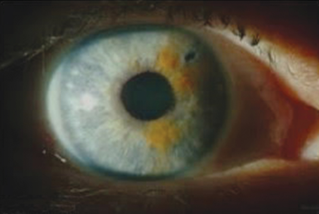
Acute angle closure should be considered in the context of a fixed mid‐dilated pupil, reduced visual acuity, and a slit‐like anterior chamber. Shining a torchlight from the temporal side of the eye will cast a shadow over the nasal iris (Box 9), highlighting possible angle closure. Urgent referral to an ophthalmologist should be sought if acute angle closure is suspected.
Conclusion
Glaucoma management varies depending on the underlying causative mechanism, with options trending towards earlier surgical intervention for both open‐angle and angle closure glaucoma. While the increased acceptance of SLT and the introduction of MIGS devices have started to change the face of glaucoma management, IOP‐lowering eye drops remain the foundation of treatment. Adherence is an ongoing treatment limitation and future therapies are being designed to diminish this.
Box 1 – Figure showing a normal left optic disc (A)* and a left optic disc with end‐stage glaucoma (B)†
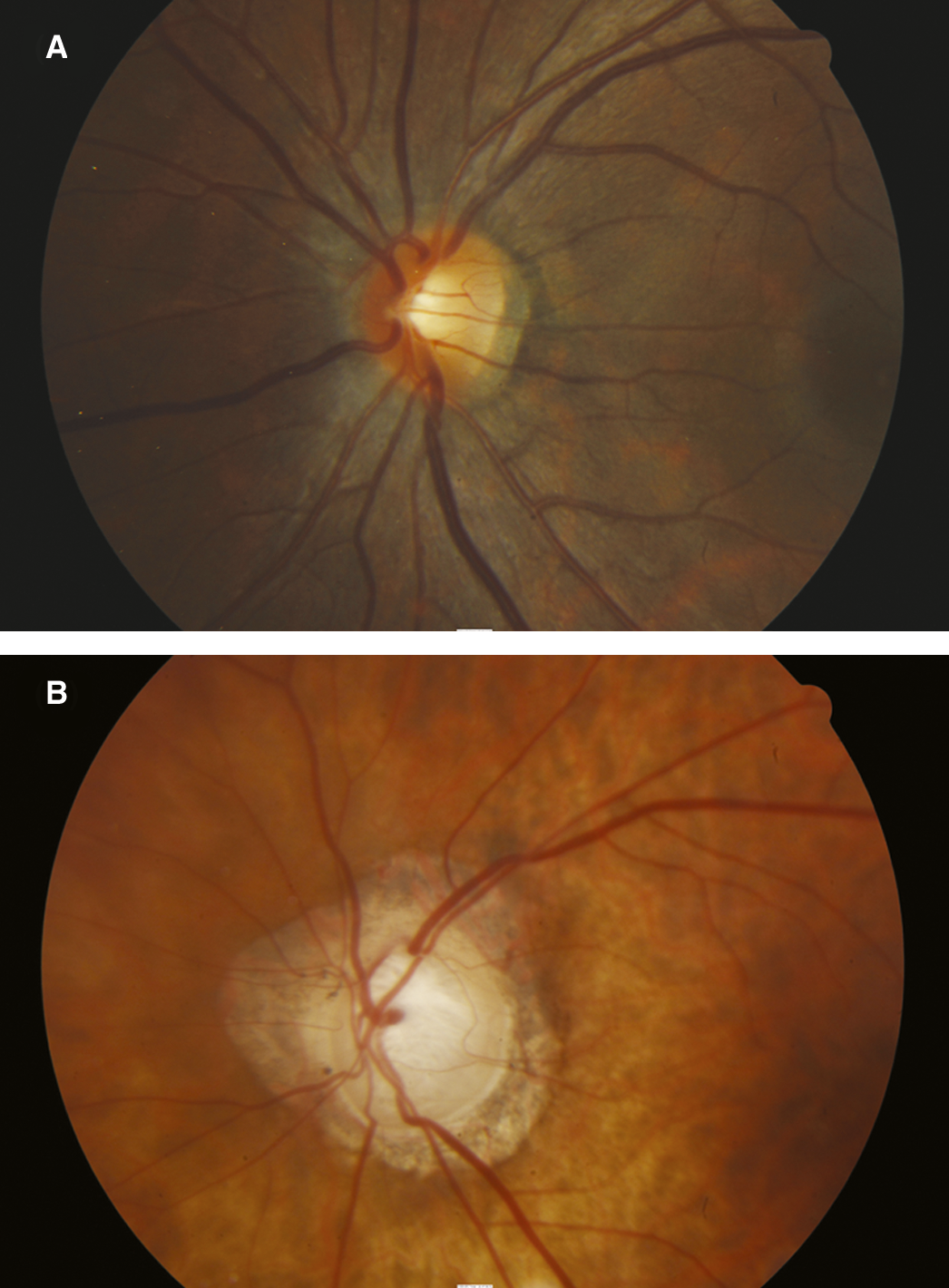
* Normal optic disc with healthy neuroretinal rim (A). † End‐stage glaucomatous optic disc with marked thinning of the neuroretinal rim, extensive cupping, and pallor (B). ◆
Box 2 – Glaucoma diagnosis flowchart
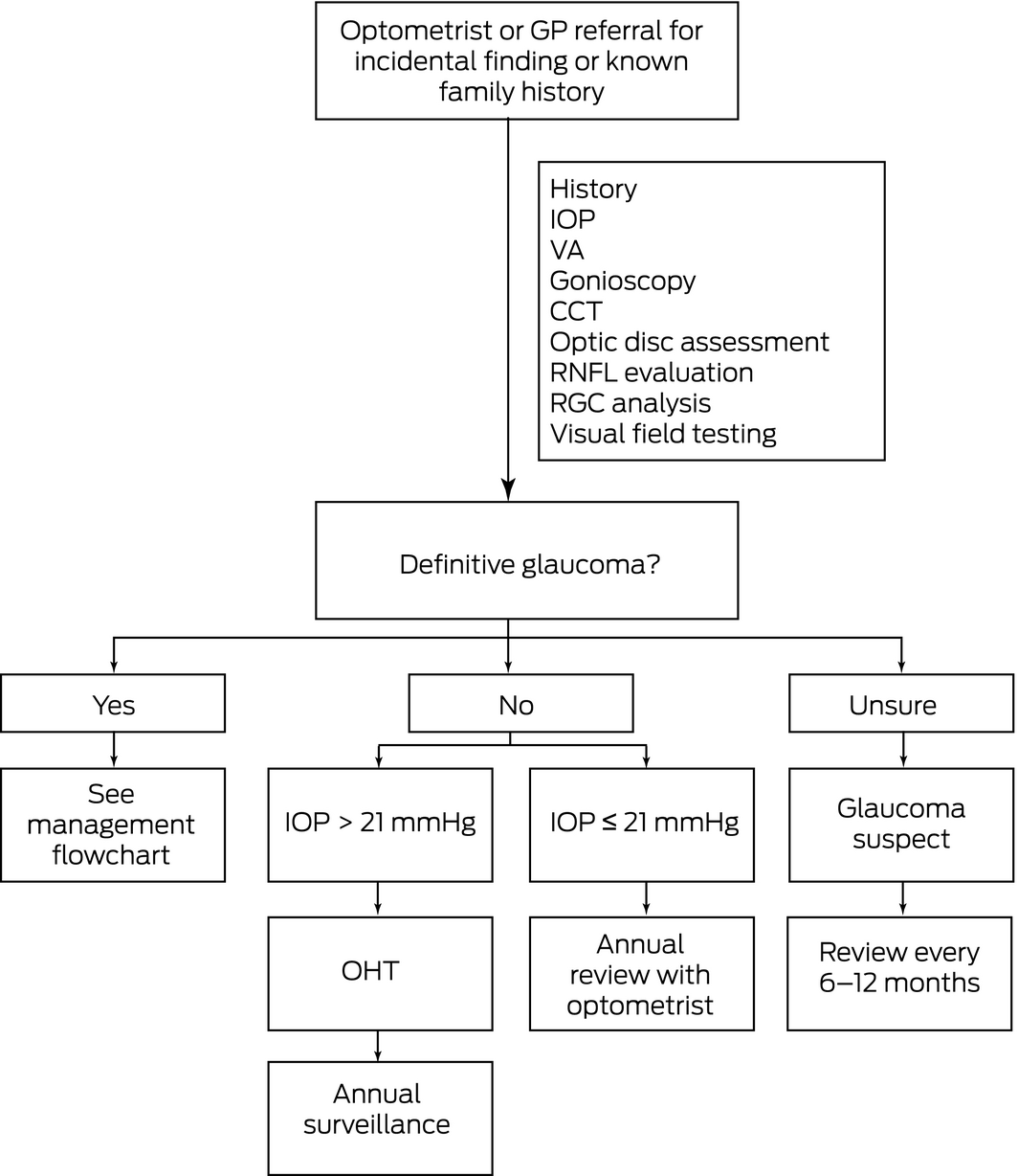
CCT = central corneal thickness. GP = general practitioner. IOP = intraocular pressure. OHT = ocular hypertension. RGC = retinal ganglion cell. RNFL = retinal nerve fibre layer thickness. VA = visual acuity. ◆
Box 3 – Glaucoma classification7
|
Open‐angle |
Angle closure |
||||||||||||||
|
Primary |
Secondary |
Primary |
Secondary |
||||||||||||
|
|
|||||||||||||||
|
Primary open‐angle glaucoma |
Pigmentary, exfoliative syndrome, uveitic, steroid‐induced, traumatic, raised episcleral venous pressure |
Primary angle closure glaucoma |
Uveitic, neovascular glaucoma, trauma, iridocorneal endothelial syndrome, neoplastic |
||||||||||||
|
|
|||||||||||||||
|
|
|||||||||||||||
Box 4 – Risk factors for glaucoma
|
Primary open‐angle glaucoma |
Primary angle closure glaucoma |
||||||||||||||
|
|
|||||||||||||||
|
Family history (first‐degree relatives) |
Race (Asian > white populations) |
||||||||||||||
|
Age (usually > 65 years) |
Age (usually middle age or older) |
||||||||||||||
|
Race (African > white > Asian populations) |
Family history |
||||||||||||||
|
Myopia |
Women > men |
||||||||||||||
|
Vasospastic conditions (migraine or Raynaud phenomenon) |
Hypermetropia |
||||||||||||||
|
Obstructive sleep apnoea |
Presence of cataract in predisposed eye |
||||||||||||||
|
Diabetes |
|
||||||||||||||
|
Nocturnal hypotension (including drug‐induced) |
|
||||||||||||||
|
Hypertension and cardiovascular disease |
|
||||||||||||||
|
|
|||||||||||||||
|
|
|||||||||||||||
Box 5 – Glaucoma management flowchart
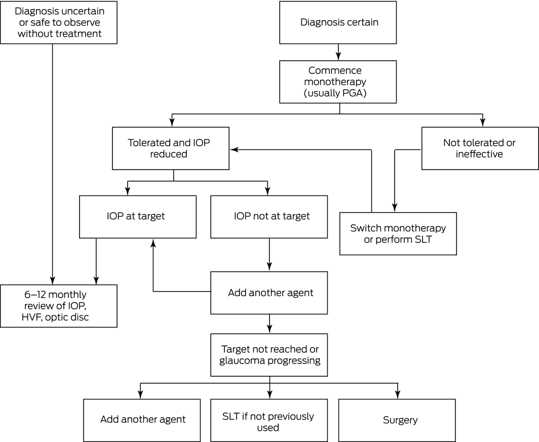
HVF = Humphrey visual field. IOP = intraocular pressure. PGA = prostaglandin analogue. SLT = selective laser trabeculoplasty. ◆
Box 6 – Commercially available intraocular pressure‐lowering therapies in Australia
|
Drug class |
Drug name |
Mechanism |
Side effects |
||||||||||||
|
|
|||||||||||||||
|
Prostaglandin analogue |
|
Increased uveoscleral outflow |
Conjunctival hyperaemia, eyelash lengthening, increased periocular and iris pigmentation, prostaglandin‐associated periorbitopathy |
||||||||||||
|
β‐Blocker |
|
Decreased aqueous humor production |
Ocular irritation, bronchoconstriction, bradyarrhythmias, hypotension |
||||||||||||
|
Carbonic anhydrase inhibitor |
|
Decreased aqueous humor production |
Topical: ocular irritation, hyperaemia, dysgeusia |
||||||||||||
|
Per oral: polyuria, anorexia, sulphur reaction, metabolic acidosis, renal failure, renal calculi |
|||||||||||||||
|
α‐2 Agonist |
|
Decreased aqueous humor production and increased uveoscleral outflow |
Conjunctival hyperaemia, allergic blepharoconjunctivitis, drowsiness, dry mouth |
||||||||||||
|
Cholinergic |
|
Increased trabecular outflow |
Blurred vision, dim vision, vitreous floaters, myopia, retinal tear or detachment, brow ache |
||||||||||||
|
|
|||||||||||||||
|
|
|||||||||||||||
Box 7 – Commercially available fixed‐combination eye drops in Australia
|
Composition |
Frequency |
||||||||||||||
|
|
|||||||||||||||
|
Bimatoprost 0.03% + timolol 0.5% |
Once daily |
||||||||||||||
|
Travoprost 0.004% + timolol 0.5% |
Once daily |
||||||||||||||
|
Latanoprost 0.005% + timolol 0.5% |
Once daily |
||||||||||||||
|
Brinzolamide 1.0% + timolol 0.5% |
Twice daily |
||||||||||||||
|
Dorzolamide 2% + timolol 0.5% |
Twice daily |
||||||||||||||
|
Brimonidine 0.2% + timolol 0.5% |
Twice daily |
||||||||||||||
|
Brimonidine 0.2% + brinzolamide 1.0% |
Twice daily |
||||||||||||||
|
|
|||||||||||||||
|
|
|||||||||||||||
Box 8 – Minimally invasive glaucoma surgery devices currently available in Australia
|
Device |
Drainage area targeted |
Common potential complications for all devices |
Specific complications |
||||||||||||
|
|
|||||||||||||||
|
iStent (Glaukos) |
Schlemm canal |
|
|
||||||||||||
|
iStent inject (Glaukos), 2 stents |
Schlemm canal |
|
|
||||||||||||
|
Hydrus microstent (Ivantis) |
Schlemm canal |
|
|
||||||||||||
|
iTrack (Ellex) viscocanalostomy |
Schlemm canal and collector channels |
|
|
||||||||||||
|
CyPass Micro‐Stent (Novartis)* |
Suprachoroidal space |
|
|
||||||||||||
|
XEN Gel Stent (Allergan) |
Subconjunctival space |
|
|
||||||||||||
|
|
|||||||||||||||
|
IOP = intraocular pressure. PAS = peripheral anterior synechiae. * Recently withdrawn by sponsor because of corneal decompensation. ◆ |
|||||||||||||||
Competing interests
No relevant disclosures.
References
- Gordon MO, Kass MA. The Ocular Hypertension Treatment Study: design and baseline description of the participants. Arch Ophthalmol 1999; 117: 573–583.
- Leske MC, Heijl A, Hyman L, et al. Early Manifest Glaucoma Trial: design and baseline data. Ophthalmology 1999; 106: 2144–2153.
- Ederer F, Gaasterland DE, Sullivan EK, et al. The Advanced Glaucoma Intervention Study (AGIS): 1. Study design and methods and baseline characteristics of study patients. Control Clin Trials 1994; 15: 299–325.
- Musch DC, Lichter PR, Guire KE, et al. The Collaborative Initial Glaucoma Treatment Study: study design, methods, and baseline characteristics of enrolled patients. Ophthalmology 1999; 106: 653–662.
- Anderson DR. Collaborative normal tension glaucoma study. Curr Opin Ophthalmol 2003; 14: 86–90.
- Leske MC. The epidemiology of open‐angle glaucoma: a review. Am J Epidemiol 1983; 118: 166–191.
- Lusthaus JA, Goldberg I. Emerging drugs to treat glaucoma: targeting prostaglandin F and E receptors. Expert Opin Emerg Drugs 2016; 21: 117–128.
- Mantravadi AV, Vadhar N. Glaucoma. Prim Care Clin Office Pract 2015; 42: 437–449.
- Kingman S. Glaucoma is second leading cause of blindness globally. Bull World Health Organ 2004; 82: 887–888.
- Tielsch JM, Sommer A, Katz J, et al. Racial variations in the prevalence of primary open‐angle glaucoma. The Baltimore Eye Survey. JAMA 1991; 266: 369–374.
- Mitchell P, Smith W, Attebo K, Healey PR. Prevalence of open‐angle glaucoma in Australia. The Blue Mountains Eye Study. Ophthalmology 1996; 103: 1661–1669.
- Wensor MD, McCarthy CA, Stanislavsky YL, et al. The prevalence of glaucoma in the Melbourne Visual Impairment Project. Ophthalmology 1998; 105: 733–739.
- Quigley HA, Broman AT. The number of people with glaucoma worldwide in 2010 and 2020. Br J Ophthalmol 2006; 90: 262–267.
- Nucci C, Martucci A, Giannini C, et al. Neuroprotective agents in the management of glaucoma. Eye 2018; 32: 938–945.
- Konstas AGP, Topouzis F, Leliopoulou O, et al. 24‐hour intraocular pressure control with maximum medical therapy compared with surgery in patients with advanced open‐angle glaucoma. Ophthalmology 2006; 113: 761.e1–765.e1.
- Stein JD, Ayyagari P, Sloan FA, et al. Rates of glaucoma medication utilization among persons with primary open‐angle glaucoma 1992 to 2002. Ophthalmology 2008; 115: 1315–1319.
- Winkler N, Fautsch MP. Effects of prostaglandin analogues on aqueous humor outflow pathways. J Ocul Pharmacol Ther 2014; 30: 102–109.
- Johnstone MA, Albert DM. Prostaglandin‐induced hair growth. Surv Ophthalmol 2002; 47 (Suppl): S185–S202.
- Sakata R, Shirato S, Miyata K, et al. Incidence of deepening of the upper eyelid sulcus in prostaglandin‐associated periorbitopathy with a latanoprost ophthalmic solution. Eye 2014; 28: 1446–1451.
- Alm A, Grierson I, Shields MB. Side effects associated with prostaglandin analog therapy. Surv Ophthalmol 2008; 53: S93–S105.
- Aptel F, Cucherat M, Denis P. Efficacy and tolerability of prostaglandin analogs: a meta‐analysis of randomized controlled clinical trials. J Glaucoma 2008; 17: 667–673.
- Lin JC. The use of ocular hypotensive drugs for glaucoma treatment: changing trend in Taiwan from 1997 to 2007. J Glaucoma 2015; 24: 364–371.
- Sambhara DS, Aref AA. Glaucoma management: relative value and place in therapy of available drug treatments. Ther Adv Chronic Dis 2014; 5: 30–43.
- Kaiser HJ, Schoetzau A, Stump D, et al. Blood flow velocities of the extraocular vessels in patients with high‐tension and normal‐tension primary open‐angle glaucoma. Am J Ophthalmol 1997; 123: 320–327.
- Lusthaus JA, Goldberg I. Brimonidine and brinzolamide for treating glaucoma and ocular hypertension; a safety evaluation. Expert Opinion on Drug Safety 2017; 16: 1071–1078.
- Arthur S, Cantor LB. Update on the role of alpha‐agonists in glaucoma management. Exp Eye Res 2011; 93: 271–283.
- Nguyen QH. Combination of brinzolamide and brimonidine for glaucoma and ocular hypertension: critical appraisal and patient focus. Patient Prefer Adherence 2014; 8: 853–864.
- Toris CB, Camras CB, Yablonski ME. Effects of brimonidine on aqueous humor dynamics in human eyes. Arch Ophthalmol 1995; 113: 1514–1517.
- Silver LH. Clinical efficacy and safety of brinzolamide (Azopt), a new topical carbonic anhydrase inhibitor for primary open‐angle glaucoma and ocular hypertension. Am J Ophthalmol 1998; 126: 400–408.
- Van der Valk R, Webers CAB, Schouten JS, et al. Intraocular pressure‐lowering effects of all commonly used glaucoma drugs: a meta‐analysis of randomized clinical trials. Ophthalmology 2005; 112: 1177–1185.
- Hu CY, Lee BJ, Cheng HF, et al. Acetazolamide‐related life‐threatening hypophosphatemia in a glaucoma patient. J Glaucoma 2015; 24: e31–e33.
- McLaren NC, Moroi SE. Clinical implications of pharmacogenetics for glaucoma therapeutics. Pharmacogenomics J 2003; 3: 197–201.
- Okeke CO, Quigley HA, Jampel HD, et al. Adherence with topical glaucoma medication monitored electronically. Ophthalmology 2009; 116: 191–199.
- Kosoko O, Quigley HA, Vitale S, et al. Risk factors for non‐compliance with glaucoma follow‐up visits. Ophthalmology 1998; 105: 2105–2111.
- McVeigh KA, Vakros G. The eye drop chart: a pilot study for improving administration of and compliance with topical treatments in glaucoma patients. Clin Ophthalmol 2015; 9: 813–819.
- Inoue K, Soeda S, Tomita G. Comparison of latanoprost/timolol with carbonic anhydrase inhibitor and dorzolamide/timolol with prostaglandin analog in the treatment of glaucoma. J Ophthalmol 2014; 2014: 975429.
- Skalicky SE, Goldberg I, McCluskey P. Ocular surface disease and quality of life in patients with glaucoma. Am J Ophthalmol 2012; 153: 1.e2–9.e2.
- Latina M, Tumbocon JA. Selective laser trabeculoplasty: a new treatment option for open angle glaucoma. Curr Opin Ophthalmol 2002; 13: 94–96.
- McAlinden C. Selective laser trabeculoplasty (SLT) vs other treatment modalities for glaucoma: systematic review. Eye 2014; 28: 249–258.
- Fan Gaskin JC, Sandhu SS, Walland MJ. Victorian trabeculectomy audit. Clin Exp Ophthalmol 2017; 45: 695–700.
- Kirwan JF, Lockwood AJ, Shah P, et al. Trabeculectomy in the 21st century: a multicenter analysis. Ophthalmology 2013; 120: 2532–2539.
- Gedde SJ, Schiffman JC, Feuer WJ, et al. Treatment outcomes in the tube versus trabeculectomy (TVT) study after five years of follow‐up. Am J Ophthalmol 2012; 153: 789–803.
- Lavia C, Dallorto L, Maule M, et al. Minimally‐invasive glaucoma surgeries (MIGS) for open angle glaucoma: a systematic review and meta‐analysis. PLoS One 2017; 12: e0183142.
- Saheb H, Ahmed II. Micro‐invasive glaucoma surgery: current perspectives and future directions. Curr Opin Ophthalmol 2012; 23: 96–104.
- Chaudhary A, Salinas L, Guidotti J, et al. XEN Gel Implant: a new surgical approach in glaucoma. Expert Rev Med Devices 2018; 15: 47–59.
- Martin KR, Broadway DC. Cyclodiode laser therapy for painful, blind glaucomatous eyes. Br J Ophthalmol 2001; 85: 474–476.
- Aquino MC, Barton K, Tan AM, et al. Micropulse versus continuous wave transcleral diode cyclophotocoagulation in refractory glaucoma: a randomized exploratory study. Clin Exp Ophthalmol 2015; 43: 40–46.
- Azuara‐Blanco A, Burr J, Ramsay C, et al. Effectiveness of early lens extraction for the treatment of primary angle‐closure glaucoma (EAGLE): a randomised controlled trial. Lancet 2016; 388: 1389–1397.
- Bacharach J, Dubiner HB, Levy B, et al. Double‐masked, randomized, dose‐response study of AR‐13324 versus latanoprost in patients with elevated intraocular pressure. Ophthalmology 2015; 122: 302–307.
- Weinreb RN, Ong T, Scassellati Sforzolini B, et al. A randomized, controlled comparison of latanoprostene bunod and latanoprost 0.005% in the treatment of ocular hypertension and open angle glaucoma: the VOYAGER study. Ophthalmology 2015; 99: 738–745.
Linked content
-
MJA Perspective: Glaucoma in perspective
-
MJA Podcast: Dr Jed Lusthaus
-
InSight+: Glaucoma: the “sneak thief of sight”
-
MJA Medical Education: Glaucoma caused by topical corticosteroid application to the eyelids
Provenance: Commissioned; externally peer reviewed.
A Diagnostic Headache
Thomas Glynn, Michelle T. Leech, Stacy K. Goergen, Emily Lin, Nadeem Toodayan, Ralph Junckerstorff
Neisseria meningitidis: an uncommon cause of conjunctivitis
Chloe Story, Tina Marinelli, Simeon Crawford, Amrita Ronnachit
Dismantling barriers to research and clinical care for individuals with a vision impairment
Eden G Robertson, Kate Hetherington, Meredith Prain, Julia Hall, Leighton Boyd AM, Rosemary Boyd OAM, Emily Shepard, Hollie Feller, Sally Karandrews, Fleur O'Hare, Kanae Yamamoto, Matthew P Simunovic, Robyn V Jamieson, Alan Ma, Lauren Ayton AM, Anai Gonzalez‐Cordero
The New South Wales Pharmacy Trial for herpes zoster: on the nose?
Christian P Pappas, Timothy R Holmes, Minas T Coroneo
Skin fragility disorder misdiagnosed as child abuse: a cautionary tale
Heather G Mack, Fred K Chen, John R Grigg
Health economic aspects of inherited retinal diseases: looking for cost‐effective treatments
Benjamin Kamien, Rachael Heath Jeffery, Fred K Chen