




Volume 210 - Issue 2
Revisiting the antinuclear antibody test with emphasis on a new pattern: anti‐DFS70 antibody
Authors: Pravin Hissaria, Andrew Broadfoot and Karl W Baumgart
Med J Aust 2019; 210 (2): 69-71. || doi: 10.5694/mja2.12103
Published online: 4 February 2019
Published online: 4 February 2019
ANA by indirect immunofluorescence remains the method of choice for screening patients suspected of having SARDs. A positive ANA test should be followed by further testing for ENA and dsDNA antibody testing for defining the ANA specificities and disease associations. The presence of anti-DFS70 antibody in absence of ENA and dsDNA antibodies effectively excludes a diagnosis of SARD.
The diagnosis of systemic autoimmune rheumatic diseases (SARDs), such as systemic lupus erythematosus (SLE), is based on a constellation of clinical features and presence of supportive specific autoantibodies. The antinuclear antibody (ANA) test is the most common first‐line test for these groups of diseases. Although several methods can detect ANA, indirect immunofluorescence remains the most widely used assay and the method of choice.1 The most significant attribute of any screening test is its high negative predictive value; that is, a negative test rules out the possibility of these diseases. The ANA test fulfils this requirement, with a negative predictive value of about 99% for SLE and 95% for other diseases such as scleroderma, Sjögren syndrome and type 1 autoimmune hepatitis.2 However, the ANA test has a very poor positive predictive value of less than 10% in a community setting because healthy people might have a positive ANA result.3 The titre or concentration of antibody in the patient's serum is determined by serial doubling dilutions, starting from a dilution of 1:40 and then 1:80 up to 1:2560.3 Therefore, the use of higher screening dilutions can improve the positive predictive value of the ANA test. The potential clinical significance of a positive ANA test can be further determined by ordering for antibodies to extractable nuclear antigens (ENAs), extended ENA tests, and tests for double‐stranded DNA (dsDNA) antibodies.
When to order an ANA test
The ANA test should be considered for patients suspected to have a SARD, including SLE, scleroderma, Sjögren syndrome and autoimmune hepatitis. It is not useful in other inflammatory diseases, such as primary antiphospholipid syndrome, systemic necrotising vasculitis, sarcoidosis and inflammatory arthritides. This test is not useful for monitoring of disease and, hence, repeat testing is strongly discouraged unless there is a change in the clinical scenario.
The significance of ANA staining patterns
Clinical laboratories report the ANA patterns detected and their endpoint titre in the patient's serum;2 for example, “ANA, positive (qualitative result) 1:2560 (endpoint dilution); homogeneous (pattern)”.
Each pattern seen correlates with different autoantibodies to distinct nuclear antigens. Box 1 lists common clinical associations of ANA patterns. Many other ANA patterns have been described which are less common and have looser associations with SARDs.
The significance of the ANA titre
Laboratories generally report titres from 1:40 or 1:80 to 1:2560. As a general rule, the clinical significance of ANA positivity rises with higher titres, implying greater amounts of antibodies. There is a high prevalence of low titre (1:40–1:160) ANAs in healthy individuals, and studies have shown that an ANA titre of 1:40 can be present in 10–30% of the healthy population.3,4
Most patients with a positive ANA test will not develop a SARD. In a study of previously unaffected relatives of patients with SLE, 89% of patients who later reached SLE classification had positive ANA levels at baseline. However, 48% of those who remained unaffected by SLE also had a positive ANA level at baseline,5 indicating that the ANA test alone has low predictive value.
Nevertheless, some patients with SARDs may have low ANA titres; therefore, clinical history and examination remain essential. SARDs are diagnosed primarily using clinical criteria, and the laboratory testing for specific autoantibodies helps support the diagnosis and can influence prognosis in some instances; for example, dsDNA antibodies are more closely correlated with lupus nephritis. The advantages and disadvantages of the ANA test are summarised in Box 2.
Further tests for autoantibodies
If an ANA is discovered, then further laboratory tests can be used to precisely determine the target antigens of these antibodies (Box 3). The most commonly performed second line tests are for ENA and dsDNA, which provide more precise diagnostic information6 (Box 4).
The DFS70 antibody: an autoantibody that rules out systemic autoimmune rheumatic diseases
ANA is usually reported according to the staining pattern observed and the titres, which are determined by doing serial dilutions of the serum. In 1994, a significant discovery described a separate pattern of an atypical homogeneous pattern, which was later designated as antidense fine speckled (DFS70) antibody and was associated with interstitial cystitis.7 Since that first report, the antigen specificity of this antibody was found to be lens epithelium‐derived growth factor (LEDGF/p75), but anti‐DFS70 continues to be more widely used in the clinical literature to describe this autoantibody. The significance of anti‐DFS70 antibody lies in its strong negative association with the presence of any SARD.8
Since the initial description, it was found that this antibody was not diagnostic of interstitial cystitis either, but was found in a range of diseases, such as atopic eczema, eye diseases, thyroiditis, prostate cancer etc.9 However, the most important finding was its presence in a significant number of healthy individuals, ranging from 2% to 21% depending on the population studied. Moreover, it was the most frequent autoantibody resulting in false positive ANA results in healthy individuals (23–50%).9
Among the SARD group, this autoantibody was found less frequently (< 1%) compared with healthy individuals, which led to the suggestion that its presence was a negative predictive marker for diagnosing SARDs, including SLE, scleroderma and autoimmune hepatitis.10 However, it should be noted that this could be seen in a proportion (up to 10% of cases) of patients with SARDs in conjunction with the presence of other disease‐specific autoantibodies. In one study, patients with positive DFS70 autoantibody were followed up for 4 years and confirmed the absence of development of any rheumatic diseases.11
The establishment of clinical significance of this autoantibody in ruling out a SARD led to its inclusion as a reportable specific autoantibody pattern in the ANA consensus conference held in Brazil in 2014.12 It was decided that there were three separate homogeneous patterns that needed to be recognised by the laboratories reporting ANA by indirect immunofluorescence. In addition to the classical ANA — which is seen as a diffuse staining in the interphase nuclei and homogeneous staining of chromatic band in the mitotic cells — two other patterns were also described, a quasi‐homogeneous pattern that shows fine speckled pattern in interphase cells but homogeneous staining of chromatin in dividing cells, and a DFS pattern, which is dense fine speckled staining observed in both interphase cells and of chromatin in metaphase cells. However, it became evident that the reporting of DFS70 antibody by indirect immunofluorescence was not sufficient and robust enough, and hence, a second solid phase test should be done for confirmation, as is the norm for all other disease‐specific autoantibodies.13 The main implication of this recommendation is that the requesting clinician needs to be aware that there can be more than one pattern of homogeneous staining. Therefore, when the clinical picture is not consistent with SARDs, they should request for specific anti‐DFS70 antibody if there is presence of high titre homogeneous ANA and speckled pattern.
In Australia, only a few laboratories perform confirmatory testing for DFS70 antibodies. However, some laboratories (50%) would report on the indirect immunofluorescence pattern if it is consistent and the supplementary ENA and anti‐dsDNA antibody testing is negative. Hence, it is possible that this pattern might be missed by few laboratories or reported as homogeneous or homogeneous and speckled patterns. Therefore, when a general practitioner or specialist sees a report of ANA with moderate or high titres and negative ENA and dsDNA antibodies, it would be worthwhile to ask for this autoantibody specifically from a reference laboratory, using either a solid phase assay or the immunoadsorption assays. This request would save unnecessary referrals and further inappropriate repeat testing in patients suspected of having a SARD. The incorporation of testing for this autoantibody has been systematically studied and proven to be cost‐effective, saving a significant amount of money and health care utilisation.14
Conclusion
ANA by indirect immunofluorescence remains the method of choice for screening patients suspected of having SARDs. A positive ANA test should be followed by further testing for ENA and dsDNA antibody testing for defining the ANA specificities and disease associations. The presence of anti‐DFS70 antibody in absence of ENA and dsDNA antibodies effectively excludes a diagnosis of SARD.
Box 1 – Antinuclear antibody patterns and their common systemic autoimmune rheumatic disease (SARD) associations
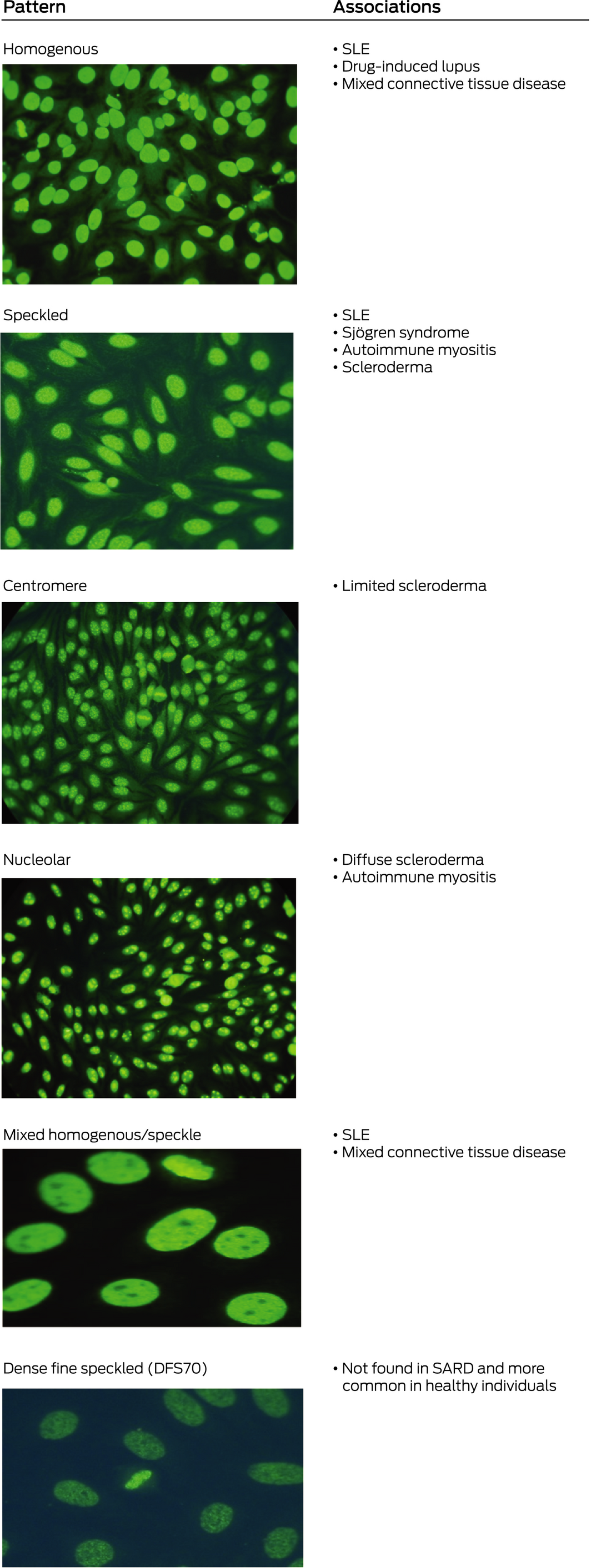
DFS = dense fine speckled. SLE = systemic lupus erythematosus. ◆
Box 2 – Clinical utility of antinuclear antibody (ANA) testing
|
Advantages |
Disadvantages |
||||||||||||||
|
|
|||||||||||||||
|
|
|
||||||||||||||
|
|
|||||||||||||||
|
CREST = limited scleroderma. DFS = dense fine speckled. SARD = systemic autoimmune rheumatic disease. ◆ |
|||||||||||||||
Box 3 – Testing for systemic autoimmune rheumatic testing
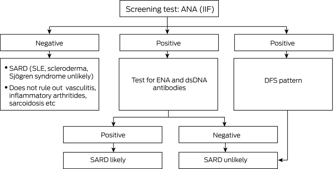
ANA = antinuclear antibody. DFS = dense fine speckled. dsDNA = double‐stranded DNA. ENA = extractable nuclear antigens. IIF = indirect immunofluorescence. SARD = systemic autoimmune rheumatic disease. SLE = systemic lupus erythematosus. ◆
Box 4 – Extractable nuclear antigen (ENA) and double‐stranded DNA (dsDNA) and their disease associations
|
ANA specificity detected by ENA and dsDNA testing |
Clinical association |
||||||||||||||
|
|
|||||||||||||||
|
SSA, SSB |
SLE, Sjögren syndrome |
||||||||||||||
|
Scl‐70 (antitopoisomerase) |
Diffuse systemic sclerosis |
||||||||||||||
|
Cenp B |
Limited scleroderma |
||||||||||||||
|
RNP |
Mixed connective tissue disease |
||||||||||||||
|
Jo‐1 |
Autoimmune myositis |
||||||||||||||
|
Ribo‐P |
SLE |
||||||||||||||
|
dsDNA |
SLE |
||||||||||||||
|
Histones |
SLE, drug‐induced lupus |
||||||||||||||
|
|
|||||||||||||||
|
ANA = antinuclear antibody. SLE = systemic lupus erythematosus. ◆ |
|||||||||||||||
Competing interests
No relevant disclosures.
References
- Solomon DH, Kavanaugh AJ, Schur P, American College of Rheumatology Ad Hoc Committee on Immunologic Testing Guidelines. Evidence‐based guidelines for the use of immunological tests: antinuclear antibody testing. Arthritis Rheum 2002; 47: 434–444.
- Abeles AM, Abeles M. The clinical utility of a positive antinuclear antibody test result. Am J Med 2013; 126: 342–348.
- Tan EM, Feltkamp TE, Smolen JS, et al. Range of antinuclear antibodies in ‘healthy’ individuals. Arthritis Rheum 1997; 40: 1601–1611.
- Arbuckle MR, McClain MT, Rubertone MV, et al. Development of autoantibodies before the clinical onset of systemic lupus erythematosus. N Engl J Med 2003; 349: 1526–1533.
- Munroe ME, Young KA, Kamen DL, et al. Discerning risk of disease transition in relatives of systemic lupus erythematosus patients utilizing soluble mediators and clinical features. Arthritis Rheumatol 2017; 69: 630–642.
- Jog RJ, James JA. Biomarkers in connective tissue diseases. J Allergy Clin Immunol 2017; 140: 1473–1483.
- Ochs RL, Stein TW, Peebles CL, et al. Autoantibodies in interstitial cystitis. J Urol 1994; 151: 587–592.
- Mahler M, Hanly JG, Fritzler MJ. Importance of the dense fine speckled pattern on HEp‐2 cells and anti‐DFS70 antibodies for the diagnosis of systemic autoimmune diseases. Autoimmun Rev 2012; 11: 642–645.
- Conrad K, Röber N, Andrade LE, Mahler M. The clinical relevance of anti‐DFS70 autoantibodies. Clinic Rev Allerg Immunol 2017; 52: 202–216.
- Mahler M, Parker T, Peebles CL, et al. Anti‐DFS70/LEDGF antibodies are more prevalent in healthy individuals compared with patients with systemic autoimmune rheumatic diseases. J Rheumatol 2012; 39: 2104–2110.
- Mariz HA, Sato EI, Barbosa SH, et al. Pattern on the antinuclear antibody‐HEp‐2 test is a critical parameter for discriminating antinuclear antibody‐positive healthy individuals and patients with autoimmune rheumatic diseases. Arthritis Rheum 2011; 63: 191–200.
- Chan EK, Damoiseaux J, Carballo OG, et al. Report of the first international consensus on standardized nomenclature of antinuclear antibody HEp‐2 cell patterns 2014–2015. Front Immunol 2015; 6: 412.
- Bizzaro N, Tonutti E, Villalta D. Recognizing the dense fine speckled/lens epithelium‐derived growth factor/p75 pattern on HEp‐2 cells: not an easy task! Comment on the article by Mariz et al. Arthritis Rheum 2011; 63: 4036–4037.
- Gundín S, Irure‐Ventura J, Asensio E, et al. Measurement of anti‐DFS70 antibodies in patients with ANA‐associated autoimmune rheumatic diseases suspicion is cost‐effective. Auto Immun Highlights 2016; 7: 10.
Provenance: Not commissioned; externally peer reviewed.
Rhabdomyolysis After COVID-19
Navya Kataria, Abel Devadass, Lana Sundac, Laura De Souza, Nabeel Sheikh, Samantha Ng, Scott Campbell, Yishen Wang, Ross S. Francis
Technological advances in the search for a CURE for asthma
Sara Quon, Timothy M Johanson, Ridhima Wadhwa, Alen Faiz, Anthony Flynn, Gary P Anderson, Amanda J Cox, Nicholas P West, Michael P Menden, Rhys S Allan
The CURE Asthma roadmap
Gary P Anderson, Anthony Flynn, Phil G Bardin, John D Blakey, Shyamali C Dharmage, Paul Foster, Peter G Gibson, Adam Jaffe, Alan James, Christine R Jenkins, Sundram Sivamalai, Peter D Sly, Guy B Marks, Vanessa M McDonald, Judy Wetttenhall
The first live term birth following uterus transplantation in Australia
Rebecca Deans, Brigitte Gerstl, Antonia W Shand, Sarah Lyons, Aaron Budden, Helen L Barrett, Grant Luxton, Mangalee Fernando, Kenneth Yong, Karen Keung, Kaushalya Arulpragasam, Henry Pleass, King Man Wan, Eva Kehag, Jana‐Emily Pittman, Mianna Lotz, Maria Fenn, Erin Nesbitt‐Hawes, Lily Byun, Katrina Tang, Mats Brannstrom, Jason Abbott
The public health impacts of mining in Australia
Javier Cortes‐Ramirez, Ruby N Michael, Leisa‐Maree Toms, Melissa Haswell
Lingual Raynaud phenomenon
Michael Taggart, Mina John