




Volume 208 - Issue 8
Parechovirus: an important emerging infection in young infants
Authors: Philip N Britton, Cheryl A Jones, Kristine Macartney and Allen C Cheng
Med J Aust 2018; 208 (8): 365-369. || doi: 10.5694/mja18.00149
Published online: 30 April 2018
Published online: 30 April 2018
Parechoviruses generally cause mild illness in children, but a current genotype 3 epidemic has been associated with severe complications, particularly in children aged under 3-6 months
Summary
- Epidemics of human parechovirus (HPeV) causing disease in young children have occurred every 2 years in Australia since 2013. HPeV genotype 3 caused the epidemic from late 2017 to early 2018.
- Most HPeV infections cause no or mild symptoms including gastroenteritis or influenza-like illness. Characteristically, young infants present with fever, irritability and on occasions a diffuse rash (“red, hot and angry” babies).
- Severe disease can manifest as meningoencephalitis, seizures or sepsis-like presentations (including septic shock), or less common presentations including signs of surgical abdomen.
- Testing for HPeV by specific molecular tests is indicated in children younger than 6 months of age with characteristic presentations without another confirmed diagnosis including febrile illnesses with other suggestive features (eg, rash, seizures), sepsis syndromes (including shock), and suspected meningoencephalitis (which may be detected by magnetic resonance imaging only).
- There are no effective antiviral therapies. Treatment is primarily supportive, including management of complications.
- Some infants with severe HPeV infection may have adverse neurodevelopment. Follow-up by a paediatrician is recommended.
In late 2017, public health warnings were issued about a national epidemic of human parechovirus (HPeV) in young children. Here, we review the epidemiology of HPeV in Australia since 2013, and provide guidance to clinicians on the management of suspected infection.
This narrative review used a PubMed search of original and review articles from 1996 to 2018, supplemented by data from the Paediatric Active Enhanced Disease Surveillance (PAEDS) network. PAEDS is a hospital-based active surveillance system that performs prospective case ascertainment for selected serious childhood conditions at sentinel paediatric hospitals, including encephalitis, vaccine-preventable diseases and adverse events following immunisation.1
Virology and epidemiology
HPeVs were formerly part of the echovirus genus in the 1960s but were assigned their own genus in the 1990s.2 Of the 17 genotypes,3,4 HPeV genotypes 1, 3 and 6 are most commonly associated with human disease.3,5 With the advent of more sensitive molecular assays, HPeV is now recognised as a small but significant cause of illness in infants aged < 3 years. HPeV was detected in 3–8% of children presenting to emergency with undifferentiated fever in European studies6,7 and appears to be more common in younger children and in children requiring hospital admission, where up to 15–20% of children with fever without source are HPeV-positive.8,9 Serological data from Europe and Japan show that 90% of infants have been infected with at least one HPeV subtype by the age of 2 years.5
More serious disease has been associated with HPeV3 infection, which was first associated with acute flaccid paralysis in a young girl in 2004.10 It is now recognised as a leading cause of sepsis-like illness and central nervous system infection, particularly in young infants.5 In Australia, an epidemic of HPeV3 was first noted in the spring and summer of 2013–2014, initially in New South Wales then nationwide.11,12 Epidemic activity was subsequently observed in the spring and summer of 2015–2016. This 2-yearly pattern is similar to that observed in the United Kingdom.13 However, in contrast to other countries, epidemics in Australia have been larger and associated with more severe disease (and poorer clinical outcomes).14,15 Recently, whole genome sequencing has shown that the first Australian epidemic was likely caused by a novel recombinant HPeV3.16
Since August 2017, PAEDS has been tracking an increased frequency of HPeV cases through the Australian childhood encephalitis study and acute flaccid paralysis surveillance systems.1,17 Increased case numbers were initially noted in Victoria, then in Queensland. In early November 2017, with cases being reported across all five PAEDS sites (Box 1), a formal notification was made to the Communicable Diseases Network Australia of a third national epidemic. In December 2017, the PAEDS network had identified > 200 cases of hospitalised HPeV infection in young infants associated with the current third epidemic.
Clinical presentation and complications
Like enteroviruses, the epidemiology and clinical syndromes associated with HPeV vary by genotype.5 HPeV1 and HPeV6 are primarily associated with milder gastrointestinal symptoms and presentation in children aged > 6 months.18,19 HPeV may be isolated from respiratory specimens in children presenting with influenza-like illness20 and in faecal specimens from children presenting with viral diarrhoea.21
HPeV3 causes a range of disease manifestations but primarily fever and irritability, and sepsis-like illness (Box 2).12,19 Rash is variable,5 but where present, infants are often described as being “red, hot, angry babies” (Box 3).12 Occasionally the illness has been mistaken for atypical Kawasaki disease with a palmar-plantar erythema.12 Young infants (< 3 months) are at highest risk of hospitalisation.3,5,12,19,22 Neonates (< 28 days) and ex-premature infants may be at higher risk of complications.14,23 HPeV3 disease can be severe with up to 20–50% of admitted patients with sepsis-like illness requiring intensive care admission.12,24,25
Severe manifestations of the acute illness include severe sepsis and meningoencephalitis, which occur in around 10% of hospitalised babies (PAEDS, unpublished data). In a series of 104 children admitted to intensive care units in the United Kingdom and Ireland, 77% required mechanical ventilation and 40% required inotropic support, with similar severe presentations reported elsewhere.26,27 Seizure activity, including central apnoea, and focal neurological signs may signal meningoencephalitis.
Magnetic resonance imaging of the brain is important to detect characteristic features of meningoencephalitis (peri-ventricular white matter diffusion restriction) that likely has prognostic significance.14,23 Head ultrasound, widely used in the assessment for meningitis in neonates, is not sensitive to diagnose white matter changes due to parechovirus.14
Other less common presentations include an acute abdomen, including intussusception, pseudo-appendicitis and bowel perforation.3,5,12 Additional unusual manifestations include HPeV3-associated haemophagocytic lymphohistiocytosis and fulminant liver failure.28,29 Chronic infection in an adolescent with agammaglobulinaemia has also been described.30
In adults, HPeV is associated with upper respiratory tract infection and mild diarrhoeal illness, and less commonly with epidemic myalgia and myocarditis.31-34 There is currently no evidence to suggest particular concern regarding infection in pregnant women or fetuses.35
Diagnosis of parechovirus
HPeV is not detected by nucleic acid testing for enteroviruses; if suspected, specific polymerase chain reaction testing is required. Testing is generally only available through specialised reference laboratories. The virus can be detected in cerebrospinal fluid (CSF), stool, respiratory secretions, serum and urine. Stool testing appears to be most sensitive (95–98%), followed by CSF testing (84–95%).12,36 Characteristically, CSF pleocytosis is mild or absent in HPeV3 central nervous system infection, in contrast to other causes of encephalitis.8,12,14,18,19,23
Currently, we recommend testing all children aged < 6 months presenting to hospital with clinical features of HPeV infection without another confirmed diagnosis. This includes young infants with sepsis syndromes (including shock), febrile illnesses with other suggestive features (eg, rash, seizures) and suspected meningoencephalitis. Specific testing on stool or CSF for HPeV nucleic acid using polymerase chain reaction should be ordered even in the absence of CSF pleocytosis in these infants. Testing is not generally indicated for children with milder illnesses presenting in the primary care setting. There are as yet insufficient data to define seasonal periods of elevated risk. HPeV infection is not a notifiable disease.
Managing parechovirus infection
There is no specific antiviral therapy for HPeV infection available at present. Empirical antibiotics should be administered according to local and national guidelines for possible bacterial sepsis or meningoencephalitis, especially in neonates, until this has been excluded by negative sterile site cultures. Once bacterial infection has been excluded, management is mainly supportive but may require close monitoring and intensive care. Although some case reports suggest benefit, there is no clear evidence to support administration of intravenous immunoglobulin.37 Physical examination and close observation should exclude acute complications such as shock, apnoea and seizures, and early management should be provided if such complications are detected. Infants with proven HPeV infection and neurological symptoms should undergo magnetic resonance imaging (and not head ultrasound) to exclude encephalitis.
HPeV is transmitted via the faecal–oral route and possibly respiratory droplets. In hospitals, contact precautions are recommended to prevent transmission to other young infants. HPeV3 has been shown to be shed in the stool for a median of 58 days,38 although the risk of transmission is probably highest during acute symptomatic illness. In the community, non-hospitalised, confirmed cases should avoid contact with other young children (including childcare and school attendance) until symptoms have fully resolved. Contacts of confirmed cases should be made aware that older children and adults may develop mild upper respiratory or diarrhoeal symptoms.39 There are cases of transmission from mildly symptomatic children to adults resulting in severe acute illness, but these appear to be uncommon.34 Hand hygiene, cough etiquette and staying away from childcare and school while unwell should be emphasised.
Follow-up studies have shown adverse neurological outcomes in patients with meningoencephalitis, and developmental concern among patients with sepsis-like illness from the 2013–2014 epidemic.14,15,23 A study of Australian infants hospitalised with HPeV3 in 2013 found that 19% showed significant concern in developmental attainment 12–16 months after infection and an additional 50% had some concern based on standardised tools.15 An earlier study documented severe neurodevelopmental sequelae, including cerebral palsy, central visual impairment and gross motor impairment.14 These findings are consistent with those from other countries, where 16% of children hospitalised with confirmed HPeV infection had significant neurological sequelae.24 These data also appear to be consistent with outcomes from other causes of encephalitis.40
Because of the evidence of adverse neurodevelopmental outcomes following severe HPeV infection, we recommend that all children hospitalised with HPeV infection should be followed up by a paediatrician at least until school entry, and preferably afterwards, to monitor development and learning, and manage complications including seizures.
Future directions
The HPeV epidemics since 2013 highlight the need for surveillance targeted at important syndromes such as encephalitis, to define the disease burden, evaluate strategies and interventions to prevent and manage cases, and to respond to the potential first signals of emerging infectious diseases. The Australian childhood encephalitis study, based on PAEDS data, has already demonstrated capacity for early detection of serious infectious epidemics in children including HPeV and enterovirus 71 (EV71).17 This would also provide information on all causes of this devastating syndrome that are potentially vaccine-preventable, such as influenza.41,42
Many unanswered questions remain regarding the epidemiology and clinical features of HPeV in Australia, and there are unique opportunities to study the evolution of the virus and disease over the three epidemic waves in Australia to date. In addition, the specific pathogenesis of HPeV3 disease needs better elucidation in order to develop specific treatments. There are currently cohort studies underway to better characterise the long term neurodevelopmental outcomes for children with central nervous system infection.
In the Asia–Pacific region, HPeV cases have mainly been reported from developed countries, such as Australia, Japan,26 Taiwan,43 Korea44 and Hong Kong.45 However, even in high resource settings, the known epidemiology of infection is likely to reflect the availability of diagnostics.46 Small case studies suggest that HPeV is an important cause of seasonal febrile illnesses in other settings but its true epidemiology in developing countries is yet to be determined.47-49
Vaccines for polio, also an enterovirus, have been highly successful in eradicating the virus from all but three countries in the world. Unfortunately, the antigenic diversity of the enterovirus/HPeV group is a barrier to the development of broadly protective vaccines. A promising strategy is to develop vaccines against specific highly pathogenic enteroviruses, such as EV71 (a cause of complicated hand, foot and mouth disease), which has led to a licenced vaccine in China.50 As HPeV seems to be associated with similar outcomes as EV71, HPeV3 may be a future target of such efforts. Although further work to define the global epidemiology of HPeV infection is required to establish the utility of vaccine development, the high morbidity in young children provides a strong case for prevention.
Box 1 – Monthly case counts of severe hospitalised parechovirus infection identified by PAEDS, 2015–2017*
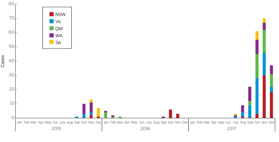
PAEDS = Paediatric Active Enhanced Disease Surveillance. * Before September 2017, surveillance only for suspected encephalitis and acute flaccid paralysis at sentinel sites; enhanced surveillance for all hospitalisations with parechovirus infection commenced in September 2017.
Box 2 – Clinical features of parechovirus infection
- Mild disease (especially in children aged > 6 months)
- Gastrointestinal symptoms: diarrhoea
- Respiratory disease: influenza-like illness
- Severe disease (especially in infants aged < 3–6 months): testing may be indicated
- Fever, irritability, rash (“red, hot, angry babies”) in infants aged < 3 months
- Sepsis-like syndromes (including shock)
- Seizures
- Meningoencephalitis, other neurological complications
- Distended abdomen
Box 3 – Appearance of a young infant with hospitalised parechovirus infection and sepsis-like disease
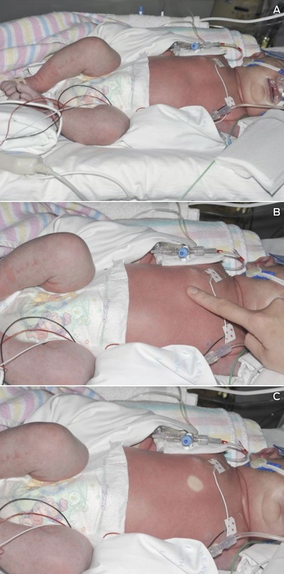
A: Confluent erythroderma of the trunk with an acral erythematous maculopapular rash of the lower limbs (note the presence of cardiorespiratory monitoring, multiple intravenous lines and nasal mask for continuous positive airway pressure ventilation support). B and C: Intense erythroderma and prolonged capillary return time. Photographs courtesy of Ameneh Khatami, Brendan McMullan and Julie Huynh.
Competing interests
No relevant disclosures.
Acknowledgements
Allen Cheng and Philip Britton are supported by NHMRC Fellowship funding. We thank the PAEDS surveillance nurses and investigators and investigators on the Australian childhood encephalitis and related studies including Julia Clark, Nigel Crawford, Jim Buttery, Christopher Blyth, Joshua Francis, Brendan McMullan, Alison Kesson, Nicole Dinsmore, Alissa McMinn, Sonia Dougherty, Carolyn Finucane and Christine Heath.
References
- Zurynski YA, McRae JE, Quinn HE, et al. Paediatric Active Enhanced Disease Surveillance inaugural annual report, 2014. Commun Dis Intell Q Rep 2016; 40: E391-E400.
- Hyypia T, Horsnell C, Maaronen M, et al. A distinct picornavirus group identified by sequence analysis. Proc Natl Acad Sci U S A 1992; 89: 8847-8851.
- de Crom SC, Rossen JW, van Furth AM, Obihara CC. Enterovirus and parechovirus infection in children: a brief overview. Eur J Pediatr 2016; 175: 1023-1029.
- Shakeel S, Dykeman EC, White SJ, et al. Genomic RNA folding mediates assembly of human parechovirus. Nat Commun 2017; 8: 5.
- Olijve L, Jennings L, Walls T. Human parechovirus: an increasingly recognized cause of sepsis-like illness in young infants. Clin Microbiol Rev 2018; 31. doi: 10.1128/CMR.00047-17.
- Cordey S, L’Huillier AG, Turin L, et al. Enterovirus and Parechovirus viraemia in young children presenting to the emergency room: unrecognised and frequent. J Clin Virol 2015; 68: 69-72.
- Vollbach S, Muller A, Drexler JF, et al. Prevalence, type and concentration of human enterovirus and parechovirus in cerebrospinal fluid samples of pediatric patients over a 10-year period: a retrospective study. Virol J 2015; 12: 199.
- Sano K, Hamada H, Hirose S, et al. Prevalence and characteristics of human parechovirus and enterovirus infections in febrile infants. Pediatr Int 2018; 60: 142-147.
- de Jong EP, van den Beuken MGA, van Elzakker EPM, et al. Epidemiology of sepsis-like illness in young infants: major role of enterovirus and human parechovirus. Pediatr Infect Dis J 2018; 37: 113-118.
- Ito M, Yamashita T, Tsuzuki H, et al. Isolation and identification of a novel human parechovirus. J Gen Virol 2004; 85(Pt 2): 391-398.
- Cumming G, Khatami A, McMullan BJ, et al. Parechovirus genotype 3 outbreak among infants, New South Wales, Australia, 2013–2014. Emerg Infect Dis 2015; 21: 1144-1152.
- Khatami A, McMullan BJ, Webber M, et al. Sepsis-like disease in infants due to human parechovirus type 3 during an outbreak in Australia. Clin Infect Dis 2015; 60: 228-236.
- Harvala H, McLeish N, Kondracka J, et al. Comparison of human parechovirus and enterovirus detection frequencies in cerebrospinal fluid samples collected over a 5-year period in edinburgh: HPeV type 3 identified as the most common picornavirus type. J Med Virol 2011; 83: 889-896.
- Britton PN, Dale RC, Nissen MD, et al. Parechovirus encephalitis and neurodevelopmental outcomes. Pediatrics 2016; 137: e20152848.
- Britton PN, Khandaker G, Khatami A, et al. High prevalence of developmental concern amongst infants at 12 months following hospitalised parechovirus infection. J Paediatr Child Health 2018; 54: 289-295.
- Nelson TM, Vuillermin P, Hodge J, et al. An outbreak of severe infections among Australian infants caused by a novel recombinant strain of human parechovirus type 3. Sci Rep 2017; 7: 44423.
- Britton PN, Dale RC, Elliott E, et al. Pilot surveillance for childhood encephalitis in Australia using the Paediatric Active Enhanced Disease Surveillance (PAEDS) network. Epidemiol Infect 2016; 144: 2117-2127.
- de Crom SC, Rossen JW, de Moor RA, et al. Prospective assessment of clinical symptoms associated with enterovirus and parechovirus genotypes in a multicenter study in Dutch children. J Clin Virol 2016; 77: 15-20.
- Watanabe K, Hirokawa C, Tazawa T. Seropositivity and epidemiology of human parechovirus types 1, 3, and 6 in Japan. Epidemiol Infect 2016: doi:10.1017/S0950268816001795 [Epub ahead of print].
- Pellegrinelli L, Bubba L, Galli C, et al. Epidemiology and molecular characterization of influenza viruses, human parechoviruses and enteroviruses in children up to 5 years with influenza-like illness in Northern Italy during seven consecutive winter seasons (2010-2017). J Gen Virol 2017; 98: 2699-2711.
- Bergallo M, Galliano I, Montanari P, et al. Molecular detection of human parechovirus in under-five-year-old children with gastroenteritis. J Clin Virol 2016; 85: 17-21.
- Harvala H, Robertson I, Chieochansin T, et al. Specific association of human parechovirus type 3 with sepsis and fever in young infants, as identified by direct typing of cerebrospinal fluid samples. J Infect Dis 2009; 199: 1753-1760.
- Verboon-Maciolek MA, Groenendaal F, Hahn CD, et al. Human parechovirus causes encephalitis with white matter injury in neonates. Ann Neurol 2008; 64: 266-273.
- Vergnano S, Kadambari S, Whalley K, et al. Characteristics and outcomes of human parechovirus infection in infants (2008-2012). Eur J Pediatr 2015; 174: 919-924.
- Selvarangan R, Nzabi M, Selvaraju SB, et al. Human parechovirus 3 causing sepsis-like illness in children from midwestern United States. Pediatr Infect Dis J 2011; 30: 238-242.
- Aizawa Y, Izumita R, Saitoh A. Human parechovirus type 3 infection: an emerging infection in neonates and young infants. J Infect Chemother 2017; 23: 419-426.
- Braccio S, Kapetanstrataki M, Sharland M, Ladhani SN. Intensive care admissions for children with enterovirus and human parechovirus infections in the United Kingdom and the Republic of Ireland, 2010-2014. Pediatr Infect Dis J 2017; 36: 339-342.
- Casas-Alba D, Martinez-Monseny A, Monfort L, et al. Extreme hyperferritinemia in dizygotic twins with human parechovirus-3 infection. Pediatr Infect Dis J 2016; 35: 1366-1368.
- Bigelow AM, Scott JP, Hong JC, et al. Human parechovirus as a cause of isolated pediatric acute liver failure. Pediatrics 2016; 138. doi: 10.1542/peds.2016-0233.
- van de Ven AA, Douma JW, Rademaker C, et al. Pleconaril-resistant chronic parechovirus-associated enteropathy in agammaglobulinaemia. Antivir Ther 2011; 16: 611-614.
- Kong KL, Lau JSY, Goh SM, et al. Myocarditis caused by human parechovirus in adult. Emerg Infect Dis 2017; 23: 1571-1573.
- Yamakawa T, Mizuta K, Kurokawa K, et al. Clinical characteristics of 17 adult patients with epidemic myalgia associated with human parechovirus type 3 infection. PloS ONE 2017; 57: 485-491.
- McGregor T, Bu’Lock FA, Wiselka M, Tang JW. Human parechovirus infection as an undiagnosed cause of adult pericarditis. J Infect 2017; 75: 596-597.
- Tanaka S, Kunishi Y. Severe human parechovirus type 3 infection in adults associated with gastroenteritis in their children. Infect Dis (Lond) 2017; 49: 772-774.
- Shinomoto M, Kawasaki T, Sugahara T, et al. First report of human parechovirus type 3 infection in a pregnant woman. Int J Infect Dis 2017; 59: 22-24.
- de Crom SC, Obihara CC, de Moor RA, et al. Prospective comparison of the detection rates of human enterovirus and parechovirus RT-qPCR and viral culture in different pediatric specimens. J Clin Virol 2013; 58: 449-454.
- Wildenbeest JG, Harvala H, Pajkrt D, Wolthers KC. The need for treatment against human parechoviruses: how, why and when? Expert Rev Anti Infect Ther 2010; 8: 1417-1429.
- Wildenbeest JG, Benschop KS, Bouma-de Jongh S, et al. Prolonged shedding of human parechovirus in feces of young children after symptomatic infection. Pediatr Infect Dis J 2016; 35: 580-583.
- Nielsen NM, Midgley SE, Nielsen AC, et al. Severe human parechovirus infections in infants and the role of older siblings. Am J Epidemiol 2016; 183: 664-670.
- Khandaker G, Jung J, Britton PN, et al. Long-term outcomes of infective encephalitis in children: a systematic review and meta-analysis. Dev Med Child Neurol 2016; 58: 1108-1115.
- Britton PN, Blyth CC, Macartney K, et al. The spectrum and burden of influenza-associated neurological disease in children: combined encephalitis and influenza sentinel site surveillance from Australia, 2013-2015. Clin Infect Dis 2017; 65: 653-660.
- Britton PN, Dale RC, Blyth CC, et al. Influenza-associated encephalitis/encephalopathy identified by the Australian Childhood Encephalitis Study 2013-2015. Pediatr Infect Dis J 2017; 36: 1021-1026.
- Huang YP, Hsieh JY, Wu HS, Yang JY. Molecular and epidemiological study of human parechovirus infections in Taiwan, 2007-2012. J Microbiol Immunol Infect 2016; 49: 321-328.
- Seo JH, Yeom JS, Youn HS, et al. Prevalence of human parechovirus and enterovirus in cerebrospinal fluid samples in children in Jinju, Korea. Korean J Pediatr 2015; 58: 102-107.
- Chiang GPK, Chen Z, Chan MCW, et al. Clinical features and seasonality of parechovirus infection in an Asian subtropical city, Hong Kong. PLoS One 2017; 12: e0184533.
- Abedi GR, Watson JT, Pham H, et al. Enterovirus and human parechovirus surveillance - United States, 2009-2013. MMWR Morb Mortal Wkly Rep 2015; 64: 940-943.
- Itta KC, Ghargi KV, Kalal S, et al. First detection of human parechovirus infection with diarrhoea, India. Infect Dis (Lond) 2017; 49: 151-152.
- Rahimi P, Naser HM, Siadat SD, et al. Genotyping of human parechoviruses in Iranian young children with aseptic meningitis and sepsis-like illness. J Neurovirol 2013; 19: 595-600.
- Chuchaona W, Khamrin P, Yodmeeklin A, et al. Detection and characterization of a novel human parechovirus genotype in Thailand. Infect Genet Evol 2015; 31: 300-304.
- Li R, Liu L, Mo Z, et al. An inactivated enterovirus 71 vaccine in healthy children. N Engl J Med 2014; 370: 829-837.
Linked content
-
MJA InSight: Be on the alert for parechovirus
-
MJA Podcast: Dr Philip Britton
Provenance: Commissioned; externally peer reviewed.
Costs of Delivering the Hep B PAST Model of Care in Remote Australia: A Retrospective Pre–Post Study
Hoa Nguyen, Anh Le Tuan Nguyen, Paula Binks, Melita McKinnon, Emily Vintour-Cesar, Karen Wills, Nicola Stephens, Benjamin Cowie, Joshua S. Davis, Yuejen Zhao, Peter Nihill, Julie A. Campbell, Andrew J. Palmer, Jane Davies, Barbara de Graaff
Buruli Ulcer Acquired From Ringtail Possum Rescue
Daniel P. O'Brien, Naomi Clarke, Sara Bonnici, Mohammad A. Hussain
Q Fever Visceral Abscesses After Contact With Native Australian Marsupials
Samantha J. Laws, Enzo Binotto, James Stewart, Jennifer Ho, Trent Yarwood, Josh Hanson, Simon Smith
Hospital-Admitted Injection-Related Infections Among Incarcerated People Who Inject Drugs in Australia: A Retrospective Cohort Study
Andrew Palmer, Matthew Carter, Jeremy Yeo, Cecilia Shim, Jason Connor, Jeremy Hayllar, Gerald Holtmann, Naomi Moy, Elliott G. Playford, Naomi Runnegar, Paul J. Clark
The Live Attenuated Influenza Vaccine in Australia: An Additional Tool for Influenza Prevention
Cyra Patel, Alexis Pillsbury, Tran Nguyen, Xia Wang, Helen E. Quinn, Clayton K. Chiu, Allen C. Cheng, Katie L. Flanagan, Zhicheng Wang
Reflex Testing for Hepatitis D Infection: A Unique Opportunity to Reduce Hepatitis D-Related Chronic Liver Disease Deaths in Australia
Jessica Howell, Lauren Andersson, Miriam T. Levy, James O'Beirne, Leon Adams, Katharine Irvine, Avik Majumdar, Golo Ahlenstiel, Kathy Jackson, Krispin Hajkowicz, Joseph Doyle, Jane Davies, Sarah Cherian, Wayne Dimech, Alexander J. Thompson