




Volume 208 - Issue 8
Developmental dysplasia of the hip: addressing evidence gaps with a multicentre prospective international study
Authors: Emily K Schaeffer, IHDI Study Group and Kishore Mulpuri
Med J Aust 2018; 208 (8): 359-364. || doi: 10.5694/mja18.00154
Published online: 7 May 2018
Published online: 7 May 2018
International studies are necessary to provide high quality evidence to guide DDH treatment and management
Summary
- There is a lack of high quality evidence available to guide clinical practice in the treatment and management of developmental dysplasia of the hip (DDH).
- Evidence has been limited by persistent confusion on diagnostic and classification terminology, variability in surgeon decision making and a reliance on single centre, retrospective studies with small patient numbers.
- To address gaps in knowledge regarding screening, diagnosis and management of DDH, the International Hip Dysplasia Institute began a multicentre, international prospective study on infants with hips dislocated at rest.
- This review discusses the current state of screening, diagnostic and management practices in DDH and addresses important unanswered questions that will be critical in identifying best practices and optimising patient outcomes.
- There is insufficient evidence to support universal ultrasound screening; instead, selective screening should be performed by 6–8 weeks of age on infants with risk factors of breech presentation, family history, or history of clinical hip instability. Follow-up of infants with risk factors and normal initial screening should be considered to at least 6 months of age.
- Brace treatment is a sensible first-line treatment for management of dislocated hips at rest in infants < 6 months of age.
- Early operative reduction may be considered as there is insufficient evidence to support a protective role for the ossific nucleus in the development of avascular necrosis.
Developmental dysplasia of the hip (DDH) is the most common paediatric hip disorder, affecting 1–3% of all infants.1,2 DDH encompasses a spectrum of conditions ranging from mild acetabular dysplasia of a reduced and stable hip, to an irreducibly dislocated hip.3 This disorder was historically referred to as congenital hip dysplasia, reflecting the pervading view that the condition was present from birth. However, as it became apparent that a number of cases were late-presenting or not diagnosed until well after birth, the term “developmental” was adopted to reflect that the condition may arise either in utero or in the post-natal period as the infant hip develops and matures.4 When left untreated or missed during early screening, DDH can lead to debilitating long term issues, including early-onset osteoarthritis of the hip, pain, limping, and the need for a total hip replacement in early adulthood.5,6 This review discusses the current state of evidence to guide screening and management of DDH. In particular, we highlight emerging data from an international prospective study that aims to identify best practices and treatment outcomes for patients globally. This narrative review is guided by a PubMed search of original and review articles along with clinical practice guidelines from the American Academy of Orthopaedic Surgeons (AAOS) and studies cited within.
Pathologies of developmental dysplasia of the hip
The hip is a ball-and-socket joint, where the femoral head sits congruently within the acetabulum. Abnormalities in acetabular morphology and/or femoral head position and shape can cause the hip to be unstable, subluxated or dislocated.7 DDH is a broad term used to describe a number of relatively distinct pathologies of the infant hip joint, which contribute to the lack of consensus for optimal screening, diagnostic and management practices.8 These pathologies represent a spectrum of severity, illustrated in Box 1. With mild acetabular dysplasia, the femoral head remains reduced and stable, but the acetabulum itself is shallow. This radiological dysplasia relies on ultrasonography or radiography for diagnosis and cannot be detected by physical examination.9 DDH also includes hip instability. A subluxable hip describes a situation when the femoral head is not centred in the acetabulum and can be more easily moved within the joint. A dislocatable hip describes a femoral head reduced at rest but able to be dislocated under stress, such as on physical examination. A dislocated hip occurs when the femoral head lies outside the acetabulum at resting position. Complete dislocations are estimated to occur in 3–4 per 1000 live births.10 The dislocation may be either reducible or irreducible on physical manipulation. Irreducible dislocations represent the most severe form of DDH and are usually the most challenging to treat by conservative or surgical methods.11-15 Although radiological dysplasia is relatively minor in comparison with more severe forms involving dislocation, each pathology along the spectrum holds clinical relevance. Radiological dysplasia is thought to account for a substantial number of hip replacements required in adulthood, with untreated dysplasia potentially leading to development of osteoarthritis of the hip.16,17 The natural history of DDH appears to relate to the severity of the condition; specifically, mild dysplasia may not manifest with clinical symptoms until adulthood, while severe dysplasia may become apparent in childhood.18 Children with an untreated dislocation may present with limb-length discrepancies, knee deformities and pain, or secondary scoliosis and back pain.6,19
Evidence to guide diagnosis, treatment and management
Despite decades of research and many studies, strong evidence to guide diagnosis, treatment and management is limited. The 2014 AAOS clinical practice guidelines included nine recommendations for screening and non-operative management of DDH before walking age.18 Of these, only two were of moderate strength, while the remainder were of limited strength. Recommendations are deemed moderate strength if there is one high quality or at least two moderate quality studies on the topic. Recommendations are considered limited strength if there is one moderate or at least one low quality study. Most existing studies on DDH are retrospective, single centre, have small sample sizes, or may include the entire spectrum of DDH severity.20-24 Compounding these issues, there is confusion in reporting of DDH diagnoses, particularly with laterality (the side affected by the condition). Many studies have examined incidence, prevalence and treatment outcomes of DDH comparing unilateral (only right or left hip involvement) and bilateral (both hip involvement) DDH,11,14,25,26 and differential success has indeed been reported in some bracing studies.11,14 However, laterality can be a complex issue considering the spectral nature of DDH (Box 1). It is typically unclear what constitutes a truly bilateral case, and with graded severity, hips may be irreducibly or reducibly dislocated, dislocatable, subluxable or dysplastic. If, for example, one hip is dislocated and one subluxable, it is unclear whether this case should be considered bilateral or unilateral. Individual studies rarely provide detailed criteria for their definition of laterality, making cross-study comparisons difficult.
Confusion regarding classification and diagnostic terminology for DDH also persists, further obfuscating available evidence.18,27,28 Diagnostic definitions are relatively subjective, creating variability between surgeons and across centres. Debate exists over whether clinical examination, ultrasound or radiography should be considered the gold-standard diagnostic method, which is likely a combination of clinical examination and radiological imaging. There will always be subjectivity in clinical examination, and even potentially objective radiological parameters such as percent coverage of the femoral head or alpha angle on ultrasound vary in interpretation. Some surgeons consider a hip dislocated only if coverage of the femoral head on ultrasound is less than 10%,29 while others make the diagnosis with coverage as high as 35%.30-32 Many studies fail to define what constitutes a dislocated hip. Improvement to available evidence requires a standardised framework for both reporting and diagnostic consistency to facilitate cross-study comparison as well as prospective studies with sound methodological design.
Screening
There has been longstanding debate on best practices for infant DDH screening. Clinical examination is universal, with all infants being tested for hip instability shortly after birth. In certain European countries and centres, ultrasound is also universally performed as a screening tool.33-37 In contrast, many North American centres employ selective ultrasound screening, whereby infants with DDH risk factors receive an ultrasound in addition to clinical examination.36,38-41 The AAOS guidelines identify that the only risk factors with sufficient evidence to warrant additional ultrasound screening are breech presentation, family history and history of clinical instability,18 recommending for these infants an ultrasound examination by the age of 6–8 weeks. The guidelines indicate insufficient evidence to support universal ultrasound screening.18 This lack of evidence led the American Academy of Pediatrics to somewhat controversially not recommend any DDH screening practices, clinical or ultrasound, in their 2016 guidelines.42
There are merits and drawbacks to both screening approaches. In screening and diagnosis, ultrasound interpretation is prone to inter- and intra-observer variability depending especially on operator skill.43-46 Universal screening has the potential to reduce missed cases presenting with more complex pathology later in infancy or childhood. However, there is also potential for overtreatment: an infant’s hips develop rapidly in this early post-natal period, and many cases of hip instability or dysplasia resolve spontaneously as the joint matures.45 Additionally, considerable cost may be associated with universal ultrasound programs, creating a burden on hospitals and families. Selective screening may reduce costs in comparison with universal programs, but may result in increased cases presenting late and requiring more extensive management.8,22,47-49 Further, even selective screening programs can be expensive and logistically difficult, especially in relatively resource-constrained settings. The AAOS clinical practice guidelines recommend against universal ultrasound screening, preferring selective screening on infants with risk factors of breech presentation, family history, or history of clinical instability.18 There is only limited to moderate evidence to support these conclusions (Box 2).18,50-53
While debate between universal and selective ultrasound screening continues, these platforms are only readily available in resource-rich settings. Ultrasound for DDH screening purposes is practically inaccessible to 80% of the world, where large populations are particularly susceptible to presentation of DDH after walking age. Some centres in North America, Europe and Australia employ either selective or universal ultrasound screening in conjunction with newborn physical examination. These regions collectively comprise a population of about 1.4 billion people.54,55 In contrast, regions including Asia, Africa and South America comprise a population of about 6.1 billion people and have limited resources for widespread ultrasound screening. A substantial proportion of the global population therefore goes unscreened during the critical early infancy developmental period, leading potentially to an overwhelming number of children presenting with DDH after walking age.56 When presenting at this late stage, extensive surgery is often required with multiple revision surgeries through to young adulthood.6,11,57-59
Despite best practice screening programs in Europe, North America and Australia, cases are still missed, resulting in children presenting later in infancy with more severe DDH. A review of the South Australian Birth Defects Register (SABDR) revealed that 3.4% of DDH cases in infants born between 1988 and 2003 were diagnosed late (> 3 months).20 Standard ultrasound metrics used to detect and diagnose DDH include the alpha angle and percent coverage of the femoral head. The alpha angle provides a measure of depth of the acetabulum, representing the angle formed between the roof of the acetabulum and the vertical cortex of the ilium. Percent coverage of the femoral head demonstrates the degree to which the femoral head is contained within the acetabulum. Although normal ranges for these metrics change rapidly in early hip joint development,60 an alpha angle greater than 60 degrees is considered normal in an infant, whereas an angle less than 45 degrees is considered severely dysplastic. These metrics are prone to measurement variability within and between observers, which may compromise timely diagnosis. Recently, existing two-dimensional ultrasound techniques have been improved through automated methods, machine learning and three-dimensional probes.43,45,46,61 Although still in the early validation stages, findings have suggested improved inter- and intra-rater reliability (Box 3). However, sophisticated ultrasound technology may have little impact in resource-poor settings unable to access such technology. There is a clear need to develop area-specific screening protocols for local providers appropriate to their own resources. Individualised care pathways will be a critical step to providing global, comprehensive management with the ultimate goal of eliminating walking-age sequelae of DDH worldwide.
Treatment and management
As for screening and diagnostic practices, DDH management depends on individual surgeon decision making, and high quality evidence identifying best management practices is lacking. Again, the absence of evidence results from widespread confusion in terminology and poor literature reporting, compounded by the broad spectrum of severity encompassed by a DDH diagnosis. Results of bracing studies may be obfuscated by including patients with reducible and irreducible dislocated hips, unstable or subluxable hips and dysplastic hips together,11,13,14,25,62 and studies comparing operative management also often fail to clearly define the included patient population.63
To attempt to identify best practice evidence in DDH management, the International Hip Dysplasia Institute (IHDI) initiated in 2010 an international, multicentre prospective study64 of infants with the most severe form of DDH — dislocated hips at rest. Infants diagnosed under 18 months of age with a dislocated hip at rest were enrolled at nine centres across North America, Europe and Australia. Diagnosis was confirmed by ultrasound or x-ray before treatment. These patients were prospectively followed for 2 years, with data collected on patient and maternal demographics, diagnosis, non-operative and operative treatment. From 2010 to 2016, 619 patients were followed, 486 of whom were diagnosed under 6 months of age and 133 between 6 and 18 months. Primary outcomes have focused on surgeon and centre variability in screening and management practices and early outcomes of operative and non-operative management.
Initial findings have shown distinct differences across contributing centres in patient demographics, DDH severity and laterality at presentation, confirming the importance of locality variation in any predictive analysis of treatment success or failure in multicentre studies.64 Additionally, infants presenting late (> 3 months of age) tend to have fewer traditional risk factors typically associated with DDH (ie, breech presentation), and late presentation may be associated with swaddling during infancy.65 The protective effect of breech presentation against late diagnosis is consistent with the findings of the SABDR study.39,66 Further investigation is needed to ascribe prognostic value or causal links between these factors and late-presenting DDH, but more rigorous examination of the efficacy of current screening processes is likely required to minimise late diagnoses. The SABDR review noted increased late-diagnosed DDH from 2003–2009 compared with 1988–2003 (11.5% v 3.5% of cases).66 The authors proposed causative factors, including lack of resources in rural birth settings and increased popularity of swaddling for infants, and suggested that promoting awareness and education to the public and to medical practitioners would reduce late diagnoses. A high quality clinical screening program may be the most important factor in reducing missed cases.66
Early outcomes of brace treatment were examined in the IHDI subgroup diagnosed under 6 months of age.15 Patients with either reducible or irreducible hip dislocations were primarily managed by either dynamic (eg, Pavlik harness) or rigid (eg, abduction) brace, and factors involved in brace failure were identified. Brace treatment was successful for 79% of all included hips, with brace failure associated with development of femoral nerve palsy, rigid brace, irreducible dislocations, right hip dislocations and treatment initiation after 7 weeks of age.15 Despite irreduciblity being a potential brace failure factor, there was still a 55% success rate with brace treatment for these hips.15 Thus, this level I evidence (levels of evidence based on the Oxford Centre for Evidence-Based Medicine hierarchy: https://www.cebm.net/2016/05/ocebm-levels-of-evidence) suggests that brace treatment is a sensible first-line treatment option in an attempt to avoid operative management (Box 2).
Early-term results of operative management were investigated for all patients diagnosed up to 18 months of age. Attempted closed reduction was successful in 91% of hips, and 91% of these initially successful reductions remained stable at a median follow-up of 22 months.51 Larger patient numbers will be necessary to identify true predictors of failure, as the study to date is likely underpowered in not finding associations between failure of closed reduction and age at reduction, clinical reducibility of the hip, or previous brace.51 Open reduction was successful at achieving and maintaining reduction in 99% of patients with a median of 23 months of follow-up, although some degree of subluxation was present in 12%.52 Despite the relatively high success of achieving and maintaining reduction in both of these patient cohorts, development of avascular necrosis (AVN) of the femoral head occurred after closed51 and open reduction52 in 25% and 26% of patients, respectively, indicating a need for more rigorous identification of AVN risk factors for prognostic and preventive purposes. Conflicting reports in the literature as to whether open or closed reduction leads to higher rates of AVN67-71 are partly clarified by these early prospective results on a more definitive cohort.
The timing of operative management in relation to the appearance of the ossific nucleus has been debated, with proponents of later intervention arguing that it is protective against the development of AVN.72-74 Advocates of early operative reduction counter that the potential for achieving and maintaining stable, concentric reduction of the hip is higher by allowing maximal acetabular remodelling.75-77 In both closed and open reduction patient cohorts in this prospective study, the presence of the ossific nucleus does not appear to have an impact on the development of AVN, although larger numbers will be necessary to confirm these preliminary findings. With no evidence in these level II studies to support waiting for the presence of an ossific nucleus, early operative reduction may be considered (Box 2).
While the IHDI study has demonstrated the power of international, prospective study groups to generate high quality evidence to guide and inform clinical practice, its limitations also highlight outstanding issues relating to DDH management. First, the IHDI study has not provided evidence for all hips with DDH — only those at the most severe end of the spectrum, with evidence and recommendations specific to hips dislocated at rest, either reducible or irreducible. Second, all bracing and surgical outcome data are preliminary, reporting on relatively small patient numbers and limited to 2 years of follow-up. AVN is an important DDH treatment complication that may lead to lifelong debilitation and pain, and follow-up to a minimum age of skeletal maturity will be required to gain a more complete understanding of the impact of various DDH management strategies.
Additionally, the IHDI study was largely limited to centres in the developed world with established screening protocols in place and relatively abundant resources. Large numbers of patients in areas such as Asia, Africa and South America who present after walking age requiring extensive surgical management were not captured in the IHDI study. Over 2017–2018, the study size and scope has been expanded into the International Hip Dysplasia Registry (IHDR). The IHDR now includes 18 centres contributing around the world, sites in India and China, and children with less severe forms of DDH (dysplasia, subluxable and dislocatable hips). With these inclusions and with follow-up to skeletal maturity, IHDR is positioned to address still-unanswered questions in DDH, with the potential to make a global impact.
Unanswered questions
There are several important questions that can be tested with methodological rigour in the near future. First, with selective ultrasound screening programs in place in North America, it will be important to understand what follow-up may be required for infants with DDH risk factors who receive a normal screening ultrasound at 6 weeks of age. A 2009 study suggested that as many as 29% of breech infants initially screened as normal show radiographic signs of dysplasia severe enough to warrant treatment at 6 months of age.50 Appropriate follow-up practice for these infants should minimise late diagnosis while preventing potentially unnecessary exposure to radiation and clinical visits. Second, in infants with radiological dysplasia and clinically stable hips, there is debate as to whether brace treatment significantly improves outcomes. Spontaneous resolution of dysplasia in early infancy is often seen, and commonly used hip ultrasound metrics may change rapidly in the first 12 weeks after birth.60 Therefore, examining whether observation alone may be as effective as bracing in this patient population will be beneficial. Third, the relative outcomes with different types of brace remain unclear. Dynamic bracing by Pavlik harness is the most common non-operative management strategy, but rigid abduction braces are also used. Until high quality comparative effectiveness studies are performed with sufficient patient numbers, choice of dynamic or rigid brace should be left to the discretion of the treating practitioner. Finally, engaging patients and families early in the study development process will be critical to identify their priorities and address their primary concerns in future research. Answering each of these questions will substantially advance the evidence available to guide early management of DDH, and possibly reduce economic and social costs on health systems and patients and families.
Future directions
As we work towards improving outcomes for children screened, diagnosed and treated for DDH, we must consider the wide variations in presentation and available resources. Efforts to improve DDH care need a global vision, with advancements applicable or feasible across resource environments. A vital first step in realising this vision is for international registries to have a broader representation of different health care systems in order to maximise impact for all patients worldwide.
Box 1 – Graded severity spectrum of developmental dysplasia of the hip
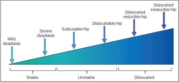
The spectrum begins with mild dysplasia (marginally abnormal acetabular morphology) of a reduced, clinically stable hip. The shallower the acetabulum, the greater the severity of dysplasia in a stable hip. When the hip is unstable, the femoral head can have abnormal movement within the acetabulum (subluxable), or it can be pushed from the acetabulum entirely (dislocatable). While there is hip joint instability in these cases, the femoral head is reduced within the acetabulum at rest. A dislocated hip occurs when the femoral head lies outside the acetabulum at rest. A dislocated hip may be reducible, whereby the femoral head can be reduced into the acetabulum during physical examination, or irreducible, remaining outside of the acetabulum even with the application of reasonable but moderate force.
Box 2 – Developmental dysplasia of the hip (DDH): evidence summary and clinical practice recommendations*
|
Topic |
Evidence |
Recommendation |
|||||||||||||
|
|
|||||||||||||||
|
Universal v selective ultrasound screening of the neonatal hip |
Moderate evidence18 suggests no statistical difference in late-presenting DDH between universal and selective screening |
Selective ultrasound screening for infants with risk factors of breech presentation, family history of DDH, or history of clinical instability18 |
|||||||||||||
|
Follow-up for infants with DDH risk factors and normal screening results |
A level III evidence50 study reported 29% of breech infants with a normal screening ultrasound required treatment for DDH at 6 months |
While possibly excessive, until strong prospective evidence can be gathered, recommend anteroposterior pelvis x-ray at 6,50 12 and 24 months† for infants with DDH risk factors |
|||||||||||||
|
Brace treatment as conservative management for dislocated hips |
Level I evidence from the IHDI study15 found a 79% brace success rate for dislocated hips in infants diagnosed < 6 months of age and 55% success rate in irreducible hip dislocations |
Brace treatment is a sensible first-line treatment choice for infants under 6 months of age diagnosed with a dislocated hip |
|||||||||||||
|
Dynamic v rigid brace |
Limited evidence18 for preference for either rigid brace or dynamic brace (ie, Pavlik harness) for treatment of an unstable (dislocated or subluxable) hip |
Until strong prospective comparative effectiveness studies have been performed, choice of dynamic or rigid brace at the treating practitioner’s discretion |
|||||||||||||
|
Early v delayed surgical reduction (closed or open) of a dislocated hip |
Level II evidence from the IHDI study51,52 found no impact on reduction success and on development of avascular necrosis in the presence or absence of the ossific nucleus |
The practitioner should balance risks of anaesthesia in an infant with the benefit of early reduction remodelling potential rather than base decision on presence of ossific nucleus |
|||||||||||||
|
Radiographic classification of DDH severity |
Evidence level not available |
The IHDI classification53 presents a more applicable and repeatable determination of DDH severity in the absence of the ossific nucleus than the Tönnis method |
|||||||||||||
|
|
|||||||||||||||
|
* Levels of evidence based on the Oxford Centre for Evidence-Based Medicine hierarchy: https://www.cebm.net/2016/05/ocebm-levels-of-evidence. Evidence and recommendations adapted and extrapolated from the current AAOS clinical practice guidelines18 and new emerging data from the IHDI study on dislocated hips at rest.15,51-53 † The 6 month x-ray is based on level III evidence;50 12 and 24 month x-rays are based on expert opinion, performed as part of the authors’ clinical practice. |
|||||||||||||||
Box 3 – Use of three-dimensional (3D) ultrasound techniques aimed at reducing variability in standard two-dimensional (2D) dysplasia metrics
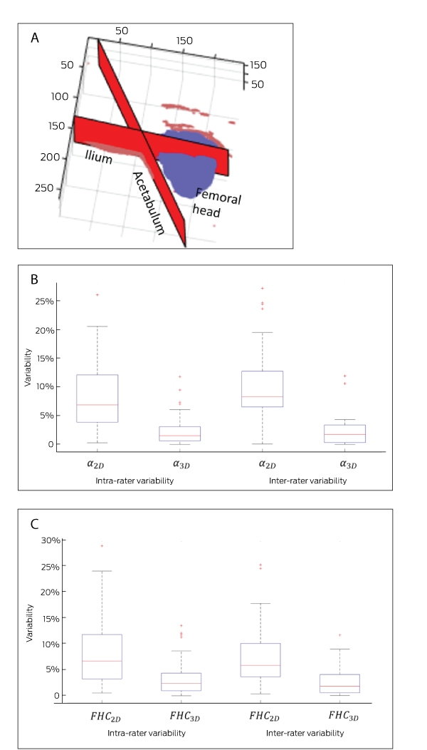
A: 3D rendering of the infant hip joint extracted from a 3D ultrasound image of an infant with a dysplastic hip: a point cloud plot showing a set of data points in 3D space from an acquired 3D ultrasound image of the infant hip. The data points in red represent the extracted ilium and acetabulum boundaries. The data points in blue represent the extracted femoral head. Axis numbers represent distance units in each dimension; 1 distance unit = 0.2 mm. B: Intra- and inter-rater variability of alpha angle measurement using 2D and 3D ultrasound techniques. The variability is represented by the standard deviation of repeated measurements of alpha angle (in degrees) by the same rater (intra-rater) and the standard deviation between two raters’ measures of the alpha angle on the same image. 2D and 3D images were collected in the same ultrasound examination session using 2D and 3D ultrasound probes. C: Intra- and inter-rater variability of percent coverage of the femoral head (FHC) using 2D and 3D ultrasound techniques.
Competing interests
We have received funding for research support from the registry seed grant, the IHDI, the Children’s Hospital Research Institute and the I'm a HIPpy Foundation.
Acknowledgements
We thank members of the IHDI Study Group for their intellectual and data contributions to the IHDI and IHDR studies; and Niamul Quader for supplying the image and data for Box 3.
References
- Godward S, Dezateux C. Surgery for congenital dislocation of the hip in the UK as a measure of outcome of screening. Lancet 1998; 351: 1149-1152.
- Tibrewal S, Gulati V, Ramachandran M. The Pavlik method: a systematic review of current concepts. J Pediatr Orthop B 2013; 22: 516-520.
- Bialik V, Bialik GM, Blazer S, et al. Developmental dysplasia of the hip: a new approach to incidence. Pediatrics 1999; 103: 93-99.
- Jackson JC, Runge MM, Nye NS. Common questions about developmental dysplasia of the hip. Am Fam Physician 2014; 90: 843-850.
- Engesæter IØ, Lie SA, Lehmann TG, et al. Neonatal hip instability and risk of total hip replacement in young adulthood. Acta Orthop 2008; 79: 321-326.
- Wedge JH, Wasylenko MJ. The natural history of congenital dislocation of the hip: a critical review. Clin Orthop Relat Res 1978; 137: 154-162.
- Karmazyn BK, Gunderman RB, Coley BD, et al. ACR Appropriateness Criteria® on developmental dysplasia of the hip—child. J Am Coll Radiol 2009; 6: 551-557.
- Shorter D, Hong T, Osborn DA. Cochrane Review: screening programmes for developmental dysplasia of the hip in newborn infants. Evid Based Child Health 2013; 8: 11-54.
- Narayanan U, Mulpuri K, Sankar WN, et al. Reliability of a new radiographic classification for developmental dysplasia of the hip. J Pediatr Orthop 2015; 35: 478.
- Tönnis D. Congenital dysplasia and dislocation of the hip in children and adults. Berlin: Springer-Verlag, 1987.
- Atalar H, Sayli U, Yavuz OY, et al. Indicators of successful use of the Pavlik harness in infants with developmental dysplasia of the hip. Int Orthop 2007; 31: 145-150.
- Harding MG, Harcke HT, Bowen JR, et al. Management of dislocated hips with Pavlik harness treatment and ultrasound monitoring. J Pediatr Orthop 1997; 17: 189-198.
- Lerman JA, Emans JB, Millis MB, et al. Early failure of Pavlik harness treatment for developmental hip dysplasia: clinical and ultrasound predictors. J Pediatr Orthop 2001; 21: 348-353.
- Viere RG, Birch JG, Herring JA, et al. Use of the Pavlik harness in congenital dislocation of the hip. An analysis of failures of treatment. J Bone Joint Surg Am 1990; 72: 238-244.
- Upasani VV, Bomar JD, Matheney TH, et al. Evaluation of brace treatment for infant hip dislocation in a prospective cohort: defining the success rate and variables associated with failure. J Bone Joint Surg Am 2016; 98: 1215-1221.
- Engesaeter LB, Furnes O, Havelin LI. Developmental dysplasia of the hip–good results of later total hip arthroplasty: 7135 primary total hip arthroplasties after developmental dysplasia of the hip compared with 59774 total hip arthroplasties in idiopathic coxarthrosis followed for 0 to 15 years in the Norwegian Arthroplasty Register. J Arthroplasty 2008; 23: 235-240.
- Dezateux C, Rosendahl K. Developmental dysplasia of the hip. Lancet 2007; 369: 1541-1552.
- American Academy of Orthopaedic Surgeons. Detection and nonoperative management of pediatric developmental dysplasia of the hip in infants up to six months of age: evidence-based clinical practice guideline. Rosemount, IL: AAOS, 2014. https://www.aaos.org/research/guidelines/DDHGuidelineFINAL.pdf (viewed Feb 2018).
- Weinstein SL. Natural history of congenital hip dislocation (CDH) and hip dysplasia. Clin Orthop Relat Res 1987; 225: 62-76.
- Azzopardi T, Van Essen P, Cundy PJ, et al. Late diagnosis of developmental dysplasia of the hip: an analysis of risk factors. J Pediatr Orthop B 2011; 20: 1-7.
- Jacobsen S, Rømer L, Søballe K. The other hip in unilateral hip dysplasia. Clin Orthop Relat Res 2006; 446: 239-246.
- Laborie LB, Markestad TJ, Davidsen H, et al. Selective ultrasound screening for developmental hip dysplasia: effect on management and late detected cases. A prospective survey during 1991–2006. Pediatr Radiol 2014; 44: 410-424.
- Rosendahl K, Markestad T, Lie RT. Ultrasound in the early diagnosis of congenital dislocation of the hip: the significance of hip stability versus acetabular morphology. Pediatr Radiol 1992; 22: 430-433.
- Abu Hassan FO, Shannak A. Associated risk factors in children who had late presentation of developmental dysplasia of the hip. J Child Orthop 2007; 1: 205-210.
- Palocaren T, Rogers K, Haumont T, et al. High failure rate of the Pavlik harness in dislocated hips: is it bilaterality? J Pediatr Orthop 2013; 33: 530-535.
- Borowski A, Thawrani D, Grissom L, et al. Bilaterally dislocated hips treated with the Pavlik harness are not at a higher risk for failure. J Pediatr Orthop 2009; 29: 661-665.
- Rosendahl K, Markestad T, Lie RT. Ultrasound screening for developmental dysplasia of the hip in the neonate: the effect on treatment rate and prevalence of late cases. Pediatrics 1994; 94: 47-52.
- Sewell MD, Rosendahl K, Eastwood DM. Developmental dysplasia of the hip. BMJ 2009; 339: b4454.
- Graf R. Classification of hip joint dysplasia by means of sonography. Arch Orthop Trauma Surg 1984; 102: 248-255.
- Holen KJ, Tegnander A, Eik-Nes SH, Terjesen T. The use of ultrasound in determining the initiation of treatment in instability of the hip in neonates. J Bone Joint Surg Br 1999; 81: 846-851.
- Terjesen T, Bredland T, Berg V. Ultrasound for hip assessment in the newborn. Bone Joint J 1989; 71: 767-773.
- Holen KJ, Tegnander A, Terjesen T, et al. Ultrasonography of clinically unstable hips: a prospective study of 143 neonates at birth and early follow-up. Acta Orthop Scand 1997; 68: 527-532.
- Bialik V, Bialik GM, Wiener F. Prevention of overtreatment of neonatal hip dysplasia by the use of ultrasonography. J Pediatr Orthop B 1998; 7: 39-42.
- Hadlow VI. Neonatal screening for congenital dislocation of the hip. A prospective 21-year survey. J Bone Joint Surg Br 1988; 70: 740-743.
- Synder M, Harcke HT, Domzalski M. Role of ultrasound in the diagnosis and management of developmental dysplasia of the hip: an international perspective. Orthop Clin North Am 2006; 37: 141-147.
- Szulc W. The frequency of occurrence of congenital dysplasia of the hip in Poland. Clin Orthop Relat Res 1991; 272: 100-102.
- von Kries R, Ihme N, Oberle D, et al. Effect of ultrasound screening on the rate of first operative procedures for developmental hip dysplasia in Germany. Lancet 2003; 362: 1883-1887.
- Mahan ST, Katz JN, Kim YJ. To screen or not to screen? A decision analysis of the utility of screening for developmental dysplasia of the hip. J Bone Joint Surg Am 2009; 91: 1705.
- Sharpe P, Mulpuri K, Chan A, Cundy PJ. Differences in risk factors between early and late diagnosed developmental dysplasia of the hip. Arch Dis Child Fetal Neonatal Ed 2006; 91: F158-F162.
- Shipman SA, Helfand M, Moyer VA, Yawn BP. Screening for developmental dysplasia of the hip: a systematic literature review for the US Preventive Services Task Force. Pediatrics 2006; 117: e557-e576.
- Tredwell SJ. Neonatal screening for hip joint instability. Its clinical and economic relevance. Clin Orthop Relat Res 1992; 281: 63-68.
- Shaw BA, Segal LS. Evaluation and referral for developmental dysplasia of the hip in infants. Pediatrics 2016; 138: e20163107.
- Jaremko JL, Mabee M, Swami VG, et al. Potential for change in US diagnosis of hip dysplasia solely caused by changes in probe orientation: patterns of alpha-angle variation revealed by using three-dimensional US. Radiology 2014; 273: 870-878.
- Cheng E, Mabee M, Swami VG, et al. Ultrasound quantification of acetabular rounding in hip dysplasia: reliability and correlation to treatment decisions in a retrospective study. Ultrasound Med Biol 2015; 41: 56-63.
- Quader N, Hodgson A, Mulpuri K, et al. Towards reliable automatic characterization of neonatal hip dysplasia from 3D ultrasound images. In: Ourselin S, Joskowicz L, Sabuncu M, et al, editors. Medical Image Computing and Computer-Assisted Intervention – MICCAI 2016. Lecture Notes in Computer Science, vol. 9900. Springer: 2016: 602-609. https://link.springer.com/chapter/10.1007/978-3-319-46720-7_70 (viewed Mar 2018).
- Quader N, Hodgson AJ, Mulpuri K, et al. A 3D femoral head coverage metric for enhanced reliability in diagnosing hip dysplasia. In: Descoteaux M, Maier-Hein L, Franz A, et al, editors. Medical Image Computing and Computer-Assisted Intervention – MICCAI 2017. Lecture Notes in Computer Science, vol. 10433. Springer: 2017: 100-107. https://link.springer.com/chapter/10.1007/978-3-319-66182-7_12 (viewed Mar 2018).
- Ilfeld FW, Westin GW, Makin MY. Missed or developmental dislocation of the hip. Clin Orthop Relat Res 1986; 203: 276-281.
- Lennox IA, McLauchlan J, Murali R. Failures of screening and management of congenital dislocation of the hip. J Bone Joint Surg Br 1993; 75: 72-75.
- Sink EL, Ricciardi BF, Torre KD, Price CT. Selective ultrasound screening is inadequate to identify patients who present with symptomatic adult acetabular dysplasia. J Child Orthop 2014; 8: 451-455.
- Imrie M, Scott V, Stearns P, et al. Is ultrasound screening for DDH in babies born breech sufficient? J Child Orthop 2010; 4: 3-8.
- Sankar WN, Gornitzky AL, Clarke NM, et al. Closed reduction for developmental dysplasia of the hip: Early-term results from a prospective, multicenter cohort. J Pediatr Orthop 2016; doi:10.1097/BPO0000000000000895 [Epub ahead of print].
- Schaeffer EK, Mulpuri K, Sankar WN, et al. Open reduction for developmental dysplasia of the hip: early outcomes from a multi-centre, international prospective cohort study [abstract]. J Child Orthop 2017; 11 (Suppl 1): S164.
- Narayanan U, Mulpuri K, Sankar WN, et al. Reliability of a new radiographic classification for developmental dysplasia of the hip. J Pediatr Orthop 2015; 35: 478-484.
- United Nations DESA/Population Division. World populations prospects 2017. Data query. https://esa.un.org/unpd/wpp/DataQuery/ (viewed Feb 2018).
- United Nations Department of Economic and Social Affairs. Statistics Division. UN data. https://unstats.un.org/sdgs/ (viewed Feb 2018).
- Rebello G, Joseph B. Late presentation of developmental dysplasia of the hip in children from southwest India – will screening help? Ind J Orthop 2003; 37: 210-214.
- Guille JT, Pizzutillo PD, MacEwen GD. Developmental dysplasia of the hip from birth to six months. J Am Acad Orthop Surg 2000; 8: 232-242.
- Heikkilä E, Ryöppy S, Louhimo L. Late diagnosis in congenital dislocation of the hip. Acta Orthop Scand 1984; 55: 256-260.
- Inoue T, Naito M, Nomiyama H. Treatment of developmental dysplasia of the hip with the Pavlik harness: factors for predicting unsuccessful reduction. J Pediatr Orthop B 2001; 10: 186-191.
- Riad JP, Cundy P, Gent RJ, et al. Longitudinal study of normal hip development by ultrasound. J Pediatr Orthop 2005; 25: 5-9.
- Quader N, Hodgson AJ, Mulpuri K, et al. Automatic evaluation of scan adequacy and dysplasia metrics in 2-D ultrasound images of the neonatal hip. Ultrasound Med Biol 2017; 43: 1252-1262.
- Swaroop VT, Mubarak SJ. Difficult-to-treat Ortolani-positive hip: improved success with new treatment protocol. J Pediatr Orthop 2009; 29: 224-230.
- Novais EN, Hill MK, Carry PM, Heyn PC. Is age or surgical approach associated with osteonecrosis in patients with developmental dysplasia of the hip? A meta-analysis. Clin Orthop Relat Res 2016; 474: 1166-1177.
- Mulpuri K, Schaeffer EK, Kelley SP, et al. What is the impact of center variability in a multicenter international prospective observational study on developmental dysplasia of the hip? Clin Orthop Relat Res 2016; 474: 1138-1145.
- Mulpuri K, Schaeffer EK, Andrade J, et al. What risk factors and characteristics are associated with late-presenting dislocations of the hip in Infants? Clin Orthop Relat Res 2016; 474: 1131-1137.
- Studer K, Williams N, Antoniou G, et al. Increase in late diagnosed developmental dysplasia of the hip in South Australia: risk factors, proposed solutions. Med J Aust 2016; 204: 240.
- Kalamchi A, MacEwen GD. Avascular necrosis following treatment of congenital dislocation of the hip. J Bone Joint Surg Am 1980; 62: 876-888.
- Kruczynski J. Avascular necrosis of the proximal femur in developmental dislocation of the hip. Incidence, risk factors, sequelae an MR imaging for diagnosis and prognosis. Acta Orthop Scand Suppl 1996; 67(S268): 3-48.
- Senaran H, Bowen JR, Harcke HT. Avascular necrosis rate in early reduction after failed Pavlik harness treatment of developmental dysplasia of the hip. J Pediatr Orthop 2007; 27: 192-197.
- Konigsberg DE, Karol LA, Colby S, O’Brien S. Results of medial open reduction of the hip in infants with developmental dislocation of the hip. J Pediatr Orthop 2003; 23: 1-9.
- Mankey MG, Arntz GT, Staheli LT. Open reduction through a medial approach for congenital dislocation of the hip. A critical review of the Ludloff approach in sixty-six hips. J Bone Joint Surg Am 1993; 75: 1334-1345.
- Salter RB, Kostuk J, Dallas S. Avascular necrosis of the femoral head as a complication of treatment for congenital dislocation of the hip in young children: a clinical and experimental investigation. Can J Surg 1969; 12: 44-61.
- Clarke NMP. Blood supply and proximal femoral epiphyseal deformity in congenital dislocation of the hip. In Uhtoff HK, Wiley JJ, editors. Behaviour of the growth plate. New York: Raven Press, 1998.
- Segal LS, Boal DK, Borthwick L. Avascular necrosis after treatment of DDH: the protective influence of the ossific nucleus. J Pediatr Orthop 1999; 19: 177-184.
- Luhmann SJ, Bassett GS, Gordon JE, et al. Reduction of a dislocation of the hip due to developmental dysplasia: Implications for the need for future surgery. J Bone Joint Surg Am 2003; 85: 239-243.
- Ogden JA. Treatment positions for congenital dysplasia of the hip. J Pediatr 1975; 86: 732-734.
- Dhar S, Taylor JF, Jones WA. Early open reduction for congenital dislocation of the hip. J Bone Joint Surg Br 1990; 72: 175-180.
Linked content
-
MJA InSight: Screen all babies for hip dysplasia
-
MJA Podcast: Dr Kishore Mulpuri and Dr Emily Schaeffer
Provenance: Commissioned; externally peer reviewed.
Decolonising Indigenous Health Research Requires Redefining Sex, Sexuality and Gender
Shakara Liddelow-Hunt, James H. L. Hill, Kate Daglas, Kaiwarr Clancy, Madi Day, Alistair Ott
Reducing Nitrous Oxide Emissions Across the Melbourne Biomedical Precinct
Ross Robertson, Andrew Downey, Daryl Williams, Bjorn Makein, Ben Dunne, Tugce Ozturk, Ying Gu, Rebecca McIntyre
Multimorbidity Clusters Among People Aged 65 Years and Over in Australia: A Nationwide Cross-Sectional Data Linkage Study
Weisi Chen, Christine Y. Lu, Sarah N. Hilmer, Alice A. Gibson, Edwin C. K. Tan
When ‘Liver Enzymes’ Are Not Hepatic: Late-Onset Pompe Disease
Shauna Madigan, Georgina England, Wayne Rankin
West Nile virus Kunjin subtype in rural NSW
Emily Gibson, Megan Whitley, Peter Murray, Linda Hueston, Jane Bennett, Raguharan Kathiresu, David N Durrheim
The impact of the Breast Screen NSW transition from film to digital mammography, 2002–2016: a linked population health data analysis
Rachel Farber, Nehmat Houssami, Katy J L Bell