




Volume 208 - Issue 11
Sarcoidosis: a state of the art review from the Thoracic Society of Australia and New Zealand
Authors: Hasib Ahmadzai, Shuying Huang, Chris Steinfort, James Markos, Roger KA Allen, Denis Wakefield, Margaret Wilsher and Paul S Thomas
Med J Aust 2018; 208 (11): 499-504. || doi: 10.5694/mja17.00610
Published online: 7 May 2018
Published online: 7 May 2018
Despite recent advances in our knowledge of sarcoidosis, it remains a diagnosis of exclusion
Summary
- Sarcoidosis is a systemic disease of unknown aetiology, characterised by non-caseating granulomatous inflammation. It most commonly manifests in the lungs and intrathoracic lymph nodes but can affect any organ.
- This summary of an educational resource provided by the Thoracic Society of Australia and New Zealand outlines the current understanding of sarcoidosis and highlights the need for further research.
- Our knowledge of the aetiology and immunopathogenesis of sarcoidosis remains incomplete.
- The enigma of sarcoidosis lies in its immunological paradox of type 1 T helper cell-dominated local inflammation co-existing with T regulatory-induced peripheral anergy.
- Although specific aetiological agents have not been identified, mounting evidence suggests that environmental and microbial antigens may trigger sarcoidosis.
- Genome-wide association studies have identified candidate genes conferring susceptibility and gene expression analyses have provided insights into cytokine dysregulation leading to inflammation.
- Sarcoidosis remains a diagnosis of exclusion based on histological evidence of non-caseating granulomas with compatible clinical and radiological findings.
- In recent years, endobronchial ultrasound-guided transbronchial needle aspiration of mediastinal lymph nodes has facilitated the diagnosis, and whole body positron emission tomography scanning has improved localisation of disease.
- No single biomarker is adequately sensitive and specific for detecting and monitoring disease activity.
- Most patients do not require treatment; when indicated, corticosteroids remain the initial standard of care, despite their adverse side effect profile.
- Other drugs with fewer side effects may be a better long term choice (eg, methotrexate, hydroxychloroquine, azathioprine, mycophenolate), while tumour necrosis factor-α inhibitors are a treatment option for patients with refractory disease.
Sarcoidosis is a systemic granulomatous disease of unknown aetiology, primarily affecting the lungs and lymphatic systems, although any organ may be involved. The presentation may be an incidental finding on chest radiology, or due to symptoms, most commonly cough or breathlessness, or relating to involvement of other organ systems such as the eyes, skin, nervous system or heart. Löfgren syndrome (fever, erythema nodosum, bilateral hilar lymphadenopathy, polyarthritis) is a distinct acute form with a good prognosis. Sarcoidosis is diagnosed by clinical and radiological findings and demonstration of non-caseating granulomas on biopsy, with exclusion of other causes. Differential diagnoses include tuberculosis, lymphoma and other causes of lung fibrosis. Few randomised, controlled studies indicate the appropriate treatment, despite increased knowledge of the immunopathological and genetic features. The management of sarcoidosis can be challenging, despite new developments in diagnostic techniques and biological agents for treatment. Here, we discuss developments in the understanding of its pathology, diagnosis and management.
For this narrative review, we searched PubMed for original papers and review articles published between 1999 and 2017 to formulate an evidence-based overview of sarcoidosis. This article is a summary of an educational resource provided by the Thoracic Society of Australia and New Zealand, available online at https://www.thoracic.org.au/documents/item/1332.
Epidemiology
Racial, demographic and ethnic variations in clinical manifestations of sarcoidosis exist. Sarcoidosis affects people of all ages regardless of race and ethnicity, with peak incidence among people aged 20–39 years, and is thought to be more common in rural communities.1,2 In Australia, the prevalence is estimated to be 4.4–6.3 per 100 000 population.3
Aetiology and pathogenesis: recent advances
The aetiology of sarcoidosis remains uncertain; however, there is improved understanding of its genetic factors, environmental associations, putative antigens and immunopathogenesis, and it probably results from the exposure of genetically susceptible individuals to specific environmental agents.4,5
Genetic factors
Familial clustering of disease, increased concordance in monozygotic twins and racial differences suggest that genetic factors are important in sarcoidosis pathogenesis, presentation and outcomes.6 The African American population has the highest incidence, with more severe and chronic disease.1 Human leucocyte antigen genotypes confer susceptibility and subtypes of sarcoidosis,7 particularly a polymorphism in the butyrophilin-like 2 receptor gene.6,7 Associations with certain disease subtypes and within populations exist, indicating that susceptibility to sarcoidosis is complex and polygenic in nature.
Mycobacteria and other putative antigens
Microbial nucleic acid analyses suggest that mycobacteria and perhaps propionibacteria play a role in the pathogenesis of sarcoidosis, with a 9- to 19-fold higher incidence in histopathological samples compared with controls, including immune responses against some microbe-derived antigens.8-10 Intracellular persistence of such bacterial antigens and failure to clear non-degradable antigen–protein complexes in patients with certain genotypes may explain chronic sarcoidosis.11
Immunological features
Initially, an antigenic peptide is presented to the T cell receptor on naive T cells via human leucocyte antigen class II molecules on antigen-presenting cells, resulting in activation with subsequent clonal proliferation and T helper (Th)1 polarisation with release of cytokines and chemokines. Th1 cells and macrophages stimulate monocytes to form non-caseating granulomas which may persist or regress. Fibrosis is an alternative outcome through a transition from a Th1 to a Th2 cytokine profile, with Th1 cytokines being important in promoting granulomatous inflammation and inhibiting fibrosis (Box 1).2,6
Unlike normal CD4+:CD8+T cell ratios of 2:1 in healthy controls, most patients with sarcoidosis have elevated ratios ranging from 3.5:1 to 15:1 at sites of inflammation.6 The Th1-dominated inflammation at the site of disease is associated elsewhere with peripheral immunological anergy, characterised by T cell lymphopenia and reduced delayed-type hypersensitivity to common recall antigens.11 This may be explained by systemic proliferation of T regulatory cells, which suppress cell-mediated immunity in the periphery but not locally at sites of disease.12,13 Recently, Th17 cell responses associated with autoimmunity and defence against extracellular pathogens have been identified, including increased interleukin-17, interferon-γ and interleukin-23R expression by CD4+ T cells in lung, peripheral blood and lymph node biopsies of patients.14,15
Diagnostic procedures
A compatible clinical–radiological presentation with histopathology showing non-caseating granulomas is diagnostic of sarcoidosis. Serum angiotensin-converting enzyme (ACE) is an indicator of the total granuloma burden but has modest sensitivity and specificity, and is elevated in other granulomatous conditions, making it of limited utility in diagnosis and monitoring.16 Other markers have yet to prove themselves superior to ACE,17 which in turn is influenced by ACE polymorphisms and the use of ACE inhibitors.16 Hypercalcaemia and hypercalciuria should be investigated as part of the diagnostic work-up and may be confounded by concomitant vitamin D and calcium supplementation. Serum 1,25-hydroxyvitamin D3 levels are also elevated while serum 25-hydroxyvitamin D3 levels may be low, leading to the erroneous diagnosis of vitamin D deficiency.16,18
Biopsy
Non-caseating granulomas on tissue biopsy and exclusion of other causes of granulomatous inflammation are required for diagnosis.19A biopsy is unnecessary in Löfgren syndrome and difficult in neurosarcoidosis.
Pulmonary function tests
Abnormal pulmonary function tests occur in about 20% of patients with mild disease, increasing to 40–70% with more advanced disease.1 Obstructive and restrictive abnormalities may occur, with functional impairment occurring with decreased forced vital capacity and diffusing capacity,1,20 although these may improve.21 Airway hyper-responsiveness (increased sensitivity to an inhaled agent) is common21 but pre- and post-bronchodilator spirometry may not show a significant change, unlike in asthma.
Radiological investigations
Chest x-ray and abdominal ultrasound provide cost-effective initial investigations. Bilateral hilar lymphadenopathy is often found incidentally, and other mediastinal lymphadenopathy and upper zone intrapulmonary abnormalities may be identified. Abdominal adenopathy as well as splenic abnormalities (such as nodular spleen or enlargement) can be seen on ultrasound. There is no consensus on whether interval chest x-ray or low dose high resolution computed tomography (CT) scanning is more appropriate for surveillance.22 An initial CT scan of the chest can assess the lung parenchyma and lymph nodes (which may calcify) and provide useful information about the liver and spleen. It can demonstrate classic parenchymal findings of nodules clustered along the bronchovascular bundles, interlobular septa and subpleural regions considered by some to be diagnostic of sarcoidosis. Nodules vary from a few in a subpleural distribution to profuse micronodules in a primarily upper lobe distribution; larger nodules may coalesce while small nodules may surround larger lesions.23 Ground glass opacification may represent profuse small nodules beyond the resolution of the CT scan. In many patients with less marked disease, resolution may occur on CT; however, in a minority, reticular opacities develop, often with an upper lobe distribution and sometimes honeycombing. If the disease progresses to fibrosis, patchy reticular changes and dense opacification become more evident in an upper zone perihilar distribution extending to the apices. Eventually, the distortion may result in traction bronchiectasis, cavity formation and the risk of aspergillomata.23 Bronchial abnormalities are relatively common, with nodular wall thickening and sometimes endobronchial lesions. Small airway obstruction by active disease or fibrosis can lead to gas trapping seen on expiratory views.
Parenchymal disease evident on CT scanning correlates with pulmonary function tests more than chest x-ray,24 and an integrated functional and morphologic staging system for sarcoidosis might allow a more confident assessment of treatment response.25 Cardiac enlargement can be seen on chest imaging, suggesting cardiac sarcoidosis or pulmonary hypertension, which in turn may be evident with right ventricular hypertrophy or enlargement of the pulmonary outflow tract.
Nuclear imaging
Nuclear medicine studies have been used to identify features of sarcoidosis and sites of disease suitable for a confirmatory biopsy, and to determine the involvement of particular organs. Sarcoidosis classically appears on gallium-67 scanning as panda and lambda signs, where salivary glands, conjunctivae and nasal passages create the impression of the facial characteristics of a panda, and mediastinal and hilar lymph nodes represent the Greek letter lambda. Fluorine-18 fluorodeoxyglucose (18F-FDG) positron emission tomography (PET) scanning has largely replaced gallium-67 scanning and has been shown to be effective26 in identifying disease activity in sarcoidosis. However, both procedures rely on pattern recognition. 18F-FDG has greater availability as it is used extensively in oncology, involves a lower radiation dose, provides better resolution, and is able to co-register scans with CT imaging.6 Currently, 18F-FDG PET is not reimbursed for sarcoidosis by national medical programs.
Bronchoscopy, bronchoalveolar lavage analysis and endobronchial ultrasound fine needle aspiration
Bronchoscopy is helpful for diagnosis and excluding other diagnoses — a transbronchial biopsy has a diagnostic yield of 70–85% and endobronchial biopsy may also demonstrate non-caseating granulomas even if radiology results are normal.27 Transbronchial biopsy has a higher rate of obtaining granulomas, at the risk of bleeding and pneumothorax.27,28 Bronchoalveolar lavage (BAL) fluid analysis can demonstrate a lymphocytosis with elevated ratios of CD4+:CD8+ cells, typically > 3.5:1, in the absence of other causes.6 BAL T lymphocyte subset analysis can identify sarcoid alveolitis and greater basal activation of BAL CD4+ T cells compared with peripheral blood lymphocytes, indicative of compartmentalisation of the immune response.29
The high diagnostic yield of endobronchial ultrasound (EBUS) allows real-time ultrasound-guided transbronchial fine needle aspiration of hilar and mediastinal lymph nodes; the technique returns a yield of up 90% and has largely replaced surgical biopsy.28,30 The choice of EBUS fine needle aspiration or bronchoscopy with transbronchial biopsy will depend on the anatomical location of lesions and whether EBUS fine needle aspiration is locally available. Sometimes both may be required if an initial EBUS fine needle aspiration with simultaneous cytology is inconclusive, but this will require two bronchoscopes.
Magnetic resonance imaging and lumbar puncture
Gadolinium-enhanced magnetic resonance imaging (MRI) can detect sarcoid of the brain, spinal cord, meninges, skull vault and pituitary lesions. In neurosarcoidosis, cerebrospinal fluid analysis may indicate non-specific lymphocytosis and elevated protein level, while CD4+:CD8+ T cell ratios and lysozyme, β2-microglobulin and ACE levels may suggest the diagnosis.31
Cardiac investigations: cardiac MRI and PET
Patients with parenchymal pulmonary sarcoidosis should receive baseline electrocardiography and transthoracic echocardiography with ambulatory electrocardiography monitoring if any conduction or rhythm disturbance is detected. Electrophysiological studies are helpful to investigate patients with syncope or dysrhythmias but cannot exclude the development of granulomatous infiltration and myocardial fibrosis.2,32 Unexplained bundle branch, heart block or frequent ventricular ectopics indicate that other investigations are required to exclude ischaemic heart disease or cardiomyopathy. Evaluation for cardiac sarcoidosis by cardiac MRI, 18F-FDG PET, or technetium-99m plus sestamibi–thallium-201 scan or gallium-67 scan should be performed depending on local availability.33,34 Delayed contrast-enhanced cardiac MRI may detect sarcoid-related cardiac damage and could be useful in patients on anti-inflammatory treatments that reduce FDG uptake.35 Nuclear medicine scans, particularly with special 18F-FDG PET protocols, may indicate increased uptake indicating cardiac inflammation associated with decreased myocardial function.36,37 Oncology-related 18F-FDG PET protocols are less useful, as glucose is avidly taken up by the normal cardiac muscle. Mantini and colleagues provide a practical guide, with the choice between PET and cardiac MRI being made based on local availability, although the investigations are not mutually exclusive.37 Endomyocardial biopsy is usually not performed as the disease has a patchy distribution.
Management of sarcoidosis
Some specialised centres for management exist, but most patients are managed according to the principal organ system involved. Respiratory physicians are able to obtain appropriate biopsy samples, but the mode of referral will be dictated by the main symptoms and organ involvement. Ocular and rheumatological symptoms are the other main presentations.
Indications for treatment
The decision to treat is guided by the aim of improving the patient’s symptoms and preventing organ damage, complications and disease progression. Ocular sarcoidosis must be treated as it may cause permanent vision loss. Sarcoid hypercalciuria requires treatment to prevent impaired renal function from nephrocalcinosis and renal calculi.1,2,6 Significant neurological and cardiac disease warrants treatment usually over a prolonged period, measured in years. Pulmonary hypertension is rare but prostacyclin analogues, endothelin receptor antagonists and phosphodiesterase-5 inhibitors may help.38 Pharmacological agents and a pacemaker or implantable cardiac defibrillator may be indicated for atrioventricular block, dysrhythmias and heart failure.
Corticosteroids
Corticosteroids have traditionally been used in acute and chronic sarcoidosis, but there are no trials to indicate the appropriate dose or duration. Although effective in gaining disease control, they may be supplemented or replaced by drugs such as methotrexate, hydroxychloroquine or azathioprine, particularly if significant steroid side effects have developed. Rheumatologists tend to use these drugs at an earlier stage to control systemic symptoms. Corticosteroids remain the recommended first line therapy for pulmonary forms of the disease,39 but methotrexate and azathioprine are also used. It is important to note that treatment acts only to suppress and not to cure sarcoidosis, as relapses are frequent. Corticosteroids do not necessarily prevent disease progression or development of pulmonary fibrosis in all patients. The 1999 international statement on sarcoidosis suggested a list of criteria for the initiation of corticosteroid therapy (Box 2).1
Australian therapeutic guidelines (https://tgldcdp.tg.org.au/etgcomplete) recommend a daily dose of 0.5 mg/kg/day of oral prednisolone for 4 weeks, with subsequent tapering or increase according to clinical and radiological response in individual patients, although this is not based on clinical trials. A protracted course is needed in patients who relapse after ceasing treatment, and if a response is observed by 3 months, the course should continue for at least 12 months.39-46 Pulmonary sarcoidosis can be very responsive, unlike neurosarcoidosis.40-42 Cutaneous and ocular sarcoidosis may respond to topical steroid-containing creams or eye drops.42 Inhaled corticosteroids may be sufficient to control cough, although a meta-analysis showed no clear benefit.43-45 Regular ophthalmological review is advisable for glaucoma or cataracts as well as sarcoid eye disease.42 Non-steroidal anti-inflammatory drugs and hydroxychloroquine may be useful in Löfgren syndrome for relief of arthralgia and myalgia.47
Immunosuppressive agents
Several immunosuppressive drugs are used as corticosteroid-sparing agents for long term treatment and to reduce steroid-related side effects40 but are contraindicated in pregnancy, breastfeeding and before conception. Risks of infections and the long term risk of malignancy need to be discussed with the patient. Box 3 provides a summary of pharmacological treatments for sarcoidosis, including the levels of evidence for the use of different pharmacological agents.43-60
Methotrexate, an inhibitor of folic acid metabolism, is the most widely used corticosteroid-sparing agent used. It has a relatively low side effect profile, with 50–70% of patients responding and about 25% weaned off corticosteroids.48 Side effects include nausea, malaise, leukopenia, hepatotoxicity, pneumonitis and an increased risk of opportunistic infections. Concomitant folic acid supplement should be given.40 Recently, multinational evidence-based recommendations have been developed.49
Azathioprine has a similar response rate but involves the risk of skin neoplasia. It has a greater inhibitory effect on cell-mediated immunity than on humoral immunity; however, no randomised controlled clinical trials have been performed.40,50,51 Levels of thiopurine methyltransferase should be measured for dose adjustment.61 Mycophenolate can be an effective treatment for neurosarcoidosis and cutaneous sarcoidosis, but data on its efficacy for other forms of sarcoidosis are limited.52,53 Likewise, some but not all studies54,55 have found leflunomide to be as effective as methotrexate in treating both pulmonary and extra-pulmonary sarcoidosis, and it may have less hepato- and pulmonary toxicity. Cyclophosphamide is considered toxic and is usually reserved for life-threatening neurological and ocular sarcoidosis that has become refractory to corticosteroids and other agents.40,42
Tumour necrosis factor-α inhibitors and other monoclonal antibodies
Logically, such agents should be beneficial, because the development of granulomas is thought to be dependent upon the cytokine tumour necrosis factor-α. Overall, tumour necrosis factor-α inhibitors (eg, infliximab, adalimumab) have modest, beneficial effects; however, cost, side effects and limited effect discourage their use and these agents are used only after other agents have failed.56 Studies to date are limited to a few placebo-controlled trials.56-60 Other diseases should be excluded that could be activated with tumour necrosis factor-α suppression (eg, multiple sclerosis, tuberculosis). Infliximab may slightly increase vital capacity in patients with active, symptomatic stage 4 disease and may be helpful for patients with refractory neurosarcoidosis; however, randomised controlled trials are required.62-65 Other agents such as anti-CD20 (rituximab) have yet to be studied in a controlled manner, but logically, an intervention that targets CD4 cells or the Th1 (and perhaps Th17) pathways would be of potential benefit. Problems include cost and availability.
Lung and other organ transplantation
Refractory and severe end-stage pulmonary sarcoidosis is an indication for consideration of lung transplantation. The subsequent immunosuppression usually protects the patient from disease recurrence, which has nonetheless been described in the donor organ.2
Prognosis
Overall, the prognosis for sarcoidosis is good, with more than 70% of patients eventually showing no evidence of disease activity, although residual changes may be seen on pulmonary radiology. A minority develop long term disease, which may prove difficult to manage, with later development of lupus pernio and other complications.
Future directions
Despite advances in our knowledge of sarcoidosis, the exact aetiology and immunopathogenesis of the disease remain ill-defined, and probably represent a reaction to several different antigens in a genetically susceptible host. It remains a diagnosis of exclusion with non-caseating granulomas on biopsy. Endobronchial ultrasound has aided diagnosis and whole body PET scanning has advanced disease localisation, but it would be ideal if the diagnosis could be made using non-invasive methods such as biomarkers that will be useful also for monitoring. There is also a need to improve the currently available treatments and to design new therapies with better efficacy and fewer side effects, especially if long term treatment is required. Better collaborative networks and a multidisciplinary approach would aid such important clinical research and trials.
Box 1 – Overview of the hypothesised immunopathogenesis of sarcoidosis*
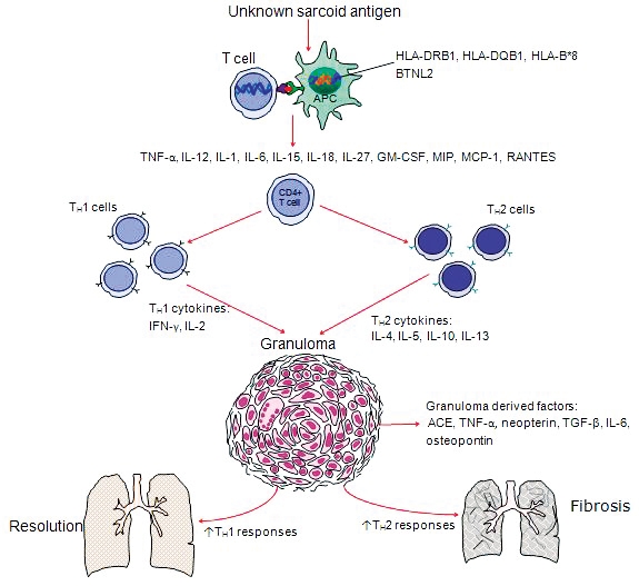
ACE = angiotensin-converting enzyme; GM-CSF = granulocyte-macrophage colony-stimulating factor; HLA = human leucocyte antigen; IFN = interferon; IL = interleukin; MCP = monocyte chemo-attractant protein; MIP = macrophage inflammatory protein; RANTES = regulated on activation, normal T cell expressed and secreted; TGF = transforming growth factor; TH = T helper; TNF = tumour necrosis factor. The inciting agent may be cleared leaving behind an undegradable antigen or it may cause a cross-reacting response with a self-antigen with strong evidence for HLA and BTNL2 alleles contributing to susceptibility. The processed antigen is presented by HLA molecules on antigen-presenting cells to sarcoid antigen-specific T cells, which express restricted Vα and Vβ regions of the T cell receptor. Ligation of co-stimulatory signals such as CD80 and CD86 on the APC binding to CD28 on the T cell is required for full T cell activation. This leads to release of a variety of cytokines and chemokines activating CD4+ T cells to become active TH1 cells that secrete IL-2 and IFN-γ that drives granulomatous inflammation yet promotes resolution. Contributions of TH2 cells and macrophage-derived cytokines, namely TGF-β, lead to development of fibrosis and chronic disease. * Diagram modified from Iannuzzietal2 and reproduced with permission from Ahmadzai.6
Box 2 – Criteria for initiating corticosteroid therapy in patients with sarcoidosis*
|
|
|||||||||||||||
|
|
|||||||||||||||
|
|
|||||||||||||||
|
* American Thoracic Society, European Respiratory Society and World Association of Sarcoidosis and other Granulomatous Disorders.1 |
|||||||||||||||
Box 3 – Pharmacological treatment for pulmonary sarcoidosis
|
Intervention |
Level of evidence* |
Comment |
|||||||||||||
|
|
|||||||||||||||
|
Inhaled corticosteroids |
A |
Conflicting results from a small number of trials but essentially ineffective for control of pulmonary disease; may be effective for cough symptom control43-45 |
|||||||||||||
|
Oral corticosteroids |
B |
No robust placebo-controlled RCT; one non-randomised trial46 |
|||||||||||||
|
(Hydroxy)chloroquine |
B |
One small RCT in pulmonary disease; may be useful as a steroid-sparing agent47 |
|||||||||||||
|
Methotrexate |
A |
Steroid-sparing agent: one small RCT in pulmonary disease48 and several case series in extra-pulmonary disease49 |
|||||||||||||
|
Azathioprine |
B |
||||||||||||||
|
Mycophenolate |
C |
||||||||||||||
|
Leflunomide |
B |
||||||||||||||
|
Infliximab |
A |
Two RCTs demonstrating small effect in pulmonary disease; underpowered to demonstrate effect on extra-pulmonary manifestations56,57 |
|||||||||||||
|
Adalimumab |
B |
One small RCT in skin disease and case series pulmonary disease; lower rates of immunogenicity and allergy symptoms58-60 |
|||||||||||||
|
|
|||||||||||||||
|
RCT = randomised controlled trial. * Level A: at least one double-blind, placebo-controlled trial with positive results with one or more case series supporting the results. Level B: majority of case series showing positive results. Level C: case series with mixed reports of effectiveness, or only a small number of cases reported. Levels of evidence as proposed by Guyatt G, Gutterman D, Baumann MH, et al. Grading strength of recommendations and quality of evidence in clinical guidelines: report from an American College of Chest Physicians task force. Chest 2006; 129: 174-181. |
|||||||||||||||
Competing interests
No relevant disclosures.
References
- Statement on Sarcoidosis. Joint Statement of the American Thoracic Society (ATS), the European Respiratory Society (ERS) and the World Association of Sarcoidosis and other Granulomatous Disorders (WASOG), adapted by the ATS Board of Directors and by the ERS Executive Committee. Am J Respir Crit Care Med 1999; 160: 736-755.
- Iannuzzi MC, Rybicki BA, Teirstein AS. Sarcoidosis. N Engl J Med 2007; 357: 2153-2165.
- Gillman A, Steinfort C. Sarcoidosis in Australia. Intern Med J 2007; 37: 356-359.
- Newman LS, Rose CS, Bresnitz EA, et al. A case control etiologic study of sarcoidosis - environmental and occupational risk factors. Am J Respir Crit Care Med 2004; 170: 1324-1330.
- Rossman MD, Kreider ME. Lesson learned from ACCESS (A Case Controlled Etiologic Study of Sarcoidosis). Proc Am Thorac Soc 2007; 4: 453-456.
- Ahmadzai H, Wakefield D, Thomas PS. The potential of the immunological markers of sarcoidosis in exhaled breath and peripheral blood as future diagnostic and monitoring techniques. Inflammopharmacology 2011; 19: 55-68.
- Spagnolo P, Grunewald J. Recent advances in the genetics of sarcoidosis. J Med Genet 2013; 50: 290-297.
- Gupta D, Agarwal R, Agarwal AN, Jindal SK. Molecular evidence for the role of mycobacteria in sarcoidosis: a meta-analysis. Eur Respir J 2007; 30: 508-516.
- Chen ES, Wahlström J, Song Z, et al. T cell responses to mycobacterial catalase-peroxidase profile a pathogenic antigen in systemic sarcoidosis. J Immunol 2008; 181: 8784-8796.
- Oswald-Richter K, Beachboard DC, Zhan X, et al. Multiple mycobacterial antigens are targets of the adaptive immune response in pulmonary sarcoidosis. Respir Res 2010; 11: 161.
- Ahmadzai H, Cameron B, Chui JJ, et al. Peripheral blood responses to specific antigens and CD28 in sarcoidosis. Respir Med 2012; 106: 701-709.
- Rappl G, Pabst S, Riemann D, et al. Regulatory T cells with reduced repressor capacities are extensively amplified in pulmonary sarcoid lesions and sustain granuloma formation. Clin Immunol 2011; 140: 71-83.
- Taflin C, Miyara M, Nochy D, et al. FoxP3+ regulatory T cells suppress early stages of granuloma formation but have little impact on sarcoidosis lesions. Am J Pathol 2009; 174: 497-508.
- Facco M, Cabrelle A, Teramo A, et al. Sarcoidosis is a Th1/Th17 multisystem disorder. Thorax 2011; 66: 144-150.
- Ten Berge B, Paats MS, Bergen IM, et al. Increased IL-17A expression in granulomas and in circulating memory T cells in sarcoidosis. Rheumatology 2012; 51: 37-46.
- Ahmadzai H, Thomas PS, Wakefield D. Laboratory investigations and immunological testing in sarcoidosis. In: Eishi Y, editor. Sarcoidosis. Intech Open Access Publishing, 2013; pp 201-237.
- Loke WS, Herbert C, Thomas PS. Sarcoidosis: immunopathogenesis and immunological markers. Int J Chronic Dis 2013; 2013: 928601.
- Bargagli E, Mazzi A, Rottoli P. Markers of inflammation in sarcoidosis: blood, urine, BAL, sputum, and exhaled gas. Clin Chest Med 2008; 29: 445-458.
- Baughman RP, Culver DA, Judson MA. A concise review of pulmonary sarcoidosis. Am J Respir Crit Care Med 2011; 183: 573-581.
- Baughman RP, Teirstein AS, Judson MA, et al. Clinical characteristics of patients in a case control study of sarcoidosis. Am J Respir Crit Care Med 2001; 164: 1885-1889.
- Moller DR. Pulmonary fibrosis of sarcoidosis: new approaches, old ideas. Am J Respir Cell Mol Biol 2003; 29(3 Suppl): S37-S41.
- Prasse A, Katic C, Germann M, et al. Phenotyping sarcoidosis from a pulmonary perspective. Am J Respir Crit Care Med 2008; 177: 330-336.
- Criado E, Sanchez M, Ramirez J, et al. Pulmonary sarcoidosis: typical and atypical manifestations at high-resolution CT with pathologic correlation. Radiographics 2010; 30: 1567-1586.
- Zappala CJ, Desai SR, Copley SJ, et al. Accuracy of individual variables in the monitoring of long-term change in pulmonary sarcoidosis as judged by serial high-resolution CT scan data. Chest 2014; 145: 101-107.
- Walsh SL, Wells AU, Sverzellati N, et al. An integrated clinicoradiological staging system for pulmonary sarcoidosis: a case-cohort study. Lancet Respir Med 2014; 2: 123-130.
- Mostard RL, van Kroonenburgh MJ, Drent M. The role of the PET scan in the management of sarcoidosis. Curr Opin Pulm Med 2013; 19: 538-544.
- Bilaçeroğlu S, Perim K, Günel O, et al. Combining transbronchial aspiration with endobronchial and transbronchial biopsy in sarcoidosis. Monaldi Arch Chest Dis 1999; 54: 217-223.
- Tremblay A, Slather DR, MacEachern P, et al. A randomized controlled trial of standard vs endobronchial ultrasonography-guided transbronchial needle aspiration in patients with suspected sarcoidosis. Chest 2009; 136: 340-346.
- Wahlström J, Berlin M, Sköld CM, et al. Phenotypic analysis of lymphocytes and monocytes/macrophages in peripheral blood and bronchoalveolar lavage fluid from patients with pulmonary sarcoidosis. Thorax 1999; 54: 339-346.
- Wong M, Yasufuku K, Nakajima T, et al. Endobronchial ultrasound: new insight for the diagnosis of sarcoidosis. Eur Respir J 2007; 29: 1182-1186.
- Ishikawa M, Nagai S, Akiguchi I. Neurosarcoidosis. In: Baughman RP, editor. Lung biology in health and disease - sarcoidosis. New York: Taylor and Francis, 2006; pp 505-514.
- Nunes H, Freynet O, Naggara N, et al. Cardiac sarcoidosis. Semin Respir Crit Care Med 2010; 31: 428-441.
- Habersberger J, Manins V, Taylor AJ. Cardiac sarcoidosis. Intern Med J 2008; 38: 270-277.
- Ohira H, Tsujino I, Ishimaru S, et al. Myocardial imaging with 18F-fluoro-2-deoxyglucose positron emission tomography and magnetic resonance imaging in sarcoidosis. Eur J Nucl Med Mol Imaging 2008; 35: 933-941.
- Patel MR, Cawley PJ, Heitner JF, et al. Detection of myocardial damage in patients with sarcoidosis. Circulation 2009; 120: 1969-1977.
- Lee NJ, Lee B, Litt H. Cardiac MRI vs. myocardial 18F-FDG PET/CT in patients with clinical concern for cardiac sarcoid. J Cardiovasc Magn Reson 2015; 17(Suppl 1): O30.
- Mantini N, Williams B Jr, Stewart J, et al. Cardiac sarcoid: a clinician’s review on how to approach the patient with cardiac sarcoid. Clin Cardiol 2012; 35: 410-415.
- Palmero V, Sulica R. Sarcoidosis-associated pulmonary hypertension: assessment and management. Semin Respir Crit Care Med 2010; 31: 494-500.
- Iannuzzi MC, Fontana JR. Sarcoidosis: clinical presentation, immunopathogenesis, and therapeutics. JAMA 2011; 305: 391-399.
- Beegle SH, Barba K, Gobunsuy R, Judson MA. Current and emerging pharmacological treatments for sarcoidosis: a review. Drug Des Dev Ther 2013; 7: 325-338.
- Paramothayan NS, Lasserson TJ, Jones PW. Corticosteroids for pulmonary sarcoidosis. Cochrane Database Syst Rev 2005; 2: CD001114.
- Lazar CA, Culver DA. Treatment of sarcoidosis. Semin Respir Crit Care Med 2010; 31: 501-518.
- Pietinalho A, Tukiainen P, Haahtela T, et al. Oral prednisolone followed by inhaled budesonide in newly diagnosed pulmonary sarcoidosis: a double-blind, placebo-controlled multicenter study. Finnish Pulmonary Sarcoidosis Study Group. Chest 1999; 116: 424-431.
- Alberts C, van der Mark TW, Jansen HM. Inhaled budesonide in pulmonary sarcoidosis: a double-blind, placebo-controlled study. Dutch Study Group on Pulmonary Sarcoidosis. Eur Respir J 1995; 8: 682-688.
- Milman N, Graudal N, Grode G, Munch E. No effect of high-dose inhaled steroids in pulmonary sarcoidosis: a double-blind, placebo-controlled study. J Intern Med 1994; 236: 285-290.
- Gibson GJ, Prescott RJ, Muers MF, et al. British Thoracic Society Sarcoidosis study: effects of long term corticosteroid treatment. Thorax 1996; 51: 238-247.
- Baltzan M, Mehta S, Kirkham TH, Cosio MG. Randomized trial of prolonged chloroquine therapy in advanced pulmonary sarcoidosis. Am J Respir Crit Care Med 1999; 160: 192-197.
- Baughman RP, Winget DB, Lower EE. Methotrexate is steroid sparing in acute sarcoidosis: results of a double blind, randomized trial. Sarcoidosis Vasc Diffuse Lung Dis 2000; 17: 60-66.
- Cremers JP, Drent M, Bast A, et al. Multinational evidence-based World Association of Sarcoidosis and Other Granulomatous Disorders recommendations for the use of methotrexate in sarcoidosis: integrating systematic literature research and expert opinion of sarcoidologists worldwide. Curr Opin Pulm Med 2013; 19: 545-561.
- Pacheco Y, Marechal C, Marechal F, et al. Azathioprine treatment of chronic pulmonary sarcoidosis. Sarcoidosis 1985; 2: 107-113.
- Müller-Quernheim J, Kienast K, Held M, et al. Treatment of chronic sarcoidosis with an azathioprine/prednisolone regimen. Eur Respir J 1999; 14: 1117-1122.
- Androdias G, Maillet D, Marignier R, et al. Mycophenolate mofetil may be effective in CNS sarcoidosis but not in sarcoid myopathy. Neurology 2011; 76: 1168-1172.
- Kouba DJ, Mimouni D, Rencic A, Nousari HC. Mycophenolate mofetil may serve as a steroid-sparing agent for sarcoidosis. Br J Dermatol 2003; 148: 147-148.
- Baughman RP, Lower EE. Leflunomide for chronic sarcoidosis. Sarcoidosis Vasc Diffuse Lung Dis 2004; 21: 43-48.
- Sahoo DH, Bandyopadhyay D, Xu M, et al. Effectiveness and safety of leflunomide for pulmonary and extrapulmonary sarcoidosis. Eur Respir J 2011; 38: 1145-1150.
- Baughman RP, Drent M, Kavuru M, et al. Infliximab therapy in patients with chronic sarcoidosis and pulmonary involvement. Am J Respir Crit Care Med 2006; 174: 795-802.
- Rossman MD, Newman LS, Baughman RP, et al. A double blinded, randomized, placebo-controlled trial of infliximab in subjects with active pulmonary sarcoidosis. Sarcoidosis Vasc Diffuse Lung Dis 2006; 23: 201-208.
- Pariser RJ, Paul J, Hirano S, et al. A double-blind, randomized, placebo-controlled trial of adalimumab in the treatment of cutaneous sarcoidosis. J Am Acad Dermatol 2013; 68: 765-773.
- Kamphuis LS, Lam-Tse WK, Dik WA, et al. Efficacy of adalimumab in chronically active and symptomatic patients with sarcoidosis. Am J Respir Crit Care Med 2011; 184: 1214-1216.
- Milman N, Graudal N, Loft A, et al. Effect of the TNF-α inhibitor adalimumab in patients with recalcitrant sarcoidosis: a prospective observational study using FDG-PET. Clin Respir J 2012; 6: 238-247.
- Lennard L. Implementation of TPMT testing. Br J Clin Pharmacol 2014; 77: 704-714.
- Baughman RP, Nunes H. Therapy for sarcoidosis: evidence-based recommendations. Expert Rev Clin Immunol 2012; 8: 95-103.
- Baughman RP, Lower EE. Medical therapy of sarcoidosis. Semin Resp Crit Care Med 2014; 35: 391-406.
- Drent M, Cremers JP, Jansen TL, Baughman RP. Practical eminence and experience-based recommendations for use of TNF-α inhibitors in sarcoidosis. Sarcoidosis Vasc Diffuse Lung Dis 2014; 31: 91-107.
- Ahmadzai H, Kiernan MC, Wakefield D, Thomas PS. Treatment of refractory neurosarcoidosis with TNF-inhibitors: what lies ahead? Intern Med J 2012; 42: 232.
Linked content
-
MJA InSight: Sarcoidosis: when to avoid Dr Google
-
MJA Podcast: Professor Paul Thomas
Provenance: Not commissioned; externally peer reviewed.
Splenic injury in severe cases of the zoonoses Q fever and rickettsial infection: diagnostic challenges
Ashleigh Drury, Philippa Harrison, Aiveen Bannan
Health and economic benefits of improving pre‐hospital identification of stroke in Australian women: a modelling study
Thomas Gadsden, Lei Si, Emily R Atkins, Cheryl Carcel, Xia Wang, Stephen Jan, Mark Woodward, Laura E Downey
Updating the diagnosis and management of elevated serum ferritin levels in the era of routine ferritin testing of blood donors by Australian Red Cross Lifeblood
Gary D Zhang, James Chen, Daniel M Johnstone, Martin B Delatycki, Katie Allen, John K Olynyk
Early cardiovascular collapse after envenoming by snakes in Australia, 2005–2020: an observational study (ASP‐31)
Geoffrey K Isbister, Katherine Z Isoardi, Angela L Chiew, Shane Jenkins, Nicholas A Buckley
Updated recommendations for warfarin reversal in the setting of four‐factor prothrombin complex concentrate
Danielle Robinson, James McFadyen, Eileen Merriman, Chee Wee Tan, Ross Baker, Huyen Tran
Updating the diagnosis and management of iron deficiency in the era of routine ferritin testing of blood donors by Australian Red Cross Lifeblood
Gary D Zhang, Daniel Johnstone, Michael F Leahy, John K Olynyk