


Clinical record
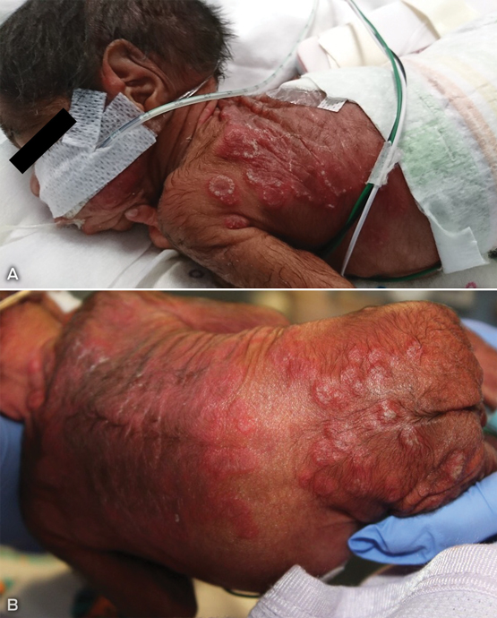
Following chorioamnionitis, a male infant from a remote Indigenous community was born by vaginal breech delivery at 26 weeks’ gestation. On Day 5, indurated, vesicular and erythematous lesions were noted on extensive areas of the back (Box, A and B). Blue light (450–475 nm) phototherapy had been commenced on Day 1 for jaundice. Further skin lesions subsequently arose on the lips, ears and scalp, and surface scale developed on established plaques. There was no major systemic disturbance.
Considered differential diagnoses were neonatal lupus erythematosus, cutaneous or systemic bacterial or fungal infection and malignant or infiltrative disorders. After a skin biopsy for histopathology and culture, bacterial swabs and skin scrapings, empiric intravenous antibiotics and amphotericin were commenced on Day 6.
Lesional skin histopathology revealed numerous fungal hyphae in the stratum corneum. Dermal fungal elements were also seen within folliculocentric collections of neutrophils, eosinophils and histiocytes. Skin scrapings showed branching septate fungal organisms on microscopy. Subsequent fungal culture from skin scrapings grew Trichophyton rubrum from the neonate.
Initial intravenous antimicrobials were ceased and treatment was limited to topical clotrimazole cream from Day 14, with rapid, dramatic improvement and clearance of cutaneous signs. His hospital progress was uneventful, with no recurrence of rashes. Maternal examination revealed extensive areas of tinea on forearms, neck and torso. She was treated with topical clotrimazole and subsequently topical terbinafine creams with resolution of her skin changes.
Despite tinea corporis being rarely reported in neonates,1,2 there have been four recent confirmed cases (unpublished) due to Trichophyton rubrum in infants while hospitalised in neonatal intensive care units. In all cases T. rubrum was also detected on their Indigenous mothers, who resided in remote Northern Territory communities. Chronic tinea corporis is endemic in these regions both in adults and children.3
It is speculated that the impaired barrier function of the skin of premature infants,4 immature microbiome and suboptimal immune responses allow aggressive dermatophyte spread during initial maternal-infant skin-to-skin contact,5 with subsequent atypical clinical manifestations. The long term consequences of this early exposure and infection on later immune responses to fungal organisms are uncertain.
Clinicians are encouraged to consider the diagnosis of tinea corporis even in the absence of typical marginated scaling or slow annular progression of neonatal rashes. When assessing defined rashes with scaling, the primary investigation undertaken should be skin scrapings taken from lesional edges for fungal analysis. Microscopy results are sensitive and specific and available within minutes. Subsequent fungal culture and speciation provides opportunity to identify the source of infection and direct contact and public health interventions. This is particularly relevant when treating pre-term infants from areas and populations where chronic fungal skin infections are endemic and often asymptomatic.
Lessons from practice
-
Tinea corporis is endemic in tropical and Indigenous communities and is often chronic and untreated.
-
Neonatal tinea acquired shortly after birth may present within days and display unusual clinical features, particularly in premature infants.
-
When this diagnosis is considered, neonatal tinea corporis is simply and rapidly diagnosed with scrapings and microscopy, and responds quickly to topical or systemic antifungal therapy.
-
Maternal tinea is readily treated and should be actively sought during antenatal assessments, particularly in populations where it is endemic.
Provenance: Not commissioned; externally peer reviewed.
- 1. Khoury S, Kidd S, Warren L. Neonatal tinea corporis due to a novel strain of Trichophyton rubrum. Australas J Dermatol 2015; 56: 311-312.
- 2. Atanasovski M, El Tal AK, Hamzavi F, Mehregan DA. Neonatal dermatophytosis: report of a case and review of the literature. Pediatr Dermatol 2011; 28: 185-188.
- 3. Koh KJ, Parker CJ, Ellis DH, et al. Use of terbinafine for tinea in Australian Aboriginal communities in the Top End. Australas J Dermatol 2003; 44: 243-249.
- 4. Kalia YN, Nonato LB, Lund CH, Guy RH. Development of skin barrier function in premature infants. J Invest Dermatol 1998; 111: 320-326.
- 5. Hubbard JM, Gattman KR. Parent–infant skin-to-skin contact following birth: history, benefits, and challenges. Neonatal Netw 2017; 36: 89-97.
No relevant disclosures.