




Medical education
Clinical skills
Volume 207 - Issue 2
How to perform the ankle brachial index test in clinical practice
Authors: Nicole M Organ and Catherine Harrison
Med J Aust 2017; 207 (2): 60-61. || doi: 10.5694/mja17.00112
Published online: 17 July 2017
Published online: 17 July 2017
A bedside test to assist clinicians in diagnosing peripheral arterial disease
A bedside test to assist clinicians in diagnosing peripheral arterial disease
The ankle brachial index (ABI), a useful bedside test, is the ratio of the highest systolic blood pressure in the lower extremity vessels at the ankle divided by the highest systolic blood pressure of the two brachial arteries. An abnormal ABI measurement is a sign of peripheral arterial disease (PAD) and also of increased risk of atherosclerotic disease elsewhere, including cardiac events, stroke and death. This increased risk is independent of a pre-existing diagnosis of vascular disease.1-3
Performing an ABI test at the bedside requires a hand-held continuous wave Doppler probe (8–10 MHz), ultrasound gel and a manual sphygmomanometer. A diagnostic vascular laboratory can provide more formal ABI testing where indicated, by performing Doppler waveform analysis, exercise testing and toe pressures. Such formal testing is important for diagnostic purposes or where bedside tests are equivocal.3,4
To perform an ABI test, the patient should be lying comfortably on an examination bed, with both head and feet resting on the bed. It is preferable that patients avoid smoking for at least 2 hours before the test — as smoking, especially within 10 minutes of the test, has been shown to decrease the ABI.5 Many publications recommend that the patient rest in this position for at least 5 minutes before obtaining measurements.3 The sphygmomanometer cuff width should be greater than 40% of the limb circumference.3
When measuring the systolic pressure at the ankle, the cuff is put around the ankle allowing room for the Doppler probe to be placed over the dorsalis pedis and posterior tibial arteries (Box 1 and Box 2). The Doppler probe should be placed at a 45–60° angle to the skin, using coupling gel between the probe and the skin. It is important to avoid excessive pressure on the probe as this would compress the artery, and to use small movements of the probe to find the strongest Doppler signal (eg, rolling slightly medial or lateral and changing the angle of the probe).3
The blood pressure cuff has to be inflated until the Doppler signal disappears completely and then inflate it for a further 20 mmHg. Afterwards, the cuff has to be slowly deflated until the signal returns. The pressure at which it first returns is the systolic pressure that should be recorded for the site. If the inflation pressure exceeds 250 mmHg, the cuff can be deflated and the vessel should be recorded as incompressible.3
All vessels should be assessed in the same manner, including both brachial arteries with the higher brachial systolic value used. If there is a variation of more than 10 mmHg, repeat the test to rule out the “white coat” effect.3 A repeatable difference of more than 10 mmHg between the two brachial systolic pressures suggests haemodynamically significant atherosclerotic disease of the arteries of the arm with the lower arterial pressure. Dividing the higher of the two ankle pressures for each limb by the higher of the brachial pressures provides the ABI.1,3 The interobserver variability of ABI testing is around 10% (range 4.7–13%).3
Nevertheless, there are some precautions that need to be taken when performing an ABI test. Wounds at the site where the cuff would normally be placed should be covered. If the wounds are extensive, they may preclude performance of the test. Patients who are unable to tolerate inflated cuff pressure or cannot lie still supine are not suitable for the test. If there is a bypass graft under the area where the cuff would be placed, ABI testing should not be attempted.3
A guide to the interpretation of ABI measurements is provided in Box 3.6,7 An abnormal test result does not always indicate a need for further testing or revascularisation. It does, however, identify those patients at a higher risk of cardiovascular disease and, in patients with diabetes, a higher risk of amputation. An abnormal ABI measurement should be a trigger for consideration of intensive medical treatment of vascular risk factors, diabetic foot care and referral to a vascular specialist where clinically indicated.2,3
An ABI measurement greater than 0.9 remains an accepted cut-off for a normal value. Specificity is high (83–99%), but sensitivity is lower (69–79%). Both are influenced by the pre-test probability. If an ABI cut-off of 1.0 or less is used, the sensitivity has been reported to be as high as 100%. Sensitivity may also be improved if the patient is referred for exercise testing in a vascular lab.3 As outlined by the American Heart Association statement, the positive predictive value of ABI testing varies with the diagnostic criteria used for diagnosing PAD and the ABI cut-off used — the positive predictive value ranges from 0.7 to 1.0, and the negative predictive value ranges from 0.74 to 0.95.3 If claudication symptoms are present, a resting ABI test may not be as accurate in diagnosing PAD and further testing may be required.8
Abnormally high or incompressible ABI may occur where there is severe vessel wall calcification, as in diabetes mellitus, medial calcinosis and end-stage renal disease. Calcification does not always coexist with occlusive or stenotic disease. It is, however, an indicator of increased risk of cardiovascular events, stroke and mortality.3
Box 1 – The posterior tibial pulse is found posterior to the medial malleolus
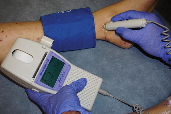
The image shows the placement of the cuff and Doppler probe and the Doppler waveform — audible Doppler is usually used in a clinical setting.
Box 2 – The dorsalis pedis pulse is found lateral to the tendon of extensor hallucis longus
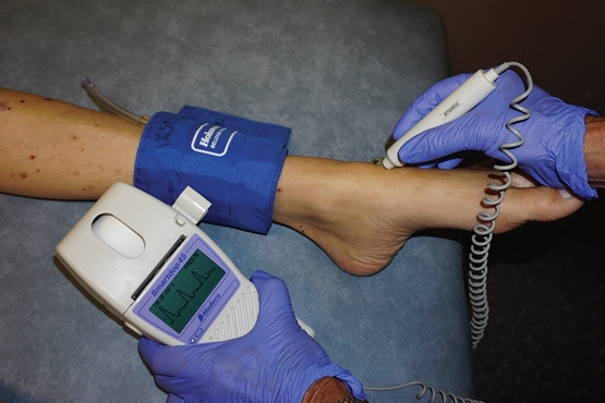
The image shows the placement of the cuff and Doppler probe and the Doppler waveform — audible Doppler is usually used in a clinical setting.
Box 3 – Guide to correlation of the ankle brachial index (ABI) and clinical status; there is significant clinical variation in symptoms3,7
|
ABI |
Clinical presentation |
||||||||||||||
|
|
|||||||||||||||
|
> 1.4 |
Vessel calcification |
||||||||||||||
|
1–1.4 |
Normal, asymptomatic |
||||||||||||||
|
> 0.9 |
Likely normal; however, if clinical suspicion, perform other tests (eg, exercise ABI or duplex US) |
||||||||||||||
|
≤ 0.9 |
PAD |
||||||||||||||
|
0.5–0.9 |
Intermittent claudication |
||||||||||||||
|
< 0.5 |
Severe disease |
||||||||||||||
|
< 0.3 |
Critical ischaemia |
||||||||||||||
|
|
|||||||||||||||
|
PAD = peripheral arterial disease. US = ultrasound. |
|||||||||||||||
Competing interests
No relevant disclosures.
References
- Yao ST, Hobbs JT, Irvine WT. Ankle systolic pressure measurements in arterial disease affecting the lower extremities. Br J Surg 1969; 56: 676-679.
- Mostaza JM, Gonzalez-Juanatey JR, Castillo J, et al. Prevalence of carotid stenosis and silent myocardial ischemia in asymptomatic subjects with a low ankle-brachial index. J Vasc Surg 2009; 49: 104-108.
- Aboyans V, Criqui MH, Abraham P, et al, American Heart Association Council on Peripheral Vascular Disease, Council on Epidemiology and Prevention, Council on Clinical Cardiology, Council on Cardiovascular Nursing, Council on Cardiovascular Radiology and Intervention, Council on Cardiovascular Surgery and Anesthesia. Measurement and interpretation of the ankle-brachial index: a scientific statement from the American Heart Association. Circulation 2012; 126: 2890-2909.
- Dachun X, Jue L, Liling Z, et al. Sensitivity and specificity of the ankle-brachial index to diagnose peripheral artery disease: a structured review. Vasc Med 2010; 15: 361-369.
- Yataco AR, Gardner AW. Acute reduction in ankle/brachial index following smoking in chronic smokers with peripheral arterial occlusive disease. Angiology 1999; 50: 355-360.
- Bailey MA, Griffin KJ, Scott DJ. Clinical assessment of patients with peripheral arterial disease. Semin Intervent Radiol 2014; 31: 292-299.
- National Institute for Health and Care Excellence. Peripheral arterial disease: diagnosis and management. Clinical guideline CG147. London: NICE; 2012. https://www.nice.org.uk/guidance/cg147 (accessed May 2017).
- Crawford F, Welch K, Andras A, Chappell FM. Ankle brachial index for the diagnosis of lower limb peripheral arterial disease. Cochrane Database Syst Rev 2016; 9: CD010680.
Provenance: Commissioned; externally peer reviewed.
The epidemiology of haemodialysis catheter infections in Australia, 2016–20: a prospective cohort study
Benjamin Lazarus, Kevan R Polkinghorne, Martin P Gallagher, Jayson Catiwa, Nicholas A Gray, Sarah Coggan, Kathryn R Higgins, Girish Talaulikar, Stephen P McDonald AM, Sradha Kotwal
30/60/90 National stroke targets and stroke unit access for all Australians: it's about time
For the 30/60/90 National Stroke Targets Taskforce
A summary of the 2023 Society of Obstetric Medicine of Australia and New Zealand (SOMANZ) hypertension in pregnancy guideline
Renuka Shanmugalingam, Helen L Barrett, Amanda Beech, Lucy Bowyer, Tim Crozier, Amanda Davidson, Marloes Dekker Nitert, Kerrie Doyle, Luke Grzeskowiak, Nicole Hall, Hicham Ibrahim Cheikh Hassan, Annemarie Hennessy, Amanda Henry, David Langsford, Vincent WS Lee, Zachary Munn, Michael J Peek, Joanne M Said, Helen Tanner, Rachel Taylor, Meredith Ward, Jason Waugh, Linda LY Yen, Ellie Medcalf, Katy JL Bell, Deonna Ackermann, Robin Turner, Angela Makris
Cough in Children and Adults: Diagnosis, Assessment and Management (CICADA). Summary of an updated position statement on chronic cough in Australia
David A Prentice
PEG‐GCSF‐induced aortitis in a patient with breast cancer: distinguishing between infective and immune‐mediated aortitis
Luigi Zolio, Prudence A Francis, Nava Ferdowsi
Catheter‐related superior vena cava syndrome: an increasing problem
John J Harvey, John I Vrazas, Elhamy Bekhit, Chris Barnes, Philip J Robinson